Truncal valve insufficiency requiring truncal valve surgery remains a major risk factor for post-operative mortality. Reference Russell, Pasquali and Jacobs1 Mechanical valve replacement is unsuitable for neonates or young infants because of patient–prosthesis size mismatch and anticoagulation difficulties. Reference Sharabiani, Dorobantu and Mahani2 Therefore, it is desirable to perform truncal valve repair sparing native leaflets as much as possible. However, native leaflet-sparing truncal valve repair, especially in neonates, could be challenging because leaflet tissues are often severely dysplastic and fragile. Recently, excellent short-term results for autologous pericardial three-leaflet reconstruction were reported in children. Reference Baird, Cooney, Chávez, Sleeper, Marx and del Nido3 However, to our knowledge, no report exists of three-leaflet reconstruction for truncal valve insufficiency in neonates. Herein, we report a successful case of three-leaflet reconstruction for severe truncal valve insufficiency in a neonate.
Case report
Written informed consent for publication of patient information and images were not obtained to protect patient identity.
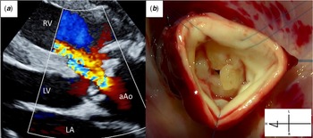
Postnatal echocardiography of a boy, born at 39 weeks of gestation and weighing 3692 g, revealed type 2 persistent truncus arteriosus with severe truncal valve regurgitation and uncontrollable heart failure. He underwent emergent surgical intervention on the second day of life. Pre-operative echocardiography revealed severe truncal valve regurgitation and moderate stenosis (peak pressure gradient = 45 mmHg). The truncal annulus diameter was 14.0 mm (z-value for aortic valve = 8.4) (Fig 1a).
Pre-operative two-dimensional transthoracic echocardiographic image (a) and operative findings (b) demonstrating severe truncal valve regurgitation and moderate stenosis. aAo=ascending aorta; H=head side; L=left side; LA=left atrium; LV=left ventricle; R=right side; RV= right ventricle.

The procedure was performed using median sternotomy and standard cardiopulmonary bypass. Autologous pericardium was harvested to reconstruct three cusps. It was treated with 0.6% glutaraldehyde solution for 5 minutes, followed by rinsing in a saline solution for 5 × 3 minutes each.
After clamping the common arterial trunk, it was transected 12 mm above the valve annulus. The truncal valve was quadricuspid, and all leaflets had severe dysplastic and myxomatous changes with fused commissures. Since the geometric height of each leaflet was short and an adequate coaptation zone was not formed, there was a large gap at the centre (Fig 1b). Therefore, tricuspidization with leaflet resection and annuloplasty would not be adequate. Native leaflet-sparing Truncal valve repair (TrVR), such as leaflet extension, was considered ineffective because severe dysplasia and myxomatous changes extended to the base of all valve leaflets. Hence, three-leaflet reconstruction using autologous pericardium was decided.
In neonates, it is difficult to measure each intercommissural width using the original-sizing device such as that in the Ozaki technique. Reference Baird, Cooney, Chávez, Sleeper, Marx and del Nido3 We measured the truncal annulus diameter using a standard sizing device and an original template that corresponded to the measured size. We created three neoleaflets of the same size. In the original template, the width reflected along the free margin length was approximately 1.4 times that of one-third of the circumference of the truncal annulus; further, leaflet height was designed to match the annulus diameter, which was one-third of the circumference of the truncal annulus. As a result, the coaptation height was expected to be approximately half the leaflet height.
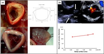
All native leaflets were excised; the annulus diameter measured 13 mm. The new leaflets were trimmed using an original template of 13-mm annular size from the glutaraldehyde-treated autologous pericardium. We sutured three pericardial patches to the native annulus with 7-0 polypropylene running sutures. Three equal new leaflets were created based on the intercoronary commissure. The new leaflet was sewn to the top of the commissure and was designed to have deep coaptation that reached up to the same horizontal plane as the commissure. The top of each commissure was secured with additional sutures to adjust the coaptation height. The intraoperative view and new leaflet design are shown in Figure 2a. Finally, ventricular septal defect closure and right ventricular outflow tract reconstruction were performed. Finally, aortic cross-clamp time and cardiopulmonary bypass time were 165 and 252 minutes, respectively.
(a) Three-leaflet reconstruction; intraoperative view and original leaflet template. Regarding original template details, the width reflected along the free margin length was approximately 1.4 times that of one-third of the circumference of truncal annulus; further, leaflet height was designed to match the annulus diameter, which is one-third of the circumference of the truncal annulus. (b) Two-dimensional transthoracic echocardiographic images (long- and short-axis view with diastolic phase) 1 year post-operatively demonstrating new leaflets with reduced mobility, reduced coaptation height, moderate/greater regurgitation, and mild stenosis and growth in neoaortic annular diameter. aAo=ascending aorta; C=circumference (= annulus diameter × 3.14); L=left coronary artery; LV=left ventricle; RCA=right coronary artery.

The patient’s post-operative haemodynamics was stable. Delayed sternal closure was performed on the fourth post-operative day. Dopamine and dobutamine administration was discontinued by the sixth post-operative day. He was extubated on the 10th post-operative day and subsequently transferred from the ICU to the general ward on the 18th post-operative day. Echocardiography at discharge revealed mild regurgitation and no stenosis (peak pressure gradient = 10 mmHg). The patient steadily gained weight, and the neoaortic annulus diameter had grown over time because of somatic growth (Fig 2b). One year post-operatively, echocardiography revealed that each neoaortic leaflet became less flexible due to sclerosis and calcification, and the coaptation height was reduced because of annular growth. This was accompanied by moderate regurgitation and mild stenosis (peak pressure gradient = 19 mmHg), but the patient’s general condition was stable (Fig 2b).
Discussion
Overall, 15–25% newborns with truncus arteriosus will show significant truncal valve insufficiency. Reference Kaza, Burch and Pinto4 Several studies have reported pre-operative truncal valve insufficiency as a risk factor for mortality in neonates. Reference Kaza, Burch and Pinto4,Reference Henaine, Azarnoush and Belli5 Neonates with moderate/greater truncal valve insufficiency often present with unstable haemodynamics, leaving no choice but to surgically intervene in the truncal valve. The vital factor in the treatment of truncal valve insufficiency is left ventricular function stabilisation, resulting in haemodynamic improvement and undisturbed growth. However, concomitant truncal valve repair in neonates is most challenging and has been associated with higher mortality.
Fujita et al obtained good results from leaflet base-preserving truncal valve repair with ethanol-treated autologous pericardium on 1-year-old and 4-month-old patients. Reference Fujita, Yamagishi and Takako6 However, there are cases wherein it is difficult to preserve the native leaflets because of dysplastic and fragile tissues in neonates with severe truncal valve insufficiency. Conversely, Baird et al reported excellent short-term outcomes from three-leaflet reconstruction for congenital aortic and truncal valve insufficiency. Reference Baird, Cooney, Chávez, Sleeper, Marx and del Nido3 Unfortunately, the main subjects of this study were older children and no mention of neonates was made with regard to three-leaflet reconstruction.
In neonates, three-leaflet reconstruction is a temporary solution providing time to identify more long-term solutions. In our case, the truncal annulus maintained its growth potential as in Baird’s report Reference Baird, Cooney, Chávez, Sleeper, Marx and del Nido3 although ours involved a short-term follow-up period. To our knowledge, no report exists of three-leaflet reconstruction for truncal valve insufficiency in neonates. Our procedure would be a life-saving technique for neonates with severe truncal valve insufficiency whose native leaflets cannot be preserved and a useful bridging technique to safe valve replacement.
Our procedure for neonates may cause worse leaflet sclerosis/calcification and earlier recurrence of regurgitation compared with other Reference Baird, Cooney, Chávez, Sleeper, Marx and del Nido3 age groups and may require reintervention. Factors possibly contributing to this include the greater promotion of sclerosis/calcification in glutaraldehyde-treated pericardium in neonates, the larger growth range in truncal annular diameter in neonates somatically growing to infants compared with in other age groups, and the relatively small geometric height of the leaflets created by truncal valve repair compared with the grown annular diameter. Therefore, further technical ingenuity is needed in the future, such as the use of ethanol-treated pericardium as reported by Fujita Reference Fujita, Yamagishi and Takako6 and leaflet designs with larger geometric heights that can tolerate mild stenosis.
In summary, although we need to evaluate more cases, three-leaflet reconstruction for neonates with severe truncal valve insufficiency might be effective as a life-saving procedure while maintaining the growth potential of the truncal annulus; however, there still are problems such as the need for reintervention in a relatively short period of time; hence, further technical ingenuity is required in the future.
Acknowledgements
The authors would like to thank all staffs in the division of paediatric cardiology, Mt. Fuji Shizuoka Children’s Hospital for providing us with accurate echocardiographic data.
Financial support
This research received no specific grant from any funding agency, commercial or not-for-profit sectors.
Conflicts of interest
None.
Ethical standards
This article does not contain any studies with human participants performed by any of the authors.