Introduction
Human infection with Opisthorchis viverrini infection is one of the most serious public health problems in Thailand. It has been estimated that at least 8.6 million people are infected by this parasite (Nithikathkul, Reference Nithikathkul2000; Nithikathkul & Puapairoj, Reference Nithikathkul and Puapairoj2002). Patients with chronic infection in highly endemic areas may suffer from cholangitis, cholangiohepatitis, liver abscess and cholangiocarcinoma (Tansurat, Reference Tansurat1971; Uptham et al., Reference Upatham, Brockelman, Viyanant, Lee, Kaengraeng and Prayoonwiwat1985). Human transmission of the disease to man is achieved through the consumption of raw freshwater cyprinid fish containing metacercariae of O. viverrini (Sadun, Reference Sadun1955). Newly excysted parasites then migrate and develop in the biliary system of the host (Apinhasmit et al., Reference Apinhasmit, Sobhon, Saitongdee and Upatham1993). In the Chinese liver fluke, the intrahepatic bile duct, gall bladder and main bile duct are the usual locations for Clonorchis sinensis to thrive. However, in most cases of heavy infection, most flukes were found in the intrahepatic bile ducts and a few in the gall bladder (Komiya, Reference Komiya1966). In hamsters, the time from the prepatency period to the appearance of eggs in faeces following infection with 50 O. viverrini metacercariae was 35 days (Flavell et al., Reference Flavell, Flavell and Field1983). The growth and development of this parasite was studied from newly juvenile to adult stages using a scanning electron microscope (SEM) (Apinhasmit et al., Reference Apinhasmit, Sobhon, Saitongdee and Upatham1993). The present paper reports the route of migration of O. viverrini from stomach to liver and gall bladder, together with studies on worm growth and development of in an experimental host, such as the Syrian golden hamster.
Materials and methods
Opisthorchis viverrini metacercariae were obtained from naturally infected cyprinid fish from endemic areas in Khon Kaen Province, northeastern Thailand. Fish were digested in a digestive solution containing 0.25% pepsin A in 0.85% NaCl and 1.5% HCl. The solution was filtered through meshes (size 80 μm). The O. viverrini metacercariae were identified and collected under a stereomicroscope. The Syrian golden hamsters, aged 6–8 weeks, were infected with 50 O. viverrini metacercariae by intragastric intubation. Five animals were anaesthetized with diethyl ether and sacrificed at periods of 1–2 min, 30 min and at 1, 2, 3, 6, 12 and 24 h post-infection for the migration study, and at 1, 1.5, 2, 3 and 4 weeks post-infection to study worm growth and development.
After sacrifice using ether, the abdomen of each hamster was incised and the internal organs divided to study the route of worm migration. Intrahepatic bile ducts, the position of the ampulla of Vater and the terminal part of the common bile duct connection to the cystic duct, were tied at each time point (1–2 min, 30 min, 1, 2, 3, 6, 12 and 24 h) to prevent the movement of worms during the operation. The stomach, duodenum, extrahepatic common bile duct, intrahepatic bile duct, gall bladder, four lobes of the liver and three sections of small intestine were removed from the host. Each organ was dissected, washed and maintained in a 0.85% sodium chloride solution for 4–6 h. Worms which moved out of each organ were collected and counted.
Five hamsters in each group that had been infected for 1, 1.5, 2, 3, 4, 6 and 8 weeks were sacrificed. The liver and gall bladder were examined and the number of worms counted, 50% of which were dried and weighed. The remaining 50% were fixed in 10% formalin stained with carmine, then used in the developmental studies.
Results and Discussion
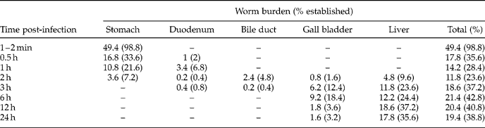
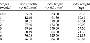
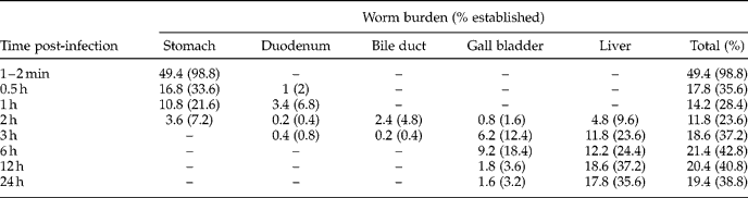
Up to 98.9% of worms were still in the stomach after 1–2 min, but after 6 h more worms had left the bile duct and migrated to the gall bladder and the liver (table 1). Worm size significantly increased as metacercariae developed into adult worms (table 2). An oral sucker appeared at the anterior end of the worm with a ventral sucker on the mid region and the characteristic oval shape of the excretory bladder at the posterior end. The intestinal bifurcation, excretory bladder, ovary and testis were present in the first week and the testis was lobulated and the uterus coiled after one and a half weeks. By two weeks, the ovary lobulated and an S-shaped excretory bladder was situated between the two testes. By the end of the third week, the extremely coiled uterus with eggs was situated between the ovary and ventral sucker. The vitelline gland was prominent. The ovary was divided into three lobes, and the testis into 4–5 lobes. The prepatent period occurred on day 24 post-infection. After four weeks, the vitelline gland was divided into eight parts, the Mehlis gland appeared and the shape and size of the body were proportional to those of 6–8-week-old worms.
Early stage biliary and intrahepatic migration of Opisthorchis viverrini within the golden hamster.
Relative increases in body size of Opisthorchis viverrini in the golden hamster from the metacercarial stage to that of the adult worm over a period of 8 weeks post-infection.
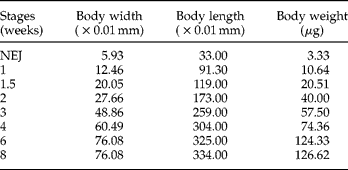
NEJ, newly excysted juvenile.
Similar studies were carried out by Komiya (Reference Komiya1966) on the growth and development of C. sinensis in guinea pigs. The ventral sucker of juveniles enlarged up to day 4 post-infection, the diameter remained the same from 5 to 7 days and the oral sucker increased in size from day 10 onwards. The rudiments of the testes became lobulated on day 5, and on day 7 testes showed lobes as in adult worms. Rudiments of the ovary also became lobulated on day 5 and the lateral loops of the uterus appeared on day 7. Egg production began on days 12–15, although vitellaria were ill-defined until day 16. The uterus was filled with eggs after 20–23 days with eggs appearing in the faeces on day 26. However, the present study has demonstrated for the first time that newly-excysted O. viverrini can migrate from the stomach to the duodenum, extrahepatic common bile duct, gall bladder and liver and worms were found in each of these organs. Mature worms may have been too large to live in the small distal bile ducts, hence causing backward migration into the larger bile duct, and into the proximal and middle regions of the bile duct. A similar finding was also found in rats infected with C. sinensis. (Hong et al., Reference Hong, Kho, Kim, Chai and Lee1993). The latter authors divided the four lobes of liver into proximal and distal portions, which were examined for the recovery and the proliferation pattern of the biliary mucosal epithelial cells at 1, 2, 5 and 15 weeks post-infection. One hundred C. sinensis metacercariae were used in each experimental rat. Worms were recovered in the distal bile duct after only two weeks post-infection, but were mainly located in the common bile duct and the right and left hepatic bile ducts. Epithelial hyperplasia developed mainly by direct and local stimulation of worms (Hong et al., Reference Hong, Kho, Kim, Chai and Lee1993). In a human autopsy study (Sithithaworn et al., Reference Sithithaworn, Tesana, Pipitgool, Kaekes, Thaiklor, Pairojkol, Sripa, Paupairoj, Sanpitak and Aranyanat1991), the distribution of O. viverrini in the right and left lobes of the liver was not significantly different with worms being found in the proximal and middle bile ducts of both lobes. Sripa & Kaewkes (Reference Sripa and Kaewkes2000) studied the localization of liver fluke antigen in hamsters and demonstrated that the O. viverrini antigen was detected in the epithelium of both the large and small bile ducts, which are not normally inhabited by adult worms. The recovery of juvenile worms in the distal bile duct in the present study probably supports the contention that the small bile duct epithelium may absorb antigen from the excretory and/or secretory products of O. viverrini, before worms move back into the large bile duct.
Further studies are required on early stage biliary, intrahepatic migration and infection of O. viverrini in the Syrian golden hamster, with particular reference to worm organogenesis.
Acknowledgements
The authors greatly appreciate the financial support received through a research grant from Khon Kaen University, and would like to extend their most heartfelt thanks to Dr Louis Royal from English Department, Faculty of Liberal Art, Huachiew Chalermprakiet University for his assistance in editing.