Implications
The vast majority of reproduction does (the mothers of rabbits raised for meat production) is housed in individual cages with a metal wire floor. Such housing is currently a topic of public debate, mainly because individual cages offer little possibility and incentive for physical and social activity. Furthermore, wire floors can harm the rabbits’ feet. Our findings suggest that semi-group housing (in which does live in groups of four for 21 days per 42-day reproduction cycle) increases bone thickness, possibly indicating increased physical activity. Plastic slatted flooring seemed to have a favourable effect on welfare by increasing foot health.
Introduction
Reproduction does (the female parent stock of rabbits raised for meat production) are commonly housed in individual wire cages of limited size (around 4000 cm2, EFSA, 2005). This type of housing is under increased scrutiny in several European countries, as it is thought to impair doe welfare. Small cages have been suggested to lead to spinal deformations (which can cause severe welfare problems such as pain and decreased mobility, Carmel, Reference Carmel2013). Their occurrence is thought to result from bone degradation because of inactivity (Drescher and Loeffler, Reference Drescher and Loeffler1996), but specific information on this topic is scarce. Another possible result of providing rabbits with only limited space is a decrease in leg bone quality because of inactivity. Short duration, high-intensity activities like running and jumping result in peak strains on the leg bones (Pearson and Lieberman, Reference Pearson and Lieberman2004) that lead to an increase in cortical bone thickness (Gordon, Reference Gordon1989). In line with this, studies on fattening rabbits (which run and jump less when housed in smaller cages, Postollec et al., Reference Postollec, Boilletot, Maurice and Michel2006) have shown that decreased cage size leads to decrease in leg bone diameter (Martrenchar et al., Reference Martrenchar, Boilletot, Cotte and Morisse2001; Buijs et al., Reference Buijs, Van Poucke, Van Dongen, Lens and Tuyttens2012). Such changes in diameter are sometimes accompanied by decreased breaking strength (Dalle Zotte et al., Reference Dalle Zotte, Princz, Metzger, Szabó, Radnai, Biró-Németh, Orova and Szendrö2009; Combes et al., Reference Combes, Postollec, Cauquil and Gidenne2010), although cortical bone thickness and breaking strength are not necessarily correlated owing to the influences of bone mineral density and microstructure (Pearson and Lieberman, Reference Pearson and Lieberman2004). The effect of space allowance on bone quality may be less expressed in breeding rabbits than in fattening rabbits, however, because the impact of activity on bone density and strength is smaller in adults than in juveniles and adolescents (Pearson and Lieberman, Reference Pearson and Lieberman2004). Furthermore, age has a direct influence on rabbit bone density and strength (Willett et al., Reference Willet, Wynnyckyj, Wang and Grynpas2011). To our knowledge, the effect of space allowance on adult rabbit bone quality has not been studied before.
One potential way of providing animals with more space and boosting their activity is to house them in groups instead of individually. Even if space allowance per animal remains constant, group housing will provide animals with a larger total area that increases the opportunity for locomotion (Postollec et al., Reference Postollec, Boilletot, Maurice and Michel2006). However, continuous group housing of reproduction does may have other undesirable effects on welfare. Aggression amongst does, and between does and other does’ kits, is common in the period around kindling and can lead to serious wounding (Andrist et al., Reference Andrist, Van den Borne, Bigler, Buchwalder and Roth2013). In addition, group housing of does can lead to pseudo pregnancy, greatly reducing the fertility of the does (Rommers et al., Reference Rommers, Boiti, De Jong and Brecchia2006). Therefore, the interest in semi-group housing is growing, in which the does are housed individually around kindling and in groups during the other part of the reproductive cycle (Maertens et al., Reference Maertens, Rommers and Jacquet2011). This is a relatively novel way of housing does, and little is known about its effects on welfare.
Another area of concern for doe welfare is the prevalence of pododermatitis. This skin condition starts with hair loss and callus formation underneath the hind feet (plantar hyperkeratosis). In severe cases the callus cracks can ultimately lead to open wounds and ulcers (Rosell and De la Fuente, Reference Rosell and De la Fuente2009; Rommers and De Jong, Reference Rommers and De Jong2011). At least in the final stage, this is a painful condition that can serve as a portal for infection, and it is a major cause for the culling of reproduction does on commercial farms (Rosell and De la Fuente, Reference Rosell and De la Fuente2013). The commonly applied wire cage flooring is known to be a major factor in the prevalence of pododermatitis, and adding a plastic footrest to such a wire floor decreases this prevalence, although not preventing it fully (Rommers and De Jong, Reference Rommers and De Jong2011; Sánchez et al., Reference Sánchez, De la Fuente and Rosell2012; Rosell and De la Fuente, Reference Rosell and De la Fuente2013). These footrests are placed on the wire floor and have slits of 11 to 12 mm wide to allow the passing of urine and faeces. The 17 to 20 mm wide slats of the footrests create much wider supports for the feet than the 2.5 to 3 mm wide wire. However, such a footrest covers only a limited part of the cage floor (usually 1000 cm2) and may thus restrict the doe to an even smaller area when attempting to avoid the wire floor. Using plastic slats throughout the cage may provide a solution for this.
In this study we compared bone quality and the prevalence of spinal deformation and pododermatitis between three different systems: individual housing on a wire floor (control group), semi-group housing on a wire floor and semi-group housing on a plastic slatted floor. Semi-group housing was expected to decrease the prevalence of spinal deformation and to improve bone quality by providing an increased possibility and incentive (social contact) for locomotion. The plastic floor was mainly expected to decrease pododermatitis, although it could also provide the does with the possibility to rest comfortably in more different postures, possibly affecting spinal deformation positively.
Method and materials
All procedures were approved by the Institute for Agricultural and Fisheries Research ethical committee for the use of animals in research. The data collectors were blinded to the treatments for all measures except pododermatitis scoring (for which the data collectors had to pick up the animals directly from their housing).
Animals and husbandry procedures
In all, 72 29-week-old Hycole does (Hycole, Marcoing, France) were allotted randomly to one of the three housing treatments, 3 days before their second kindling.
The does remained in their treatment for four consecutive reproduction cycles, although they were moved to another experimental room each cycle at the time of weaning (32 days post-kindling), where they were housed in clean cages or pens of the same type. Does were inseminated 11 days after kindling and 3 days before the next kindling non-pregnant does were exchanged with pregnant does from a spare compartment (within treatment). Conditions in the spare compartment were the same as in the experimental room (semi-group housing or individual housing on wire or plastic according to each doe’s experimental treatment, same space allowance and light and temperature regime). In total, 31 such replacements were necessary, although not all because of infertility (some does were replaced because it was not possible to regroup them with three unfamiliar conspecifics). In all, 8, 9 and 14 replacements were made in the individual cages, semi-group housing pens with wire floors and semi-group housing pens with plastic floors, respectively. Data on does that spent more than one reproduction cycle in the spare compartment (two, one and four does from individual cages, semi-group housing pens with wire floors and semi-group housing pens with plastic floors, respectively) were excluded from the analyses. In total, 48 out of the 72 does that were placed in the systems at the start of the experiment completed the experiment with four consecutive kindlings and were never replaced.
Three days prepartum, all does were moved to a different cage or pen to create new groups of unfamiliar does in each pen each cycle (animals in individual housing were moved only to avoid the incorrect attribution of the effect of moving itself to the housing system). This regrouping of all does in each cycle was performed to prevent heterogeneity in activity between groups resulting from the replacement of non-pregnant does in some of the groups. The does had ad libitum access to a commercial pelleted rabbit feed (17.0% CP, 16.2% crude fibre and 10.3 MJ digestible energy). However, non-pregnant, non-lactating animals in the spare compartment were limited to 140 g/day to prevent obesity. Water and a simple cage enrichment (a wooden gnawing block fixed to the side wall of the cage or pen) were available continuously to all does. Underpressure ventilation and a central heating system were used to achieve a stable climate (mean temperature 16.9±2.1°C s.d., mean rH 56±9% s.d.).
Housing treatments
Three housing treatments were included in the experiment: individual cages with a wire floor, semi-group pens with a wire floor and semi-group pens with a plastic slatted floor (Figure 1). In all, 24 does were housed in each housing system. BW at the start and end of the experiment did not differ significantly between the treatments (start: P=0.475, mean: 4.7±0.4 kg s.d., end: P=0.263, mean 4.8±0.3 kg s.d.). The housing types were homogeneously distributed over each experimental room.
Housing systems. Top: a semi-group pen divided into four individual units (as used from 3 days before 18 days after kindling), middle: a semi-group pen with grouped does (as used from 18 days after kindling to 3 days before next kindling), bottom: a row of individual cages.

Each individual cage (Meneghin Srl, Povegliano, Italy) had a floor area of 38×104 cm. However, from 3 days before kindling until weaning at 32 days post-kindling, an area of 29×38 cm in the front of the cage was separated from the rest of the cage and used as a nest box, as is commercial practice. Although does were able to enter their nest box freely, most spent little time in the nest box once they had kindled, and in any case the insertion of the separator segmented the available cage area. In addition to the floor level, an elevated platform of 38×30 cm was available to the does. The height of the cage varied: 28 cm in the nest box, 28 cm underneath the platform, 35 cm above the platform and 63 cm in the back of the cage. The floor and platform were made of 2.5 mm wide metal wires spaced 13 mm apart. A plastic footrest (Meneghin Srl) of 25×40 cm (17 mm wide slats separated by 12 mm wide slits) was mounted on the wire floor in the middle of the cage.
The semi-group pens (Van der Vinne, Brucht, the Netherlands) each housed four does and measured 100×200 cm. In addition, a plastic slatted platform of 200×30 cm was available to the does. Three days before each kindling three walls were added, separating each pen into four equal units of 50×100 cm plus 1500 cm2 of platform. As such, the space allowance per doe was greater in these semi-group pens than in the individual cages (6500 v. 5092 cm2 per doe, including platforms). Furthermore, the nest box (33×24×28 cm length×width×height) was external in the semi-group pens, thus not taking up any pen space. Pens were roofless and the only height limitation in these systems occurred underneath the platform mounted 30 cm above floor level. Eighteen days after kindling the three walls were removed again and all four does could use the entire pen area. However, the does were barred from access to the nest boxes from this day on (to provide a safe haven for the kits in case of doe–kit aggression) until 3 days before the next kindling. The wire floor semi-group pens were equipped with the same floor and footrest as the individual cages (1000 cm2 footrest/doe). The plastic floor of the other semi-group pens had 13 mm wide slats separated by 12 mm wide slits.
At the end of cycle 4 all does were euthanized by an i.v. injection of T-61® (Intervet International, Boxmeer, the Netherlands) preceded by anaesthesia with an i.m. injection of xylazine (Xyl-M 2%®; VMD, Arendonk, Belgium) and ketamine (Anesketin®; Eurovet, Heusden-Zolder, Belgium).
Measurements
Pododermatitis
At the end of the 1st, 2nd and 4th cycle, the hind feet of the does were scored for pododermatitis on a 0 to 4 scale, previously described by Rommers and De Jong (Reference Rommers and De Jong2011): (0: footpads intact and fully haired, 1: patch of bald, callused skin (hyperkeratosis) shorter than 2.5 cm, 2: patch of bald, callused skin of 2.5 cm or longer, 3: cracked callus and 4: wound). When the score differed between both feet of the same individual, the foot with the higher score was used.
Spinal deformation
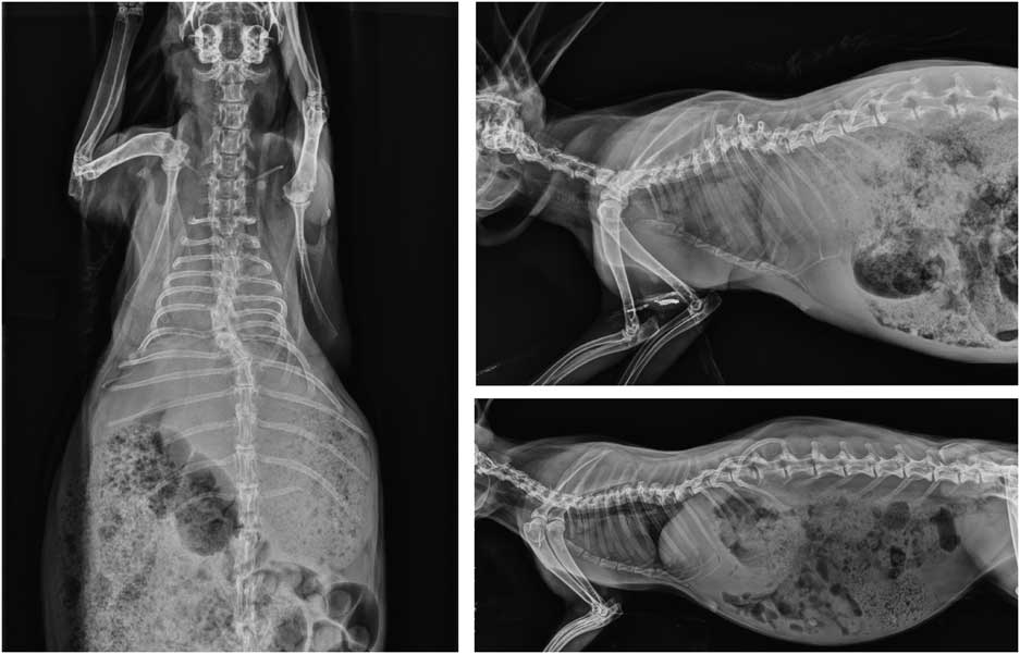
After euthanasia, two X-rays were made per doe (one lateral and one ventrodorsal). Three types of spinal deformation were assessed from these X-rays: scoliosis, kyphosis and lordosis (lateral, dorsal and ventral deviation of the spine, respectively, examples are shown in Figure 2).
X-rays of spinal deformities observed in the present study. Left: ventrodorsal view, scoliosis in thoracic vertebrae 5 to 9, top right: lateral view, kyphosis in thoracic vertebrae 6 to 8, bottom: lordosis in thoracic vertebrae 7 to 10.

Bone quality
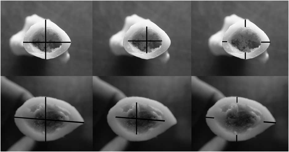
The hind legs of the does were separated from the carcasses and stored at −20°C until analysis, when they were thawed overnight. When fully thawed, most soft tissue was removed from the tibia and femur. The right tibia and femur were subsequently boiled for 15 min in 95°C water, defleshed further and dried at 25°C for 24 h. Subsequently, the maximum shear force until initial structural failure (i.e. the breaking of the bone) was determined using a Mecmesin BFG 25000 N force gauge (Mecmesin, Slinfold, UK) at a speed of 12 mm/min (ASAE, 2004). The left tibia and femur were submerged in 65°C water with 60 g NaBO3.4H2O per litre for 24 h, after which any remaining flesh was removed. Subsequently, bone length was determined, after which the bones were sawed in half 1 mm below the fibula (tibia) or at the mid-diaphysis (femur). Outer width (two measures), inner width (two measures) and cortical thickness (four measures) were determined using digital callipers (Figure 3). After all measures were acquired, the measurement process was repeated. During this second round of measurements, the data collector was blinded to the results of the first round of measurements. All outer width measures were averaged per animal, as were inner width measures and cortical thickness measures.
Outer width (left), inner width (middle) and cortical thickness (right) as determined on the tibia (top) and femur (bottom).

Statistical analysis
Owing to the low occurrence of scores >1, pododermatitis was analysed as a binomial parameter (presence of scores ⩾1) using a logistic regression model with housing (individual on wire v. semi-group on wire v. semi-group on plastic) as the only fixed factor. Spinal deformation was analysed using the same model. All bone quality measures were checked for approximate normality and subsequently analysed by one-way ANOVA with housing as a fixed factor. All analyses were conducted in R 3.0.1.
Results
Bone quality
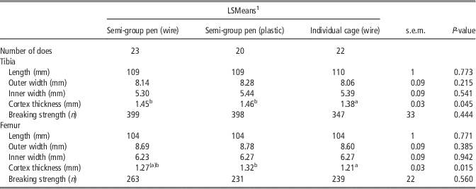
Does housed in individual cages had (or tended to have) ~5% thinner tibia and femur cortices than those in semi-group housing (Table 1). The decreased cortical thickness was the result of a numerical decrease in outer width and a numerical increase in inner width, although neither of these separate measures differed significantly between the housing systems. Breaking strength was not significantly influenced by housing either (P=0.444 and P=0.560 for the tibia and femur, respectively), although there was a numerical decrease of ~10% for tibia breaking strength in the individually caged does. The relative standard deviation (s.d./mean×100) was much higher for breaking strength measures than for cortical thickness measures (39% v. 9% and 39% v. 10% for the tibia and femur, respectively).
The effect of housing treatment on bone quality

1 Values with different superscripts differ significantly (P<0.05), values with the same superscript between brackets tend to differ (P<0.1).
Spinal deformation
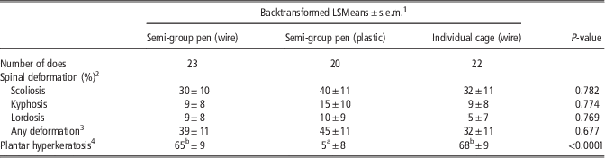
Neither the prevalence of the separate types of spinal deformation (scoliosis, kyphosis or lordosis) nor the prevalence of any of these deformations was influenced by the housing system (Table 2).
The effect of housing treatment on spinal deformation and plantar hyperkeratosis
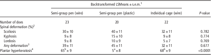
1 Values with different superscripts differ significantly (P<0.05).
2 Percentage of does with a certain type of spinal deformation.
3 Percentage of animals with scoliosis, kyphosis, lordosis or a combination of these.
4 Percentage of animals with a pododermatitis score ⩾1 (i.e. those with at least a patch of bald, callused skin).
Pododermatitis
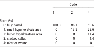
In the 1st cycle, all does had fully haired feet (score 0). Even at the end of the last experimental cycle, the occurrence of scores 2 and 3 was only 11% and 1%, respectively, and no open wounds (score 4) were observed (Table 3). Therefore, the 0 to 4 scale was changed to reflect only the absence (score 0) or presence (score 1 to 4) of plantar hyperkeratosis at the end of the last cycle (Table 2). The percentage of does with plantar hyperkeratosis was much lower on the plastic floor (5% for semi-group housing on plastic) as compared with the wire floor (65% and 68% for semi-group housing on wire and individual housing on wire, respectively, P<0.0001).
The percentage of does per pododermatitis score per experimental cycle (72, 70 and 70 does assessed in cycles 1, 2 and 4, respectively)
Discussion
In line with our expectations, semi-group housing had a favourable effect on some aspects of bone quality, whereas a plastic slatted floor greatly reduced the percentage of does with plantar hyperkeratosis. In contrast to our expectations, no relation between spinal deformation and the housing treatments was found, although its prevalence was high overall.
Does housed in individual cages had thinner bone cortices than those housed in semi-group housing that provided more space, especially during the half of each cycle that the semi-group housing does were grouped (although it needs to be remarked that semi-group housing does also had more space per individual in the other half of the cycle, when they were housed separately). The decrease in bone cortex thickness is in line with previous findings that fattening rabbits have a decreased outer tibia width when housed in smaller cages (Martrenchar et al., Reference Martrenchar, Boilletot, Cotte and Morisse2001; Dalle Zotte et al., Reference Dalle Zotte, Princz, Metzger, Szabó, Radnai, Biró-Németh, Orova and Szendrö2009; Buijs et al., Reference Buijs, Van Poucke, Van Dongen, Lens and Tuyttens2012). Increased activity leads to increased bone width (Gordon, Reference Gordon1989), and the semi-group housing provided both more space and more incentive for activity. Thus, the improved bone quality in the semi-group systems may be explained by increased activity. As restriction of locomotion is thought to be greatly detrimental to the welfare of caged rabbits (EFSA, 2005), the increased cortical thickness in the semi-group housing systems could be interpreted as a positive sign. However, increased activity may also be a result of animals having to flee aggressive pen mates, and aggression and unrest are likely to impact negatively on doe welfare. Although this situation would occur only during half of the reproduction cycle, further study of the behaviour of group housed does will be necessary to determine if possible changes in activity should be interpreted in a negative or a positive way.
Although individually caged does had thinner bone cortices, the breaking strength of these bones did not differ significantly. Again, this in line with at least some fattening rabbit studies (Martrenchar et al., Reference Martrenchar, Boilletot, Cotte and Morisse2001; Buijs et al., Reference Buijs, Van Poucke, Van Dongen, Lens and Tuyttens2012). This lack of a difference in breaking strength may have been caused by the decreased accuracy of the breaking strength measure, as the relative standard deviation was much higher for breaking strength than for cortical thickness. In our protocol, the accuracy of the bone thickness measure was optimized by averaging a total of eight measurements. Breaking strength, in contrast, can only be determined once per bone and is thus prone to greater variation.
Some form of spinal deformation was found in 38% of the does, in line with the occurrence found by Drescher and Loeffler (Reference Drescher and Loeffler1996) for similarly aged, cage-housed does (40%). These authors suggested that spinal deformations were caused by an increased occurrence of ‘flat sitting’ and decreased activity in low or small cages. However, the 40 cm high cages used by Drescher and Loeffler were considerably lower than those used in the present study (cage housing: 60 cm high in some parts of the cage, semi-group housing: no height limitation), whereas the prevalence of spinal deformation was similar. Thus, our results do not lend further support to the suggestion that low cages cause spinal deformations. Although does seemed to be more active in the semi-group housing systems (as suggested by their increased bone cortex thickness), the prevalence of spinal deformation was not decreased in such systems. As such, we did not find further evidence for the suggestion that spinal deformations in rabbit does are related to their activity. Severe spinal deformations can cause pain, reduced mobility and perineal scalding owing to irritation by urine and faeces (Carmel, Reference Carmel2013), and thus represent a major welfare issue. However, the vast majority of the deformations found in the present study was mild and none of these deformations were clinically visible. As such, it seems unlikely that the observed deformations had a major impact on doe welfare, although a complementary study of movement and behaviour could give a more objective evaluation.
The percentage of does with plantar hyperkeratosis was 65% to 68% for does housed on a wire floor with a plastic footrest. In the vast majority of the cases this meant that the animals had small bald callused patches only (score 1). Larger or cracked calluses were rare and wounds and ulcers were absent. The absence of hyperkeratosis during the first lactations and the high prevalence in later lactations is consistent with previous results for cages equipped with a footrest (Rosell and De la Fuente, Reference Rosell and De la Fuente2009). More surprisingly, using a floor that was completely made of plastic slats reduced hyperkeratosis greatly compared with the wire floor with a footrest. This suggests that either the does housed on wire did not restrict themselves to the 1000 cm2/doe that was covered by the footrest, or that even limited contact with the wire floor was enough to cause hair loss and callus formation. Although the greater prevalence of hyperkeratosis is a clear sign that the wire floor used in this study was more abrasive than the plastic floor, it is not fully clear how this impacts on rabbit welfare. A Spanish on-farm study (Rosell and De la Fuente, Reference Rosell and De la Fuente2009) found that 15% of the does housed on a wire floor with a footrest developed ulcerative pododermatitis by the fifth lactation. Although this prevalence was far lower than the 65% these authors reported for does housed without a footrest, the installation of a footrest is thus not enough to prevent the painful end stage of pododermatitis fully. As plantar hyperkeratosis is considered as a very early stage of pododermatitis (Rosell and De la Fuente, Reference Rosell and De la Fuente2009), plastic floors seem to slow down the development of pododermatitis, thus impacting positively on welfare. A more prolonged experiment than the present one might be needed to see if the calluses observed on the wire floor will actually progress into wounds or ulcers over time, in the absence of a similar pattern in does housed on a plastic floor.
In conclusion, our research suggests that housing rabbit does on plastic slatted floors instead of wire floors with a footrest influences their welfare positively. Compared with individually housed does, semi-group housed does had thicker leg bone cortices, suggesting increased activity. Interpretation of this last finding in terms of welfare requires further study of which type of activity was increased (spontaneous locomotion or fleeing aggressive pen mates).
Acknowledgements
This study was funded by the Belgian Federal Public Service of Health, Food Chain Safety and Environment through the contract (RT 11/11 RABBITRY). The authors thank the staff of the ILVO – Animal Sciences Unit (Jolien Vander Linden, Dimitri van Grembergen, André Vermeulen and Virginia Sánchez Gallego) for technical assistance and for taking care of the rabbits, and the staff of the ILVO – Technology and Food Science Unit for building experimental equipment.