The liver is the largest internal organ of the body and is composed of two types of epithelial cells, hepatocytes and cholangiocytes (Ref. Reference Alpini, Prall, LaRusso and Arias1). Hepatocytes account for approximately 70% and cholangiocytes for 3–5% of the endogenous liver cell population (Refs Reference Alpini, Prall, LaRusso and Arias1, Reference Racanelli and Rehermann2). Cholangiocytes line the intrahepatic and extrahepatic bile duct system of the liver (Ref. Reference Alpini, Prall, LaRusso and Arias1). The bile ductules and ducts comprise a branched system of interconnected tubes, which collects bile secreted at the canalicular membranes of hepatocytes (Ref. Reference Nathanson and Boyer3), and delivers it to the gallbladder or the duodenum (Refs Reference Alpini, Prall, LaRusso and Arias1, Reference Alpini4). Although cholangiocytes represent a small proportion of the cells of the liver, they have an important pathophysiological role in the modification of the composition of bile during transit in the bile ducts (Refs Reference Alpini, Prall, LaRusso and Arias1, Reference Alpini4, 5 Reference Alvaro6, Reference Glaser7, Reference Glaser8, Reference Kanno9, Reference Tietz10). This process involves the secretion and absorption of water, electrolytes and other organic solutes from hepatocellular bile.
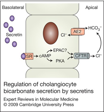
One of the most important and well-studied functions of cholangiocytes is the excretion of bicarbonate into bile, which is stimulated by secretin. Secretin receptors belong to the family of G-protein-coupled receptors (Ref. Reference Ulrich, Holtmann and Miller11), which signal through the activation of adenylyl cyclase and protein kinase A (PKA) (Ref. Reference Ulrich, Holtmann and Miller11). In the liver, secretin receptors are expressed only on cholangiocytes, on the basolateral membrane (Refs Reference Farouk12, Reference Alpini13). Secretin stimulates ductal bile secretion by a series of coordinated events (Fig. 1). First, secretin binds to the basolateral secretin receptors, which induces elevation of intracellular cyclic adenosine monophosphate (cAMP) leading to the activation of PKA (Ref. Reference Alvaro, Mennone and Boyer14). Subsequently, PKA phosphorylates the cystic fibrosis transmembrane conductance regulator (CFTR) triggering the opening of this Cl– channel leading to secretion of Cl– at the apical membrane of cholangiocytes, which results in membrane depolarisation (Ref. Reference McGill15). The Cl– efflux from CFTR creates a Cl– gradient that favours activation of the apically located Cl–/HCO3– exchanger (Ref. Reference Martinez-Anso16), which results in secretin-stimulated bicarbonate-enriched bile (Refs Reference Alpini, Prall, LaRusso and Arias1, Reference Alpini4, Reference Alvaro6, Reference Glaser8, Reference Tietz10). In addition to PKA, another downstream target of cAMP, EPAC (exchange proteins activated directly by cyclic AMP), can also regulate Cl– channel function independently of PKA (Fig. 1) (Ref. Reference Holz17). This has not been directly demonstrated to have a role in the cAMP-dependent activation of CFTR in cholangiocytes. However, it was recently shown that EPAC isoform 2 is involved in the mechanism regulating purinergic-receptor-induced cAMP signalling, which regulates the chemosensory functions of cholangiocyte primary cilia, suggesting that EPAC participates in the regulation of Cl– efflux (Ref. Reference Masyuk18).
Regulation of cholangiocyte bicarbonate secretion by secretin. Cholangiocytes are the only cell types in the liver expressing the basolateral secretin receptor. Secretin binds to the G-protein-coupled secretin receptor (SR), stimulating increased intracellular cAMP levels, which results in the activation of protein kinase A (PKA). Subsequently, PKA phosphorylates cystic fibrosis transmembrane conductance regulator (CFTR), stimulating Cl– efflux from the apical domain of cholangiocytes thereby activating the Cl–/HCO3– exchanger (AE2) and secretion of bicarbonate into the bile.

A number of studies have suggested that secretin receptor expression is linked to cholangiocyte proliferative responses in animal models of biliary hyperplasia, such as BDL, partial hepatectomy, chronic feeding of bile acids (e.g. taurocholic acid) and cirrhosis induced by administration of high levels of carbon tetrachloride (CCl4) (Refs Reference Alpini, Prall, LaRusso and Arias1, Reference Alpini4, Reference LeSage19, Reference Alpini20, Reference Alpini21, Reference Alpini22). These models of cholangiocyte hyperplasia are closely associated with increased secretin-stimulated choleresis, which is characterised by increased secretin-receptor gene expression, elevated secretin-stimulated cAMP levels, enhanced Cl–/HCO3– exchanger activity and amplified secretin-stimulated bicarbonate secretion (Refs Reference Alpini, Prall, LaRusso and Arias1, Reference Alpini4, Reference Glaser7, Reference Alpini22). Thus, the response to secretin and secretin-receptor expression is a valuable pathophysiological tool (Refs Reference Alpini, Prall, LaRusso and Arias1, Reference Alpini4, Reference Glaser8, Reference Tietz10, Reference LeSage19, Reference Alpini22, Reference Francis23, Reference Glaser24, Reference LeSage25, Reference LeSage26, Reference LeSage27) that can be used to evaluate cholangiocyte proliferation. However, any role for secretin itself in the regulation of cholangiocyte proliferation has yet to be explored.
Research over the past decade has greatly improved our knowledge and understanding of the cellular and molecular factors regulating biliary proliferation (Refs Reference Alpini, Prall, LaRusso and Arias1, Reference Alvaro28). In particular, several papers have demonstrated that proliferating cholangiocytes display neuroendocrine phenotypes, and hence secrete and respond to a number of hormones, neuropeptides and neurotransmitters (Refs Reference Alpini, Prall, LaRusso and Arias1, Reference Alvaro28, Reference Marzioni29, Reference Gigliozzi30, Reference Gaudio31, Reference Marzioni32). During the course of cholestasis, cholangiocytes undergo neuroendocrine transdifferentiation regulated by a number of neuroendocrine hormones. In support of the concept of neuroendocrine transdifferentiation, proliferating cholangiocytes acquire phenotypic features of neuroendocrine epithelium (Ref. Reference Alvaro28), including expression of neuroendocrine markers, such as chromogranin A, glycolipid A2-B4, S-100 protein, neural cell adhesion molecule, and the addition of neuroendocrine granules (Refs Reference Roskams33, Reference Desmet, Roskams and Van Eyken34). Activation of the neuroendocrine phenotype in cholangiocytes (Ref. Reference Alvaro28), which is similar to that of hepatic stellate cells during liver disease development (Ref. Reference Ebrahimkhani, Elsharkawy and Mann35), might have a key role in the progression of biliary fibrosis during cholestatic liver diseases [such as primary biliary cirrhosis (PBC) and primary sclerosing cholangitis (PSC)], which target cholangiocytes, resulting in increased biliary proliferation and/or apoptosis in these disease states (Refs Reference Alpini, Prall, LaRusso and Arias1, Reference Lazaridis, Strazzabosco and LaRusso36). In the following sections, we explore the factors that regulate cholangiocyte proliferation in relation to their acquired neuroendocrine phenotype and factors controlling the potential contribution of the biliary system to liver fibrosis.
Regulators of cholangiocyte proliferation
Gastrointestinal hormones
The role of secretin in the regulation of biliary physiology has been thoroughly studied (Refs Reference Alpini, Prall, LaRusso and Arias1, Reference Alpini4, Reference Alvaro6, Reference Glaser8, Reference Kanno9). The gastrointestinal hormones somatostatin and gastrin inhibit cholangiocyte responses to secretin by inhibiting secretin-induced intracellular cAMP levels and bicarbonate excretion into bile (Refs Reference Glaser8, Reference Kanno9, Reference Tietz10). Somatostatin and gastrin also inhibit cholangiocyte proliferation (Refs Reference Alpini22, Reference Glaser24, Reference Glaser37, Reference Tracy38). The antiproliferative actions of somatostatin occur upon its interaction with the somatostatin receptor subtype 2 (SSTR2) (Ref. Reference Alpini22) and have been observed in both in vivo and in vitro models (Refs Reference Tietz10, Reference Alpini22, Reference Tracy38). The somatostatin analogue octreotide decreases cholangiocyte proliferation and periportal extracellular matrix (ECM) deposition (i.e. fibrosis) in response to biliary obstruction (Ref. Reference Tracy38). In isolated large cholangiocytes from rats with BDL, somatostatin inhibited both secretin receptor expression and proliferation as evaluated by measurement of DNA synthesis and H3 histone expression (Ref. Reference Alpini22). Recently, in a preclinical study in polycystic kidney (PCK) rats (a model of autosomal recessive polycystic kidney disease), the somatostatin analogue octreotide inhibited cholangiocyte proliferation and reduced liver fibrosis (Ref. Reference Masyuk39). This study provides strong evidence for the use of octreotide in the treatment of patients with polycystic liver diseases and also indicates that octreotide might reduce biliary fibrosis in other liver disease paradigms (Ref. Reference Masyuk39).
Similarly to somatostatin, gastrin inhibited secretin-induced ductal secretion and secretin-induced cAMP levels by interacting with cholecystokinin-B (CCK-B) receptors (Ref. Reference Glaser8). Chronic administration of gastrin has also been shown to inhibit cholangiocyte proliferation in BDL rats (Ref. Reference Glaser24). Interestingly, when gastrin was administered to rats with established BDL a reversal of cholangiocyte proliferation was also observed (Ref. Reference Glaser37).
The importance of intracellular cAMP signalling as a regulator of cholangiocyte proliferation was also demonstrated by chronic administration of forskolin (an adenylate cyclase activator) (Ref. Reference Francis23) to normal rats (Ref. Reference Francis23). Compared with control animals, forskolin administration to normal rats increased the number of bile ducts, intracellular cAMP levels and secretin-induced choleresis, as observed in animals with BDL (Ref. Reference Francis23). Forskolin also stimulated the proliferation of isolated cholangiocytes, which was blocked by the inhibition of the PKA–Src–MEK–ERK1/2 pathway providing the first evidence that this signalling pathway was critical for cholangiocyte proliferation (Ref. Reference Francis23).
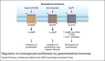
A recent study demonstrated that glucagon-like peptide 1 (GLP1) modulates cholangiocyte adaptive responses to cholestasis (Ref. Reference Marzioni29). In addition to having a role in the modulation of glucose homeostasis, GLP1 induces the acquisition of neuroendocrine phenotypes by pancreatic ductal cells – cells that share similar origins and features as cholangiocytes (Refs Reference Drucker40, Reference Bulotta41). GLP1 and its receptor antagonist extendin-4 both have similar effects whereby they stimulate cholangiocyte proliferation in control rats as well as in isolated cultures from control and BDL rats (Ref. Reference Marzioni29). Expression of the GLP1 receptor (GLP1R) protein was significantly upregulated in BDL rats compared with sham-operated animals (Ref. Reference Marzioni29). Cholangiocytes from BDL animals express mRNA for preproglucagon, the precursor of GLP1, unlike cells from normal rats (Ref. Reference Marzioni29). This finding suggests that GLP1 is important in biliary growth during cholestasis. In fact, administration of exendin-4 significantly decreased ductal mass and biliary functional activity in BDL rats (Ref. Reference Marzioni29). The proproliferative effect of GLP1 is mediated through phosphoinositide 3-kinase, cAMP–PKA and Ca2+–CAMKIIα signalling mechanisms (Ref. Reference Marzioni29), and is yet another example of the importance of cAMP signalling in the regulation of biliary proliferation during cholestasis. A summary of the cholangiocyte proliferative responses regulated by gastrointestinal hormones is shown in Fig. 2.
Regulation of cholangiocyte proliferation by gastrointestinal hormones. Cholangiocytes express CCKBR, SSTR2 and GLP1R G-protein-coupled receptors on their basolateral membranes. The gastrointestinal hormones gastrin and somatostatin both inhibit cholangiocyte proliferation through mechanisms that inhibit intracellular cAMP levels. In addition, the somatostatin analogue octreotide inhibits hepatic fibrosis in the polycystic kidney (PCK) rat model and in rats after bile duct ligation, indicating the potential to limit hepatic fibrosis associated with biliary proliferation via the targeting of SSTR2. Glucagon-like peptide 1 (GLP1) stimulates cholangiocyte proliferation by increasing intracellular cAMP levels with the concomitant activation of PI3K and Ca2+-CAMKIIα. Abbreviations: CCKBR, cholecystokinin-B receptor; GLP1R, glucagon-like peptide 1 receptor; SSTR2, somatostatin receptor subtype 2.

Bile acids
Bile acids accumulate during cholestasis, resulting in greater exposure of cholangiocytes to their effects (Ref. Reference Alpini20). Bile acids also have varied effects on biliary function, apoptosis and growth. In vitro, both taurocholate and taurolithocholic acid (TLC) stimulated cholangiocyte proliferation and increased the secretin-induced cAMP response and Cl–/HCO3– exchanger activity in isolated rat cholangiocytes (Ref. Reference Alpini42). We, and others, have demonstrated that cholangiocytes express the apical Na+-dependent bile acid transporter (ASBT; official symbol, NTCP2; gene symbol, Slc10a2) providing a mechanism to mediate bile acid uptake (Refs Reference Alpini43, Reference Lazaridis44). The concept that bile acids stimulate biliary proliferation was expanded to an in vivo model. Normal rats were fed a diet containing taurocholate or TLC (1%) for 1–4 weeks in order to mimic the bile acid accumulation observed during cholestasis (Ref. Reference Alpini20). Following bile acid feeding, there was an increase in cholangiocyte proliferation, secretin receptor gene expression and secretin-induced cAMP levels, similar to levels found in animals with BDL (Ref. Reference Alpini20). Cholangiocytes from these mice displayed secretin-stimulated bile flow and bicarbonate secretion that was not observed in control animals, but which was again similar to that in animals with BDL (Refs Reference Alpini4, Reference Tietz10, Reference Alpini20). Bile acid feeding (with taurocholate and TLC) was also shown to increase cholangiocyte Slc10a2 expression in a PKC-dependent mechanism, indicating a link between proliferation and bile acid accumulation during cholestasis (Ref. Reference Alpini45). Finally, ursodeoxycholate and taurodeoxycholate have been shown to inhibit cholangiocyte proliferation of BDL cholangiocytes, both in vitro and in vivo (Ref. Reference Alpini46). Secretin has been demonstrated to stimulate colchicine-sensitive ASBT translocation to the cholangiocyte plasma membrane, which was associated with increased taurocholate uptake, greater-than-expected biliary lipid secretion and bile flow, and the prolongation of the biliary transit time of taurocholate (Ref. Reference Alpini47). This work provides evidence that secretin stimulates cholehepatic shunting of bile acids through increased ASBT levels on the cholangiocyte apical membrane (Ref. Reference Alpini47). These findings are consistent with studies demonstrating that alternative splicing of the Slc10a2 mRNA results in the formation of a truncated form termed t-ASBT (Ref. Reference Lazaridis48). Transport studies have revealed that t-ASBT can function as a bile acid efflux protein on the basolateral membrane of cholangiocytes (Ref. Reference Lazaridis48).
Angiogenic factors
The peribiliary vascular plexus (PBP) stems from the hepatic artery, nourishes the biliary epithelium, and sustains a countercurrent of substances, such as VEGF (vascular endothelial growth factor) and other angiogenic factors that are reabsorbed from bile towards the hepatocytes (Ref. Reference Gaudio49). VEGF is secreted by a number of epithelia and modulates cellular functions by both autocrine and paracrine mechanisms (Ref. Reference Sartelet50). VEGF is a key regulator of biliary proliferation during cholestasis (Ref. Reference Gaudio49). Gaudio and colleagues elegantly demonstrated that proliferation of cholangiocytes precedes the expansion of the PBP in the intrahepatic biliary tree (Ref. Reference Gaudio49). This finding suggests a crosstalk mechanism between cholangiocytes and vascular cells – an interaction that mediates the adaptive changes of biliary cells and the microvascular system in cholestatic liver diseases (Ref. Reference Gaudio49). Cholangiocytes express VEGF receptors VEGFR2 and VEGFR3, and respond to VEGF-A and VEGF-C with increased proliferation (Ref. Reference Gaudio31). In the BDL model, cholangiocytes have upregulated VEGFR2 and VEGFR3 protein expression and secrete increased levels of VEGF compared with normal cholangiocytes (Ref. Reference Gaudio31). Indeed, immunoneutralisation of VEGF-A and VEGF-C decreases biliary proliferation during BDL (Ref. Reference Gaudio31). The importance of these growth factors during biliary proliferation was also revealed by increased proliferation of cholangiocytes in normal rats chronically treated with recombinant VEGF-A and VEGF-C (Ref. Reference Gaudio31). Furthermore, in BDL rats, interruption of the flow of the hepatic artery by ligation (HAL) induced: (1) the disappearance of the PBP; (2) increased cholangiocyte apoptosis and impaired biliary proliferation and secretin-stimulated ductal secretion; and (3) decreased cholangiocyte VEGF secretion (Ref. Reference Gaudio51). HAL effects on the PBP and cholangiocyte functions were prevented by administration of r-VEGF-A, which, by maintaining the integrity of the PBP and cholangiocyte proliferation, prevents apoptosis and functional damage of bile ducts following ischaemic injury (Ref. Reference Gaudio51). In contrast to the gastrointestinal hormone GLP1, VEGF induced cholangiocyte proliferation by activation of the inositol-(Reference Alpini, Prall, LaRusso and Arias1,Reference Alpini4,Reference Alvaro5)-trisphosphate (IP3)–[Ca2+]i–PKCα pathway and phosphorylation of Src and ERK1/2 (Ref. Reference Gaudio31). These findings indicated that VEGF regulates cholangiocyte proliferation in an autocrine mechanism through the upregulation of VEGF secretion induced by cholestasis, which is another example of the differentiation of proliferating cholangiocytes to a neuroendocrine phenotype.
Furthermore, another important study evaluated the expression and the effect of angiogenic factors in cholangiocytes from autosomal dominant polycystic kidney disease (ADPKD) patients and from a ADPKD mouse model (Pkd2 WS25/–) (Ref. Reference Fabris52). Cholangiocytes stained positively for VEGF, VEGFR1, VEGFR2 and Ang-2 in ADPKD and Caroli disease, and for Ang-1 and Tie-2 in ADPKD (Ref. Reference Fabris52). VEGF stimulated the growth of normal and ADPKD (at higher extent) cholangiocytes (Ref. Reference Fabris52). VEGF expression on cholangiocytes positively correlated with microvascular density and the expression of VEGF, VEGFR, Ang-1 and Tie-2 was higher in cholangiocytes from ADPKD (Ref. Reference Fabris52). VEGF and Ang-1 have an autocrine proliferative effect on cholangiocyte growth and a paracrine effect on portal vasculature, thereby promoting the growth of the cysts and their vascular supply (Ref. Reference Fabris52). Moreover, VEGF receptor inhibition by SU-5416 blocks liver cyst growth in Pkd2 WS25/– mice (Ref. Reference Amura53), further supporting the concept that the VEGF signalling pathway is an important therapeutic target in the treatment of liver cysts in ADPKD (Ref. Reference Amura53). Cholangiocytes are a major source of hepatic endothelin-1 (ET-1) production during the development of hepatopulmonary syndrome after BDL, indicating that cholangiocyte-derived ET-1 may be an important endocrine mediator of experimental hepatopulmonary syndrome (Ref. Reference Luo54). Further studies into the role of angiogenic factors such as angiopoietin and ET-1 on the growth and neuroendocrine nature of cholangiocytes are necessary.
Nerve growth factor, neuropeptides and neurotransmitters
Cholangiocytes express the M3 acetylcholine (ACh) receptor, and ACh, by acting on M3 receptor subtypes, induces a Ca2+-calcineurin-mediated potentiation of the secretin-induced adenylyl cyclase activity (Ref. Reference Alvaro5). The role of ACh and the cholinergic system in the regulation of cholangiocyte proliferation was also explored in the model of total vagotomy in rats with BDL (Ref. Reference LeSage55). Vagotomy impairs cholangiocyte proliferation and enhances apoptosis, leading to decreased ductal mass in response to BDL (Ref. Reference LeSage55). Similarly, secretin-induced choleresis of BDL rats was virtually eliminated by vagotomy in association with decreased cholangiocyte cAMP levels (Ref. Reference LeSage55). Maintenance of cholangiocyte cAMP levels by forskolin administration prevents the effects of vagotomy on cholangiocyte proliferation, apoptosis, and secretion, which highlights the importance of cholinergic innervation in the regulation of biliary mass and, as mentioned earlier, the dependence of cholangiocyte proliferation on intracellular cAMP-dependent signalling mechanisms (Ref. Reference LeSage55).
Previous studies have demonstrated that sympathetic innervation is necessary for both hepatocyte and cholangiocyte proliferation during liver regeneration (Ref. Reference Iwai and Shimazu56). Adrenergic denervation of BDL rats via the administration of a single intraportal injection of 6-hydroxidopamine (6-OHDA): (1) inhibits cholangiocyte proliferation, and the number of bile ducts; (2) decreases secretin-stimulated choleresis and cholangiocyte cAMP levels; and (3) increases the number of cholangiocytes undergoing apoptosis (Ref. Reference Glaser57). Chronic administration of clenbuterol (a β2-adrenergic agonist) and dobutamine (a β1-adrenergic agonist) prevents the decrease in cAMP levels and secretion induced by 6-OHDA-induced denervation, maintains cholangiocyte proliferation and decreases cholangiocyte apoptosis (Ref. Reference Glaser57). The induction of cholangiocyte apoptosis induced by adrenergic denervation by 6-OHDA was prevented by taurocholate feeding (Ref. Reference Marzioni58), which also restored cholangiocyte proliferation and secretin-stimulated ductal secretion that was decreased after adrenergic denervation (Ref. Reference Marzioni58). These finding suggest that adrenergic innervation might have a key role in regulating cholangiocyte proliferation during regeneration and proliferation associated with cholestasis and could be a therapeutic target area.
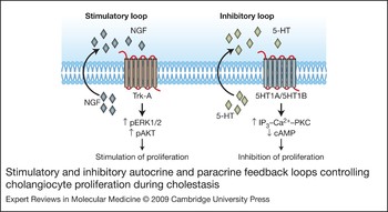
We have recently demonstrated that the neuroendocrine hormone serotonin has a key role in the autocrine regulation of cholangiocyte proliferation (Refs. Reference Marzioni59). Serotonin is thought to play a role in the origins of pruritus and fatigue, which are classical clinical features of PBC (Ref. Reference Swain and Maric60, Reference Jones and Bergasa61). Cholangiocytes express the serotonin 1A and 1B receptors and secrete serotonin (Ref. Reference Marzioni59). Stimulation of these receptors markedly inhibited the growth of rat cholangiocytes during cholestasis via IP3–Ca2+–PKC signalling mechanism resulting in the downregulation of the cAMP-dependent proliferative signalling (Ref. Reference Marzioni59). Of great importance, hyperplastic cholangiocytes isolated from BDL rats secrete serotonin, and cholangiocyte proliferation in response to cholestasis was enhanced both in vitro and in vivo, if serotonin secretion was immunoneutralised (Ref. Reference Marzioni59). Serotonin is postulated to counterbalance the excessive proliferation responses of biliary epithelium during cholestasis (Ref. Reference Marzioni59). Recent studies also demonstrated that cholangiocytes secrete nerve growth factor (NGF) (Ref. Reference Gigliozzi30). In vitro, NGF stimulated the proliferation of cholangiocytes via AKT- and ERK1/2-dependent mechanisms (Ref. Reference Gigliozzi30). In vivo, immunoneutralisation of NGF during BDL in rats resulted in decreased biliary mass and reductions in proliferation and enhanced apoptosis (Ref. Reference Gigliozzi30). We propose that regulation of cholangiocyte proliferation by stimulatory and inhibitory autocrine or paracrine loops such as serotonin and NGF during cholestasis has an important role in the pathogenesis of cholestatic liver diseases. Examples of the stimulatory and inhibitory autocrine or paracrine loops are illustrated in Figure 3.
Stimulatory and inhibitory autocrine and paracrine feedback loops controlling cholangiocyte proliferation during cholestasis. During cholestasis, cholangiocytes secrete higher levels of NGF, which can stimulate cholangiocyte proliferation in autocrine or paracrine mechanisms via activation of Trk-A on the basolateral membrane of cholangiocytes through increases in ERK1/2 and AKT activity. By contrast, during cholestasis, cholangiocytes secrete higher levels of 5-HT, which negatively regulates cholangiocyte proliferation via activation of IP3–Ca2+–PKC-dependent signalling mechanisms, resulting in the downregulation of intracellular cAMP levels. Abbreviations: 5-HT, 5-hydroxytryptamine (serotonin); 5HT1A/B, 5-HT receptor 1A/B; IP3, inositol triphosphate; NGF, nerve growth factor; pAKT, phosphorylated AKT; pERK1/2, phosphorylated extracellular-regulated kinase 1/2; PKC, protein kinase C; Trk-A, neurotrophic tyrosine kinase receptor type 1.

The aminergic peptide and neurotransmitter histamine is responsible for many functions in the body, such as neurogenic functions, inflammatory and allergic responses, and gastric secretion (Refs Reference Hou62, Reference Jancso63, Reference Parsons and Ganellin64). Normal and BDL cholangiocytes express all of the G-protein-coupled histamine receptor subtypes (HRH1, HRH2, HRH3 and HRH4) (Refs Reference Francis65, Reference Francis66); however, the expression of H3R is significantly increased in proliferating cholangiocytes following BDL (Ref. Reference Francis65). Activation of H3R by chronically administering the agonist (R)-(α)-(−)-methylhistamine dihydrobromide (RAMH) to rats for 7 days after BDL resulted in a decrease in the growth of the biliary tree with no difference in the rate of apoptosis, suggesting that H3R activation slows the rate of proliferation rather than reduces the number of cholangiocytes by a cell-death mechanism (Ref. Reference Francis65). In addition, administration of histamine to this animal model of cholestasis also resulted in a decrease in cholangiocyte proliferation, and blocking histamine actions by using the selective H3R antagonist thioperamide maleate resulted in a partial reversal of these effects (Ref. Reference Francis65).
We have very recently demonstrated that sensory hepatic innervation and biliary expression of the α-type calcitonin gene-related peptide 1 (α-CGRP) regulates cholangiocyte proliferation during cholestasis induced by BDL (Ref. Reference Glaser67). CGRP is a potent vasodilator peptide (Refs. Reference Tippins68, Reference Brain69) that participates in the regulation of vascular tone and regional organ blood flow (Refs. Reference Wimalawansa70, Reference DiPette71) and knockout of α-CGRP decreases intrahepatic bile duct mass in BDL mice (Ref. Reference Glaser67). In vitro, both α- and β-CGRP stimulated proliferation of BDL cholangiocytes by activation of PKA and CREB (Ref. Reference Glaser67). These studies indicate that sensory innervation is important for regulation of biliary proliferation, and other sensory neuropeptides such as substance P could have a role in the chronic inflammation observed in certain cholangiopathies (Refs Reference Bang72, Reference Bang73, Reference Neuhuber and Tiegs74).
Steroid hormones
Steroid hormones such as oestrogen and progesterone have been shown to be proproliferative in a number of cell types (Refs Reference Galtier-Dereure75, Reference Chalbos76). Since PBC largely affects middle-aged women (when oestrogen and progesterone levels have dropped) (Ref. Reference Beckmann77), a number of studies have evaluated the roles of oestrogen and progesterone in the regulation of biliary proliferation, in particular during cholestasis. Cholangiocytes express both the oestrogen (estrogen) receptor (ER)-α and ER-β subtypes, with a corresponding increase in the expression of ER-β in cholangiocytes isolated from rats with BDL for 3 weeks compared with control animals (Ref. Reference Alvaro78). When cholangiocytes were stimulated in vitro with 17-β-estradiol, proliferation was significantly increased by ER-dependent activation of Src–Shc–ERK1/2 signalling mechanisms (Ref. Reference Alvaro79). When BDL male rats were treated in vivo with anti-oestrogens such as tamoxifen or ICI182,760, or when BDL female rats were subjected to ovariectomy, the growth of the biliary tree was dramatically reduced, and cholangiocytes underwent cell death by apoptosis (Refs Reference Alvaro78, Reference Alvaro80). Based on these studies, the current working hypothesis is that oestrogens might delay the evolution of cholangiopathies into ductopaenia (Ref. Reference Svegliati-Baroni81). ER expression in cholangiocytes is markedly reduced in patients with late-stage PBC and ER modulators improve serum parameters of cholestasis in PBC patients (Refs Reference Alvaro82, Reference Invernizzi83).
Recently, it has been demonstrated that the steroid hormone progesterone stimulates the proliferation of both male and female cholangiocytes (Ref. Reference Glaser84). Cholangiocytes express the PR-B nuclear receptor and several membrane receptors for progesterone (PRGMC1, PRGMC2 and mPRα) (Ref. Reference Glaser84). In vivo, progesterone increases the number of bile ducts of normal rats, whereas an antiprogesterone antibody inhibits cholangiocyte growth stimulated by BDL. Interestingly, normal and BDL cholangiocytes expressed the biosynthetic pathway (i.e. STAR, 3β-HSD, p450scc) produce and secrete progesterone (Ref. Reference Glaser84). In vitro studies performed in a normal rat cholangiocyte cell line (NRC) revealed that: (1) progesterone increased cholangiocyte proliferation; (2) supernatants collected from normal and BDL cholangiocytes increased cholangiocyte proliferation, which was partially inhibited by preincubation with antiprogesterone; and (3) inhibition of progesterone steroidogenesis with aminoglutethimide prevented cholangiocyte proliferation (Ref. Reference Glaser84). These findings further support the concept that neuroendocrine, autocrine or paracrine mechanisms have a key role in the modulation of cholangiocyte proliferative responses to cholestasis.
Other factors
Alvaro and colleagues have shown that cholangiocytes are a target cell type for the growth hormone (GH)–insulin-like growth factor-1 (IGF1) pathway (Ref. Reference Alvaro85). They demonstrated that GH induces IGF1 expression and release in isolated cholangiocytes, which is associated with the consequent stimulation of cell growth by IGF1 (Ref. Reference Alvaro85). In addition, a recent study (Ref. Reference Gatto86) has demonstrated the expression of IGF1 isoforms in rat hepatocytes and cholangiocytes, and their involvement in the protection against cholestatic liver injury. Specifically, the authors have shown that the ‘locally acting’ IGF1 isoforms (expressed by hepatocytes and cholangiocytes) decreased during cell damage and increased during cell proliferation (Ref. Reference Gatto86). Furthermore, the ‘locally acting’ IGF1 was more active than the ‘circulating’ isoform in protecting cholangiocytes from glycochenodeoxycholate-induced cytotoxicity (Ref. Reference Gatto86). These findings indicate the presence of a stimulatory autocrine or paracrine loop in cholangiocytes.
The interaction of CD44 and hyaluronic acid was also among the proproliferative factors for cholangiocytes in cholestatic livers (Ref. Reference He87). CD44 is a multifunctional cell adhesion molecule, which takes part in cell–cell and cell–matrix interactions (Refs Reference Bajorath88, Reference Bartolazzi89). Hyaluronic acid, the main component of the ECM, is the primary ligand of CD44 (Ref. Reference Tammi, Day and Turley90). High levels of hepatic CD44 expression have been observed in patients with PSC and cholangiocarcinoma (Refs Reference Mikami91, Reference Xu92). Compelling evidence that the proliferative cholangiocytes lining the intrahepatic ducts are a prime source of hepatic CD44 was reported when CD44-positive cholangiocytes were found closely associated with extracellular hyaluronan accumulated in the portal tracts of BDL livers (Ref. Reference He87). In vitro, cholangiocyte proliferation was stimulated by hyaluronan treatment, and blocked by siRNA against CD44 or anti-CD44 antibody (Ref. Reference He87). The interaction between CD44 and hyaluronan might have a pathogenic role in the development of cholestatic liver diseases by enhancing biliary proliferation (Ref. Reference He87).
Secretion of profibrogenic factors by cholangiocytes
Several studies have demonstrated that proliferating cholangiocytes secrete profibrotic factors. During biliary fibrosis, proliferating bile duct epithelial cells are the predominant source of the profibrogenic connective tissue growth factor (CTGF) (Ref. Reference Sedlaczek93). In addition to hepatic stellate cells, activated bile duct epithelial cells are an important source of profibrogenic CTGF during biliary fibrosis (Ref. Reference Sedlaczek93). One study aimed to localise the cellular sources of the collagens excessively deposited in the liver in the course of secondary biliary fibrosis (Ref. Reference Milani94). Epithelial cells of newly formed bile ducts express mRNA for α1(IV) procollagen, indicating that proliferating cholangiocytes are a source of hepatic collagen during fibrosis (Ref. Reference Milani94). Transforming growth factor-β2 (TGF-β2) expression is also a specific property of proliferating bile duct epithelial cells, and its expression is thought to be related to the formation of specialised periductular connective tissue during bile duct proliferation (Ref. Reference Milani95).
Laminin synthesis occurs in mesenchymal, endothelial and bile duct epithelial cells in the liver (Ref. Reference Milani96). In addition, platelet-derived growth factor (PDGF) is expressed in proliferating cholangiocytes during experimental biliary fibrosis in rats (Ref. Reference Grappone97). Pentoxifylline exerts an antifibrogenic effect by reducing PDGF-induced ERK-dependent signalling and proliferation of ECM-producing cells (Ref. Reference Pinzani98). Recently, it was demonstrated that the oral administration of an endothelin-A receptor antagonist blocks collagen synthesis and deposition in a rat model of liver fibrosis (Ref. Reference Cho99). Indeed, proliferating cholangiocytes are the major source of hepatic ET-1 (Ref. Reference Luo54), implicating cholangiocytes once again the process of hepatic fibrosis. The targeting of the profibrogenic programme that is activated in proliferating cholangiocytes, and the profibrogenic factors they secrete should enable the development of novel therapies for liver diseases that are associated with biliary fibrosis.
Evidence for biliary epithelium involvement in fibrosis associated with chronic liver diseases
Studies observing ductular reaction, which involves activation of the oval cell compartment, have pointed to the potential involvement of the biliary epithelium in fibrosis associated with other chronic liver diseases (Ref. Reference Lowes100). Although this area remains somewhat controversial as to cell origin (i.e. oval cell or cholangiocyte) and contribution, ductular reaction has also been shown to be associated with the severity of nonbiliary chronic liver diseases (Ref. Reference Lowes100). There is an association between the severity of liver disease and an increase in the number of oval cells (i.e. ductular reaction), consistent with the hypothesis that oval cell proliferation is associated with an increased risk of hepatocellular carcinoma in chronic liver disease (Ref. Reference Lowes100). Patients that underwent liver transplantation for chronic hepatitis B virus infection were shown to have a fibrosing cholestatic hepatitis demarcated by thin, perisinusoidal bands of fibrosis extending from portal tracts to surround plates of ductular-type epithelium (Ref. Reference Davies101). Ductular reaction was also present in livers with fibrosis due to alcoholic liver disease, hepatitis C infection and nonalcoholic steatohepatitis (Refs. Reference Ray102, Reference Clouston103, Reference Richardson104).
Cholangiocyte proliferation and activation of portal fibrosis
Liver fibrosis during acute and chronic cholestasis involves the stepwise process of ‘ductular reaction’, which refers to an increasing number of ductules (i.e. cholangiocyte proliferation), accompanied by polymorphonuclear leukocytes, and an increase in matrix, leading to periportal fibrosis and eventually biliary cirrhosis (Ref. Reference Roskams and Desmet105). A number of studies have suggested that proliferating cholangiocytes have a role in the induction of fibrosis, either directly via epithelial–mesenchymal transition (EMT), or indirectly via activation of other liver cell types. A number of very recent articles have implemented biliary EMT in the process of liver fibrosis. EMT refers to the process in which mature epithelial cells lose the cell–cell contacts and protein expression patterns characteristic of epithelia and acquire the phenotypic characteristics of mesenchymal cells. EMT has been shown to be an important mechanism in the pathogenesis of renal fibrosis by providing a source of fibrogenic myofibroblasts (Refs. Reference Robertson106, Reference Burns, Kantharidis and Thomas107). Recently, EMT has been implicated as a key mechanism in the pathogenesis of liver fibrosis. In an elegant study of human samples from a variety of liver diseases, Diaz and colleagues present convincing histological data revealing that EMT occurs in human liver fibrosis, particularly in disease associated with prominent bile ductular proliferation, such as biliary atresia and PBC (Ref. Reference Diaz108). They observed significant colocalisation between CK19 (a cholangiocyte-specific epithelial marker) and other markers of EMT (vimentin, Snail and fibroblast-specific protein 1) in biliary atresia and PBC by using a multispectral imaging system. Interestingly, no evidence of hepatocyte EMT was observed in biliary atresia (Ref. Reference Diaz108). Robertson and co-workers also demonstrated that biliary EMT occurs during post-transplantation recurrence of PBC (Ref. Reference Robertson109). The biliary EMT was associated with cholangiocyte expression of S100A4 (a key marker of early fibroblast lineage), vimentin and pSMAD 2/3, and was driven by TGF-β (Ref. Reference Robertson109). S100A4 expression appears to occur before the appearance of other features of recurrent PBC, which suggests that EMT may be an initiating event and could explain the loss of bile duct epithelia in this disease (Ref. Reference Robertson109). Similar findings have been demonstrated by studies of rodents and humans, in which hepatic stellate cells and hepatic epithelial progenitor cells coexpress epithelial and mesenchymal markers, indicating that EMT occurs in adult livers (Ref. Reference Sicklick110). Rygiel and colleagues have also clearly demonstrated that EMT contributes to portal tract fibrosis (Ref. Reference Rygiel111). Their work shows that cholangiocytes forming small and medium-sized bile ducts, which then respond with ductular reaction, undergo EMT during chronic liver disease, which results in the formation of invasive fibroblasts (Ref. Reference Rygiel111). The accumulating evidence indicates that EMT probably has a critical role in the process of portal fibrosis during chronic liver diseases.
In addition to EMT, studies have also demonstrated the potential for cholangiocytes to participate in crosstalk with other cell types in the liver, such as resident portal fibroblasts and injury-activated myofibroblastic hepatic stellate cells (Ref. Reference Omenetti112). Hedgehog (Hh)-mediated mesenchymal–epithelial interactions have a role in modulation of the responses of both cell types during cholestasis induced by bile duct ligation (Ref. Reference Omenetti112). Mesenchymal cells produce Hh ligands that enhance the viability and proliferation of cholangiocytes, which produce Hh ligands that promote the growth of myofibroblast cells. The authors of this study postulated that adult livers resurrect developmental signalling systems such as the Hh pathway to guide the remodelling of the biliary tree and stromal environments during cholestasis (Ref. Reference Omenetti112). It has also been shown that proliferating cholangiocytes release PDGF-BB during biliary injury, stimulating the fibrogenic process (Refs Reference Kinnman113, Reference Kinnman114). PDGF-BB has been shown (1) to induce resident myofibroblast gene expression; (2) to promote the proliferation of lobular hepatic stem cells; and (3) to attract lobular myofibroblastic hepatic stellate cells into the portal tracts (Refs Reference Kinnman113, Reference Kinnman114). Other factors that stimulate cholangiocyte proliferation, such as steroid hormone and cAMP, might also contribute the crosstalk occurring during cholestasis. Further investigation of EMT and crosstalk interactions between cholangiocytes and myofibroblastic cells warrants further investigation and could provide novel therapeutic interventions for the fibrosis associated with chronic liver diseases.
Reducing fibrosis by targeting proliferating cholangiocytes
Several recent studies have demonstrated that the selected targeting of signalling mechanisms such as via αvβ6 integrin and peroxisome proliferator-activated receptor-γ (PPARγ) in cholangiocytes retards the progression of biliary fibrosis. Integrins are cellular receptors consisting of an α- and a β-subunit that form at least 24 different dimers, which mediate cell–cell and cell–ECM interactions (Ref. Reference Luo and Springer115). Two recent studies have demonstrated that targeting αvβ6 integrin expressed by proliferating biliary epithelia might provide a novel antifibrotic therapy (Refs Reference Patsenker116, Reference Popov117). The αvβ6 integrin is strongly upregulated in the proliferating biliary epithelium in animal models such as BDL, thioacetamide and Mdr2(Abcb4)–/– mice, and in human models of chronic hepatitis C (Refs Reference Patsenker116, Reference Popov117). This integrin also drives fibrogenesis via adhesion to fibronectin and stimulates autocrine or paracrine TFG-β1 activation (Ref. Reference Patsenker116). Most importantly, a single dose of a small molecule αvβ6 integrin inhibitor in vivo induced expression of antifibrogenic and profibrolytic genes, reduced activated cholangiocyte proliferation and reduced adhesion to fibronectin (Refs Reference Patsenker116, Reference Popov117).
Troglitazone, an antidiabetic drug that activates PPARγ, inhibits bile duct proliferation and fibrosis during BDL in rodents (Ref. Reference Marra118). The development of liver fibrosis was reduced in rats receiving troglitazone, as indicated by significant decreases of procollagen type I gene expression and liver hydroxyproline levels (Ref. Reference Marra118). In addition, accumulation of cells expressing α-smooth muscle actin surrounding newly formed bile ducts after BDL, as well as total hepatic levels of SMA were partially inhibited by troglitazone treatment, indicating the presence of a reduced number and/or activation of hepatic stellate cells and myofibroblasts (Ref. Reference Marra118). These findings suggest that limiting cholangiocyte proliferation might contribute to the reduced scarring seen in this system. Another area of interest for the development of future therapies may be targeted at the disruption of cholangiocyte secretion of profibrogenic factors, such as CTGF, or the blockade of their downstream signalling mechanisms in hepatic stellate cells.
Future perspectives
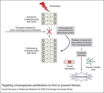
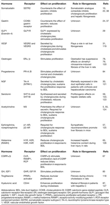
As our understanding of the autocrine and paracrine neuromodulators that regulate cholangiocyte proliferation during the progression of cholestatic liver diseases increases, so does our potential for the development of therapeutic strategies. The factors regulating cholangiocyte proliferation during cholestasis, which represent potential therapeutic targets are summarised in Table 1. Cholangiocyte proliferation is closely associated with a transdifferentiation of biliary epithelia to express neuroendocrine phenotypes and might provide unique signalling mechanisms that can be targeted for drug development (Ref. Reference Alvaro28). Preventing or limiting cholangiocyte proliferation and the activation of expression of profibrotic genes and secretion of profibrotic factors during the progression of cholestatic liver diseases represent a novel first line defence to control and/or prevent fibrogenesis (Fig. 4). In addition, further studies addressing the interaction of proliferating cholangiocytes with other cell types, such as portal myofibroblasts and hepatic stellate cells, are needed. The potential connection between the switching on of the neuroendocrine phenotype in proliferating cholangiocytes and activation of liver fibrotic processes by fibroblasts should reveal potential novel signalling mechanisms that could be simultaneously targeted for therapeutic benefit. The challenge ahead will be to consistently and properly modulate these many signalling systems and the interactions between various cell types to implement successful pharmacological therapies.
Targeting cholangiocyte proliferation to limit or prevent fibrosis. Cholangiocyte proliferation activated by cholestasis stimulates the neuroendocrine transdifferentiation of cholangiocytes. Cholangiocytes release autocrine or paracrine factors that regulate proliferative responses and also activate fibrogenic responses of portal fibroblasts and hepatic stellate cells, resulting in activated myofibroblasts and fibrogenesis. In addition, cholangiocytes can undergo epithelial–mesenchymal transition and increase the number of fibrogenic cells in the portal areas. Therapies targeted to prevent and/or reduce cholangiocyte proliferation might play a novel role in the prevention and/or limitation of liver fibrosis.

Neuroendocrine and other factors affecting cholangiocyte proliferation

Abbreviations: BDL, bile duct ligation; CCKB, cholecystokinin B; CGRP, calcitonin gene-related peptide; CLR, calcitonin-receptor-like receptor; ER, oestrogen (estrogen) receptor; GH, growth hormone; GLP1, glucagon-like peptide 1; IGF1, insulin-like growth factor 1; PPARγ, peroxisome proliferator-activated receptor-γ; PR, progesterone receptor; RAMH, α-methyl histamine; RAMP1, receptor activity-modifying protein 1; RCP, receptor component protein; SSTR2, somatostatin receptor subtype 2; Trk-A, neurotrophic tyrosine kinase receptor type 1; VEGF, vascular endothelial growth factor.
Acknowledgements and funding
Portions of the studies discussed here were supported partly by a grant award from Scott & White to S.G., by the Dr Nicholas C. Hightower Centennial Chair of Gastroenterology from Scott & White; the VA Research Career Scientist Award, a VA Merit Award and the NIH grants DK76898, DK58411 and DK062975 to G.A. Alpini; by University and Federate Athenaeum funds from University of Rome “La Sapienza” to Eugenio Gaudio; and the MIUR grant PRIN #2007, prot. 2007HPT7BA_003 to D.A. The authors would like to thank the peer reviewers for their time and comments on the manuscript.