Tetralogy of Fallot is the most common congenital cyanotic heart disease beyond infancy, accounting for ~10% of all congenital cardiac defects.Reference Bashore 1 “Corrective” intra-cardiac repair has been performed for more than four decades with good results.Reference Murphy, Gersh and Mair 2 Nevertheless, long-term follow-up studies have reported reduced right and left ventricular function.Reference Abd El Rahman, Abdul-Khaliq and Vogel 3 , Reference Ghai, Silversides, Harris, Webb, Siu and Therrien 4 Atrial function is a crucial determinant of ventricular filling,Reference Piotrowski, Goch, Wlazlowski, Gawor and Goch 5 , Reference Prioli, Marino, Lanzoni and Zardini 6 and the atria may play an important role during the course of evolving heart failure.Reference Piotrowski, Goch, Wlazlowski, Gawor and Goch 5 , Reference Kono, Sabbah, Rosman, Alam, Stein and Goldstein 7 Data on the atrial function in such patients is sparse, contradictive,Reference Hui, Abd El Rahman and Dsebissowa 8 – Reference Luijnenburg, Peters and van der Geest 10 and to the best of our knowledge there are no data available on the interaction between the two atria.
Speckle tracking echocardiography-derived strain and three-dimensional echocardiography for volumetric assessment of atrial volumes are reproducible methods that have been used in the assessment of atrialReference Aune, Baekkevar, Roislien, Rodevand and Otterstad 11 , Reference Cameli, Lisi and Focardi 12 and ventricular performance.Reference Mueller, Rentzsch and Hoetzer 13
The purpose of this study conducted in a cohort of patients after surgical correction of tetralogy of Fallot was, first, to assess atrial performance using these novel techniques, and second, to clarify the presence of inter-atrial, inter-ventricular, and atrio-ventricular interactions.
Methods
Patient population
A total of 34 asymptomatic patients after corrective surgery of tetralogy of Fallot were included in the study. The exclusion criteria were the presence of patent foramen oval, residual ventricular septal defect, the presence of significant tricuspid or mitral regurgitation, and the absence of sinus rhythm.
The mean±SD age of the patients was 20.88±9.22 years. Their mean±SD age at repair was 4.65±4.95 years. The mean±SD age of follow-up was 16.03±6.99 years. During surgical repair, the right ventricular outflow tract was reconstructed using a valved homograft in 19 patients and right ventricular outflow patch in 15 patients. Of the patients, six (17%) underwent a reoperation because of significant pulmonary regurgitation (n=4), right ventricular outflow aneurysm (n=1), and significant right ventricular outflow tract obstruction (n=2). Of the 34 patients, 30 (88%) were in NYHA functional class I, whereas four patients (12%) were in class II.
On the same day, all patients underwent electrocardiogram, two-dimensional speckle tracking echocardiography, and real-time three-dimensional echocardiography to evaluate ventricular and atrial function.
Ultrasound data from the patient group were compared with those from 35 age-matched healthy controls, who were examined for an innocent murmur. Their mean±SD age was 16.86±7.05 years. The study protocol was approved by the local ethics committee, and informed written consent was obtained from the patients or their parents.
Electrocardiogram examination
All patients had a 12-lead surface electrocardiogram performed with Siemens recorder (Siemens, Erlangen, Germany) at a speed of 50 mm/s and 1 mV/cm standardisation. Maximal QRS width in any lead was measured from the first to the last sharp vector crossing the isoelectric line.
Conventional echocardiographic examination
All patients were examined by echocardiography using GE M4S or GE M5S-D phased-array transducer with a Vingmed Vivid 9 ultrasound system (General Electrics, Fairfield, Connecticut, United States of America). Initially, routine diagnostic imaging was performed, including colour flow mapping and continuous Doppler. The severity of pulmonary regurgitation was assessed by two experienced paediatric cardiologists. Severe pulmonary regurgitation was recognised by the presence of wide colour Doppler regurgitation jet at the level of the pulmonary artery, with reverse flow extending up to the level up to the pulmonary bifurcation and/or pulsed Doppler pulmonary regurgitation jet lasting less than two-thirds of diastolic duration.Reference Hatle 14 Patients not fulfilling these criteria were considered to have mild-to-moderate pulmonary regurgitation. The presence or absence of restrictive physiology defined as the presence of antegrade pulmonary flow in late diastole coinciding with atrial systole throughout the respiratory cycle was determined.Reference Gatzoulis, Clark, Cullen, Newman and Redington 15
Speckle tracking echocardiography
The cardiac chambers were scanned using a conventional four-chamber view in the left lateral decubitus position. The gain was set to achieve optimal differentiation of the myocardium and endocardium. Then a loop of three cardiac cycles was recorded and digitally stored as cine loops and transferred to an image database via a web data program for further two-dimensional speckle tracking analysis. All images were recorded at a frame rate of 50–80 frames/second to allow reliable operation of the two-dimensional speckle tracking software (EchoPac; General Electrics, Fairfield, Connecticut, United States of America).
For the ventricular speckle tracking analysis, regions of interest were placed in the apical four-chamber view of a two-dimensional image in the free lateral wall of the right and left ventricle as well as the inter-ventricular septum. Two-dimensional speckle tracking-derived longitudinal mean deformation (strain) was calculated from the basal, middle, and apical region of each ventricular free wall and inter-ventricular septum.
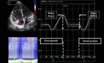
For atrial speckle tracking (Fig 1), the atrial endocardial surface was manually traced by a point and click approach. An epicardial surface tracing was then automatically generated by the system, thus creating the region of interest. Care was taken to exclude the pulmonary veins and coronary sinus from the tracking. After manual adjusting the width and shape of the regions of interest, the software divides the regions of interest into six segments, and the resulting tracking quality for each segment was automatically scored as either acceptable or non-acceptable, with the possibility of further manual correction. Segments in which no adequate imaging quality could be obtained were rejected by the software and excluded from the analysis. The beginning of the QRS was set as the reference from which the software starts to calculate atrial deformation. The software generated the longitudinal strain curves for each segment and a mean curve of all segments (Fig 1). The peak atrial longitudinal strain, measured at the end of the reservoir phase, that is, end of atrial filling, and peak atrial contraction strain, measured just before the start of active contractile phase, were calculated by averaging values observed in all atrial segments.Reference Cameli, Lisi, Righini and Mondillo 16
Peak atrial longitudinal strain and peak atrial contraction strain derived from atrial speckle tracking echocardiography in a patient with repaired tetralogy of Fallot. PACS=peak atrial contraction time; PALS=peak atrial longitudinal strain.

The recordings for atrial strain, volumes, and ejection fraction were analysed by two independent examiners to assess the inter-observer variability. In addition, the atrial measurements were analysed by one observer in two separate occasions to assess the intra-observer variability. Reproducibility of ventricular measurements was not assessed because the methods used are more established when compared with that of atrial measurements.
Assessment of atrial and ventricular volumes using real-time three-dimensional echocardiography
Real-time three-dimensional echocardiography was performed with an active matrix 4D-volume phased-array probe (4 V-D, 1.5–4.0 MHz, GE Fairfield, Connecticut, United States of America). Images were obtained from an apical view. Data acquisition was carried out using the multi-beat full-volume mode, in which sequential acquisitions of narrow subvolumes (4–6) were scanned during three consecutive heart beats without moving the transducer and during breath holding. These subvolumes were subsequently stitched together to form an entire pyramidal data set. The temporal resolution of the images was 25–30 frames/second, which can be manually adjusted from the machine.
To include the entire ventricular and atrial cavity within the three-dimensional pyramidal volume, each chamber was scanned separately. During acquisition of the right ventricle, the probe was positioned laterally and tilted anteriorly to visualise the right ventricular outflow tract.
Acquisitions were stored in a DICOM format and transferred to a separate workstation for offline data analysis. Ventricular and atrial volumes were analysed, respectively, with left and right ventricular dedicated software (Tom Tec Imaging Systems, Munich, Germany), as previously described.Reference Cameli, Lisi, Righini and Mondillo 16 , Reference Seguela, Hascoet, Brierre, Bongard and Acar 17
For assessment of the atrial volume, the atrial endocardial border in the three planes was manually traced – pulmonary veins and coronary sinus, and the appendages were excluded – starting the measurements in the frame with the largest atrial dimension, corresponding to ventricular end-systole, just before the opening of the atrio-ventricular valves. Then the frame with the smallest atrial dimension – at ventricular end-diastole, just before the opening of the atrio-ventricular valves – is selected and the same manual tracing is applied. Atrial maximum and minimum volumes are obtained, and atrial ejection fraction is derived from the two volumes according to the formula: Atrial ejection fraction=(Atrial (volume maximum)−Atrial (volume minimum))/(Atrial (volume maximum)).Reference Cameli, Lisi, Righini and Mondillo 16
Statistical analysis
Statistical analyses were performed using SPSS 15.0 (SPSS Inc., Chicago, Illinois, United States of America). All continuous variables were tested for normality using the Kolmogorov–Smirnov test. The data are shown as mean±SD. The student t-test was used to assess the differences between two unpaired groups. Correlations were evaluated using the Pearson correlation. A p-value<0.05 was considered statistically significant. Inter- and intra-observer variability was assessed by the Bland–Altman plot.Reference Bland and Altman 18
Results
Conventional electrocardiogram and echocardiographic examination
All patients after tetralogy of Fallot repair had right bundle branch morphology. The mean±SD of the QRS duration among them was 151.21±26.59 ms. Of the patients, 16 patients (47%) had a peak gradient >40 mmHg across the right ventricular outflow tract. In all, 28 patients (82%) had mild-to-moderate pulmonary regurgitation, whereas six patients (17%) had severe pulmonary regurgitation. Doppler pattern of right ventricular restrictive physiology – antegrade pulmonary flow in late diastole coinciding with atrial systole throughout the respiratory cycle – was detected in nine patients (26%). The right ventricular outflow gradient did not differ significantly among patients with restrictive and non-restrictive right ventricular physiology (43.21±20.94 versus 36.96±17.90 mmHg, p=0.40).
The ventricular performance and the effect of pulmonary regurgitation and right ventricular restrictive physiology
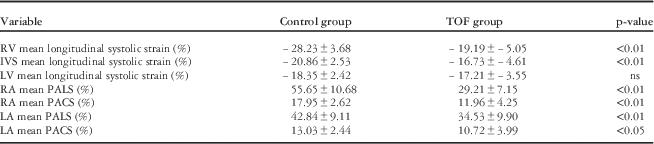
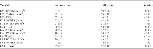
The speckle tracking and three-dimensional echocardiography data among patients and controls are summarised in Tables 1 and 2.
Two-dimensional speckle tracking data among TOF patients and control group.

IVS=inter-ventricular septum; LA=left atrium; LV left ventricle; PACS=peak atrial contraction strain; PALS=Peak atrial longitudinal strain; RA=right atrium; RV=right ventricle; TOF=tetralogy of Fallot
Real-time three-dimensional echocardiography data of the TOF and control group.

BSA=body surface area; EDV=end-diastolic volume; EF=Ejection fraction; ESV=end-systolic volume; LA=left atrium; LV=Left ventricle; RA=right atrium; RV=right ventricle; TOF=tetralogy of Fallot
Compared with patients with mild-to-moderate pulmonary regurgitation, patients with severe pulmonary regurgitation had significantly increased right ventricular end-diastolic (116±25 versus 92±24 ml/m2, p<0.05), end-systolic (67±18 versus 49±16 ml/m2, p<0.01) volume corrected to the body surface area, longer QRS duration (175.40±22.67 versus 140.69±22.67 ms, p<0.01), and reduced right ventricular ejection fraction (40±7 versus 46±6%, p<0.05). A weak negative correlation was found between the right ventricular ejection fraction and the gradient across the right ventricular outflow tract (r=−0.41, p<0.05).
Patients with restrictive right ventricular physiology when compared with patients with non-restrictive physiology had a significantly reduced mean right ventricular systolic strain (14.48±2.95 versus 19.96 versus 4.51, p<0.05). On the other hand, no significant difference was found between the two groups regarding the right ventricular end-diastolic (96±22 versus 108±36 ml/m2), end-systolic (61±24 versus 52±15 ml/m2) volume corrected to the body surface area and right ventricular ejection fraction (45±9 versus 44±6%).
In the patient group, a significant correlation was seen between the QRS duration and the right ventricular end-diastolic (r=0.60, p<0.01) and end-systolic (r=0.64, p<0.01) volume both corrected to the body surface area.
The inter-ventricular interaction
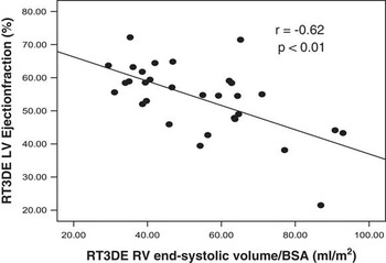
Among the tetralogy of Fallot patients, a significant negative correlation between left ventricular ejection fraction and the right ventricular end-systolic (r=−0.62, p<0.01, Fig 2) and end-diastolic (r=−0.47, p<0.01) volume corrected to the body surface area was observed. A positive association was obtained among the tetralogy of Fallot patients between the left ventricular ejection fraction and right ventricular ejection fraction (r=0.51, p<0.01), as well as between the left ventricular ejection fraction and the QRS duration (r=0.59, p<0.01).
Correlation between the right ventricular end-systolic volume corrected to the body surface area and left ventricular ejection fraction among tetralogy of Fallot patients after surgical correction. BSA=body surface area; LV=left ventricle; RT3DE=real-time three-dimensional echocardiography; RV=right ventricle.

The atrial performance and the effect of right ventricular pressure, volume load, and right ventricular restrictive physiology on the right atrial performance
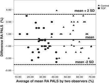
The mean difference for inter-observer agreement (95% limits of agreement) regarding right atrial peak longitudinal strain (Fig 4), peak contraction strain, and ejection fraction was 0.11 (−4.04 to +4.26%), 0.15 (−2.31 to +2.61%), and 0.15 (−1.69 to +1.99%), respectively, whereas that of left atrial peak longitudinal strain, peak contraction strain, and ejection fraction was 0.62 (−4.06 to +5.3%), 0.43 (−2.11 to +2.97%), and 0.52 (−2.64 to +3.68%), respectively.
The mean difference for intra-observer agreement (95% limits of agreement) regarding right atrial peak longitudinal strain, peak contraction strain, and ejection fraction was 0.11 (−3.35 to +3.57%), 0.12 (−2.04 to +2.4%), and 0.22 (−1.43 to +1.84%), respectively, whereas that of left atrial peak longitudinal strain, peak contraction strain, and ejection fraction was 0.47 (−2.41 to +3.35%), 0.33 (−1.58 to +2.23%), and 0.52 (−2.24 to +3.28%), respectively.
The right and left atrial peak longitudinal, contraction strain, and ejection fraction were significantly reduced among patients when compared with controls (Tables 1 and 2).
Patients with an right ventricular outflow gradient >40 mmHg showed a significantly reduced right atrial peak longitudinal strain (25.60±6.95 versus 32.21±5.55%, p<0.01), increased right atrial end-systolic (67±38 versus 30±8 ml/m2, p<0.01), and end-diastolic (46±35 versus 16±6 ml/m2, p<0.01) volume corrected to the body surface area, and reduced right atrial ejection fraction (37±15 versus 49±12%, p<0.05) when compared with patients with a right ventricular outflow gradient <40 mmHg.
Compared with patients with mild-to-moderate pulmonary regurgitation, patients with severe pulmonary regurgitation had significantly increased right atrial end-diastolic (48±38 versus 21±19 ml/m2, p<0.05) and end-systolic (65±42 versus 38±24 ml/m2, p<0.05) volume corrected to the body surface area, and significantly reduced right atrial ejection fraction (33±18 versus 48±9%, p<0.01) and right atrial peak longitudinal strain (24.83±7.52 versus 30.86±6.41%, p<0.05). No significant difference was found between the two groups regarding the right atrial peak contraction strain (11.00±5.87 versus 12.32±3.55%).
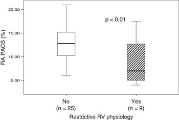
Patients with restrictive right ventricular physiology when compared with patients with non-restrictive physiology had a significantly increased right atrial end-systolic (81±43 versus 33±12 ml/m2, p<0.01) and end-diastolic (62±38 versus 17±7 ml/m2, p<0.01) volume corrected to the body surface area, reduced right atrial ejection fraction (27±14 versus 50±7%, p<0.01), reduced right atrial peak longitudinal strain (22.36±6.68 versus 31.40±5.88%, p<0.01), and reduced right atrial peak contraction strain (8.74±4.95 versus 12.99±3.52%, p<0.01, Fig 3).
Box-plot compares right atrial right peak atrial contraction strain in postoperative tetralogy of Fallot patients with and without right ventricular restrictive right ventricular physiology. PACS=peak atrial contraction strain; RA=right atria; RV=right ventricle.

The right atrio-ventricular coupling
Among the tetralogy of Fallot patients, the right atrial end-systolic volume corrected to the body surface area correlates negatively with right atrial peak longitudinal (r=−0.76, p<0.01) and contraction (r=−0.66, p<0.01) strain. A significant negative correlation between right atrial ejection fraction and right ventricular end-systolic volume corrected to the body surface area (r=−0.54, p<0.01) was elicited. Meanwhile, a positive correlation existed between the right atrial ejection fraction and the right ventricular ejection fraction (r=0.47, p<0.01). In addition, the right atrial peak longitudinal strain correlates with the mean strain of the right ventricle (r=0.64, p<0.01). In the healthy group, no right atrio-ventricular coupling was found.
Bland–Altman plot of inter-observer right atrial peak atrial longitudinal strain measurement. PALS=peak atrial longitudinal strain; RA=right atrium.

The left atrio-ventricular interaction
Among the patients, no correlation was found between the left atrial ejection fraction and left ventricular end-systolic or end-diastolic volume corrected to the body surface area. A weak correlation existed between the left atrial ejection fraction and that of the left ventricle (r=0.41, p<0.05). The left atrial peak longitudinal strain correlated with the mean strain of the inter-ventricular septum (r=0.41, p<0.05); on the other hand, no correlation was found between the left atrial longitudinal and the mean strain of the lateral wall of the left ventricle.
The inter-atrial interaction
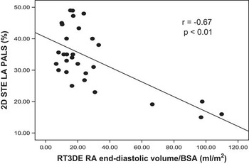
In the tetralogy of Fallot group, a negative correlation was found between right atrial end-diastolic volume corrected to the body surface area and left atrial peak longitudinal strain (r=−0.67, p<0.01, Fig 5), left peak contraction strain (r=−0.67, p=0.0001), and left atrial ejection fraction (r=−0.68, p<0.01). No correlation was found between the left atrial ejection fraction and left ventricular end-systolic or end-diastolic volume corrected to the body surface area.
Atrial interaction is also detectable among tetralogy of Fallot patients. An enlarged right atrium hampers adequate distension and filling of the left atrium. BSA=body surface area; LA=left atria; PALS=peak atrial longitudinal strain; RA=right atria; RT3DE=real-time three-dimensional echocardiography; 2D STE=two-dimensional speckle tracking echocardiography.

Discussion
Our data showed that biatrial dysfunction is detectable among postoperative tetralogy of Fallot patients using two-dimensional Speckle tracking and three-dimensional Echocardiography. In this cohort, interaction exists not only on the ventricular but also on the atrial level, and a robust right atrio-ventricular coupling is present on the right side.
The ventricular systolic function and inter-ventricular interaction
Although surgical correction of tetralogy of Fallot may reconstruct the anatomy, right and left ventricular dysfunction is still evident.Reference Murphy, Gersh and Mair 2 Therefore, monitoring the ventricular function in these patients in daily clinical practice requires non-invasive, bedside, robust, and easily applied modalities. The best diagnostic tool that can be applied to fulfil these requirements is still echocardiography. Assessment of the right ventricle and abnormally shaped left ventricle using conventional echocardiographic modalities is challenging, as geometrical assumptions are frequently needed. The two-dimensional speckle tracking-derived strain and volumetric assessment of the ventricular ejection fraction via real-time three-dimensional echocardiography does not need any geometrical assumption, and therefore might be useful in this clinical setting to reflect better the global and regional ventricular function. Our study demonstrated a reduced two-dimensional speckle tracking echocardiography-derived longitudinal deformation of the right ventricular free wall, inter-ventricular septum, and the ejection fraction of both the ventricles. Accordingly, we can conclude that after tetralogy of Fallot repair, not only the right ventricle but also the left suffers from regional as well global systolic abnormalities, as has been shown in other studies.Reference Abd El Rahman, Abdul-Khaliq and Vogel 3 , Reference Ghai, Silversides, Harris, Webb, Siu and Therrien 4 The left ventricular dysfunction is most likely multi-factorial, including intraoperative myocardial ischaemic injuryReference Hausdorf, Hinrichs, Nienaber, Schark and Keck 19 or myocardial injury related to chronic hypoxia before repair.Reference Krymsky 20 The strong negative correlation observed in our study between the left ventricular ejection fraction and right ventricular end-systolic volume corrected to body surface area (Fig 2), as well as the positive correlation with right ventricular ejection fraction confirms previous assumptions that part of left ventricular dysfunction is related to right ventricular dilatation and dysfunction.Reference Lange, Onnasch, Bernhard and Heintzen 21 Such interaction is most probably mediated via a paradoxically working inter-ventricular septum.Reference Abd El Rahman, Hui and Dsebissowa 22 The reduced mean inter-ventricular longitudinal strain found in our study among the tetralogy of Fallot group (Table 1) supports this hypothesis. Pulmonary regurgitation in this setting plays an important role;Reference Lange, Onnasch, Bernhard and Heintzen 21 however, other factors may also operate and should not be ignored. In the present study, patients with severe pulmonary regurgitation had significantly reduced right and left ventricular ejection fraction, emphasising the role of pulmonary regurgitation in this setting. In addition, the correlation found between the QRS duration, a marker for ventricular asynchrony, and the left ventricular ejection fraction points that the ventricular interaction might also be mediated via ventricular electrical conduction abnormalities as well.Reference Ghai, Silversides, Harris, Webb, Siu and Therrien 4
The atrial function, inter-atrial, and atrio-ventricular interactions
Atrial function is a crucial determinant of ventricular filling, especially among patients with impaired relaxation, and the atria play an important role during the course of evolving heart failure.Reference Piotrowski, Goch, Wlazlowski, Gawor and Goch 5 , Reference Kono, Sabbah, Rosman, Alam, Stein and Goldstein 7 In postoperative tetralogy of Fallot patients, data regarding atrial function are sparse and contradictive.Reference Hui, Abd El Rahman and Dsebissowa 8 – Reference Luijnenburg, Peters and van der Geest 10 One of the causes for paucity of the data is the absence of a modality that can be used in daily practice. Recently, two new echocardiographic modalities have been described for the evaluation of atrial function, namely, two-dimensional speckle tracking echocardiography and three-dimensional assessments of the atrial volumes and ejection fraction.
Using speckle tracking echocardiography, two main novel parameters could be obtained, namely, the peak atrial longitudinal strain, which reflects passive atrial deformation, that is, reservoir function, and peak atrial contraction strain representing the active atrial deformation, that is, pump function.Reference Cameli, Lisi, Righini and Mondillo 16 Our study shows that peak longitudinal strain of the right and left atria were reduced among the tetralogy of Fallot patients when compared with controls (Table 2). The reduced peak longitudinal strain of the right atrium can be explained by reduced right ventricular systolic function. This is supported by the negative correlation found in our study between the right ventricular end-systolic volume corrected to the body surface area and the right atrial peak longitudinal strain. The positive correlation demonstrated in our study between the right atrial ejection fraction and right ventricular ejection fraction can provide additional proof for this assumption. Accordingly, we may hypothesise that a robust atrio-ventricular interaction is present on the right side. In contrast, an atrio-ventricular coupling on the left side was not observed in our study. Similar to our findings, Bazaz et alReference Bazaz, Edelman, Gulyasy and Lopez-Candales 23 reported in a variety of adult patients that the coupling of ventricular ejection fraction and atrial filling is more pronounced for the right side compared with the left side, a finding that supports our observations. In the present study, the left side atrio-ventricular interaction was not obvious, as the left atrial peak longitudinal strain correlated significantly to the mean strain of the inter-ventricular septum but not to that of the lateral wall of the left ventricle. The latter was not significantly different from normal controls (Table 1). Accordingly, measuring the left ventricular strain of the lateral wall of the left ventricle is not sufficient to reflect the reservoir function of the left atrium, as the walls of the left ventricle are not uniquely affected.Reference Abd El Rahman, Hui and Yigitbasi 24 The negative correlation between the right atrial volume and left atrial peak longitudinal strain (Fig 5) found in our study may point to an additional factor responsible for the reduced reservoir of the left atrium, namely, an inter-atrial interaction. A distended right atrium reduces the compliance of the left atrium and thus hampers its adequate filling and pumping. Such interaction might be a reflection of the observed inter-ventricular interaction. Further studies using MRI are needed to confirm these suggestions.
Data regarding right atrial pump function is contradictive. Similar to our results, Hui et alReference Hui, Abd El Rahman and Dsebissowa 8 and Riesenkampff et alReference Riesenkampff, Mengelkamp and Mueller 9 found a reduced atrial pump function, whereas Luijnenburg et alReference Luijnenburg, Peters and van der Geest 10 found an enhanced right atrial pump function in the tetralogy of Fallot patients in a manner that coincides with abnormal relaxation. Difference in the right atrial after-load – right ventricular end-diastolic pressure and right ventricular end-systolic volume – in these studies might be a good explanation for the discrepancy. The negative correlation found in our study between the right atrial peak contraction strain and the right ventricular end-systolic volume corrected to the body surface area favours this speculation. In addition, it is not surprising to find in the present study that patients with restrictive right physiology had reduced right atrial performance when compared with others without restrictive physiology, as, among patients with restrictive physiology, the right ventricular myocardium is extremely stiff with reduced right ventricular compliance. Consequently, atrial contribution in ventricular filling, especially in late diastole, is reduced or even absent.Reference Lam, Kaya, Goktekin, Gatzoulis, Li and Henein 25 Invasive measurement of the right ventricular end-diastolic pressure is needed to prove our speculations. Another possible explanation for the reduced right atrial performance is the effect of atrial scarring because of transatrial approach. Theoretically, investigating this factor necessitates the elimination of pressure or volume load on the right ventricle. Among the studied cohort, this is impossible as variable degrees of right ventricular pressure or volume load after the corrective surgery are inevitable. Studying patients after surgical closure of atrial septal defect can help to clarify this issue and should be an objective for further studies.
The left atrial peak contraction strain reflecting left atrial pump function was reduced in our study (Table 2). The same observation was found by Luijnenburg et alReference Luijnenburg, Peters and van der Geest 10 The previously reported reduced compliance of the left ventricle in the postoperative tetralogy of Fallot patients either secondary to myocardial fibrosisReference Hausdorf, Hinrichs, Nienaber, Schark and Keck 19 and/or right ventricular enlargementReference Abd El Rahman, Abdul-Khaliq and Vogel 3 , Reference Lange, Onnasch, Bernhard and Heintzen 21 might a good explanation for reduced left atrial pump function.
Limitations
Currently, there is no widely accepted standard for atrial function assessment. For this issue, we used the novel speckle-derived strain and three-dimensional echocardiography. Both methods have been validated in previous studies.Reference Aune, Baekkevar, Roislien, Rodevand and Otterstad 11 , Reference Cameli, Lisi, Righini and Mondillo 16 Tracking the thin-walled atria requires reduction in the width of the region of interests, which may result in the reduction of the lateral resolution of the technique and convert it into an angle-dependent one. In postoperative tetralogy of Fallot patients, the atrial wall is usually not thin as pressure overload exists; accordingly not much reduction in the region of interests width is required, which will consequently improve the speckle tracking technique.
The relatively small-sized sample (34 patients) is another limitation that may affect the correlation coefficient, especially if more patients with much reduced right ejection fraction were included in the study. In addition, it may affect the comparison within the subgroups of severe pulmonary regurgitation (n=6) and right ventricular restrictive physiology (n=9). Further multicentre studies are needed to get a definite clue regarding the effect of right ventricular volume load and restrictive physiology on the right atrial performance.
Information about invasive pressure measurements is lacking as our study was non-invasive. Invasive pressure measurements could have given more insight into the ventricular and atrial functional parameters and its implications.
Our study has been conducted on young adults operated upon according to current surgical strategies. Our result may therefore be not representative for older tetralogy of Fallot patients.
Conclusion
In tetralogy of Fallot patients with moderately enlarged right ventricle and mildly impaired biventricular systolic function, biatrial function abnormality exists and can be quantified via two-dimensional speckle tacking and real-time three-dimensional echocardiography. Different forms of interactions on the atrial and ventricular levels are evident in such a cohort.
Acknowledgements
The authors thank Anne Gale of the Deutsches Herzentrum Berlin for editorial assistance.
Financial Support
This research received no specific grant from any funding agency, commercial, or not-for-profit sectors. It was supported by the Competence Network for Congenital Heart Defects funded by the Federal Ministry of Education and Research (BMBF).
Conflicts of Interest
None.
Ethical Standards
The authors assert that all procedures contributing to this work comply with the ethical standards of the Helsinki Declaration of 1975, as revised in 2008, and has been approved by the institutional committee of Homburg University.