Percutaneous treatment of aortic coarctation is a widely used option. Covered stents have increased the profile of efficacy and safety of this procedure.Reference Butera, Piazza and Chessa 1 – Reference Tzifa, Ewert and Brzezinska-Rajszys 6
Sometimes, anatomy of aortic coarctation or recoarctation closely involves the subclavian or carotid arteries. In this setting, a covered stent may create an iatrogenic obstruction of the important branching vessels. On the contrary, bare stents can allow the blood to flow through the branching vessels. However, bare stents are not the preferred choice in case of associated wall aneurysm.
Here we report on a 32-year-old woman with a significant aortic recoarctation close to the origin of both the subclavian arteries associated with aortic wall aneurysm.
We manually modified a covered stent to provide both covering of the stenotic/aneurysmatic area and an unobstructed free flow to the branching vessel.
Patient and procedure
A 32-year-old woman affected by the deletion of chromosome 21 came to our unit for clinical evaluation. At the age of 5 months, she had undergone end-to-end anastomosis of the aortic arch because of aortic coarctation.
During follow-up, she developed persistent arterial systemic hypertension poorly controlled by anti-hypertensive drugs.
Clinical evaluation showed a weight of 41 kg, height of 145 cm, arterial systemic pressure of 160/80 mmHg, reduced femoral pulses, and a systolic gradient of 30 mmHg between the arms and legs.
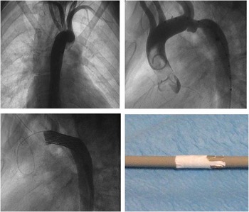
Magnetic resonance imaging showed an abnormal branching of the aortic neck vessels with an absent innominate artery. The first vessel was the right common carotid artery followed by the left common carotid artery, the right subclavian artery, and, as last vessel, the left subclavian artery (Fig 1 right and left upper). The subclavian arteries were located close to the recoarctation. A reduction of 60% of the lumen of the aortic arch was shown. Finally, an aneurysm of the aortic wall was present at the level of the recoarctation.
Right upper: Ascending aortography in anteroposterior view showing abnormal neck vessel branching. Left upper: Ascending aortography in the lateral view showing abnormality of the neck arterial vessels, aortic recoarctation and aneurysm of the wall both occurring in an area close to the origin of both the sublavian arteries. Right bottom: A 34-mm 8 zig Cheatham platinum stent used, partially inflated and with part of the covering partially removed, paying attention to leave in place about 1 mm expanded poly-tetra-fluoro-ethylene tissue at the level of the points where the covering is welded to the stent. Left bottom: Ascending aortography in the lateral view showing the stent properly placed, aortic coarctation dilated, aneurysm sealed, and the flow through the subclavian arteries maintained.

After discussion with the surgical team, a percutaneous strategy was proposed to the patient and her family. Because of a significant degree of neuropsychological retardation, informed consent for the procedure was signed by her parents.
The procedure was performed under general anaesthesia and orotracheal intubation. A right femoral artery access was obtained by using a 6 Fr sheath. Heparin 100 International units per kilogram and cefazoline 1 g were administered. Catheterisation was performed by using a 6 Fr multipurpose catheter and a 0.035 inches Terumo guidewire. The catheter was exchanged for a 6 Fr pigtail catheter and angiographies performed in anteroposterior, 45° left anterior oblique, and lateral views.
Pressure evaluation showed a peak-to-peak systolic gradient across the aortic arch of 55 mm of mercury, with systolic pressure in the ascending aorta of 140 mmHg.
Abnormality of the neck arterial vessels was confirmed on angiography (Fig 1 right and left upper). The recoarctation occurred in an area close to the origin of both the subclavian arteries (Fig 1, left upper). Furthermore, it was associated with an aneurysm of the aortic wall (Fig 1, left upper). Bare or covered stent implantation were the available options.
The last option was the preferred one. However, the covering of the stent would have significantly interfered with the flow in the subclavian arteries.
It was decided to manually and partially uncover the proximal part of the stent to have a modified stent that could act as a bare stent at the level of the origin of the subclavian arteries and as a covered stent at the level of the aneurysm (Fig 1, right bottom).
First, we chose the best angiographic view to delineate the aortic coarctation and the branching arterial neck vessels. The lateral view gave us the best imaging of the area.
Second, we obtained the following measurements: horizontal aorta diameter: 12 mm; diameter of the coarctation: 5 mm; diameter of the aorta at the diaphragm: 13 mm; distance between the subclavian arteries and the origin of the aneurysm: 10 mm; and length of the area to be stented: 30 mm.
A 34-mm 8 zig Cheatham platinum stent (NuMED Inc., Hopkintom, New York, United States of America) was chosen. Then, the stent was partially inflated on an 8-mm Tyshak II balloon (NuMED Inc.) and we manually removed 1 cm of expanded poly-tetra-fluoro-ethylene covering by using a surgical scissor. This part of the procedure was performed paying attention to leave in place about 1 mm poly-tetra-fluoro-ethylene tissue at the level of the points where the covering is welded to the stent (Fig 1, right bottom). This was done to avoid the rolling up of the covering when the stent was advanced inside the Mullins sheath. After this preparation, the stent was crimped down on a Crystal Balloon 12×40 (Balt, Montmorency, France) mm and implanted by using a 10 Fr Mullins sheath (William Cook ApS Europe, Bjaeverskov, Denmark) on a 0.035-inch, 260-cm long, J-tip, extra-stiff Amplatzer guidewire (Boston Scientific Corp., Natick, Massachusetts, United States of America).
The stent was properly placed. The coarctation was dilated, the aneurysm sealed, and the flow through the subclavian arteries maintained (Fig 1, left bottom). No residual gradient was measured. The fluoroscopic time was 10 minutes.
At the end of the procedure, the Mullins sheath was removed by keeping the guidewire in place. This was done to exchange the Mullins sheath for a 10 Fr femoral sheath in order to check for femoral access. Suddenly, the patient experienced hypotension and the Mullins sheath was promptly repositioned over the wire with rapid recovery of arterial pressure. An 8×20-mm Crystal balloon (Balt) was placed through the Mullins sheath in the abdominal aorta and multiple hand angiographies were performed while retrieving the Mullins sheath. A lesion of the right iliac artery was shown with contrast extravasation.
It was treated by implanting a 7×60-mm-long Viabhan prothesis (Gore, Flagstaff, United States of America) that was post-dilated by using a 6×30-mm Fox Cross peripheral angioplasty balloon. Furthermore, the post-procedural course was uneventful. The patient was hospitalised for 5 days. At physical examination, femoral pulses were symmetric, and arterial systemic blood pressure was 125/70 mmHg without gradient between the upper and lower part of the body. At echocardiographic evaluation, no signs of recoarctation were seen. The patient was discharged under acid acetylsalicylic acid treatment.
Follow-ups were carried out at 1, 3, 6, 9, 12, and 24 months. The latest evaluation confirmed good results both at physical examination and at echocardiographic study.
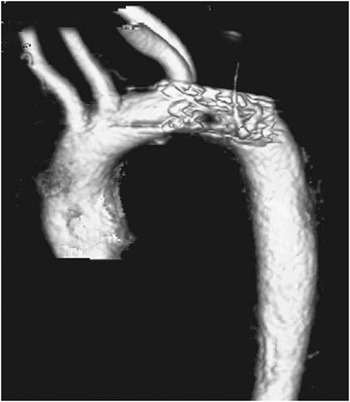
Thoracic computerised tomography scan showed no residual flow inside the aneurysm and no abnormalities of the flow through the subclavian arteries (Fig 2).
A three-dimensional reconstruction of thoracic scan showing no residual flow inside the aneurysm and no abnormalities of the flow through the subclavian arteries.

Discussion
Covered stent introduction in clinical practice has significantly widened the field of application of transcatheter techniques in the treatment of aortic coarctation.Reference Butera, Piazza and Chessa 1 – Reference Tzifa, Ewert and Brzezinska-Rajszys 6
In fact, atretic or near-atretic lesion, post-surgical recoarctation, and stenosis associated with the aortic wall aneurysm can now be treated by using covered stents.Reference Butera, Piazza and Chessa 1 – Reference Butera, Marini and Carminati 8
However, the risk associated with the use of covered stents is the potential for side branch occlusion. In our case, multiple problems were simultaneously present: aortic recoarctation, atrial wall aneurysm, anomaly of the neck vessels with independent origin of the four vessels, and finally, close proximity of the area of recoarctation of the aneurysm to the left and right subclavian arteries.
Use of a bare stent would have had the advantage of allowing free blood flow towards the subclavian arteries. However, the risk of aortic wall injury related to the previous surgical history, and the presence of aortic wall aneurysm, were considered to be too high.
Recently, Cheatham et alReference Tsai, Hill and Cheatham 9 have described a method to create a hole across the expanded poly-tetra-fluoro-ethylene covering of a covered stent, straddling the left subclavian artery.
This would have been an option; however, we thought to develop another alternative in order to avoid the need to obtain two other arterial accesses that would have increased the vascular risks.
We obtained careful measurements of the target area and we manually removed 10 mm of distal-expanded poly-tetra-fluoro-ethylene covering by using surgical scissors. This procedure took a few minutes. We took care to leave in place a 1-mm sleeve of expanded poly-tetra-fluoro-ethylene where it is welded with the stent. This was decided to avoid possible roll up of the stent when advanced inside the Mullins sheath. Furthermore, we thought that even if it had partially straddled across the subclavian artery lumen, it would not have impaired the blood flow because it was almost as thick as the stent strut.
Another option would have had been to remove all the expanded poly-tetra-fluoro-ethylene segment fixing the remaining covering with 7.0 surgical stitches.
Positioning allowed us to exclude the aneurysm and relieve the stenosis without any interference, with the blood flow towards the subclavian arteries.
Another important technical point that needs to be underlined is the importance of testing for iliac/femoral artery integrity when removing the introducer while leaving the guidewire in place.
Obviously, the operator should compress the femoral access at the level of the groin to avoid significant blood loss. In this way, it is possible to test whether any injury occurred at the level of femoral-iliac axis. If this occurs, when having the wire in place, it is possible to advance the Mullins sheath again with its dilator to temporarily seal the lesion. Implantation of an endoprothesis may solve the vascular problem.
Conclusions
New tools are needed to deal with complex aortic coarctation. In particular, development of fenestrated or partially covered stents should be considered.
Aknowledgement
None.
Financial Support
This research received no specific grant from any funding agency, commercial or not-for-profit sectors.
Conflicts of Interest
None.