Implications
Zinc oxide (ZnO) is used in piglets diets at 2000 to 3000 mg/kg to reduce diarrhea and improve performance in the post-weaning phase (Poulsen, Reference Poulsen1989). We here report that microencapsulated ZnO added to weaning diets at a low concentration (400 mg/kg) is as effective as pharmacological ZnO (3000 mg/kg) in promoting an healthy intestinal mucosa in the 2 weeks post-weaning, and in maintaining the same feed efficiency in the 6 weeks post-weaning. This would eventually allow a reduction of ZnO inclusion rates and consequent environmental impact of excreted zinc.
Introduction
The post-weaning syndrome is one of the main causes of the economic losses associated with pig production, as at weaning environmental and stress factors can compromise the gut functionality and cause a decrease in absorptive capacity, diarrhea onset, and, eventually, growth retard. The mucosal integrity is essential for gut barrier function as a first line of protection against harmful exogenous agents, such as pathogens and bacterial products, and, within the gut barrier components, tight junctions (TJ) proteins seem to have a preponderant role in preventing barrier disruption (Lallès, Reference Lallès2010). Several extra-cellular molecules continuously modulate TJ status, among which inflammatory cytokines are considered as major mediators of intestinal epithelial permeability (Lallès, Reference Lallès2010). Especially in the ileum, which is the site where the Peyer’s patches are located and the microbial pressure is the highest, these inflammation mediators are always stimulated and can cause an increase in para-cellular permeability, thereby increasing bacterial translocation (Piè et al., Reference Pié, Lallès, Blazy, Laffitte, Sève and Oswald2004).
Despite a fragmented regulatory framework, which varies from country to country, zinc oxide (ZnO) at 2000 to 3000 mg/kg (pharmacological dose) has been generally accepted as a tool to reduce diarrhea, improve gut health and growth performance in the first weeks post-weaning (Poulsen, Reference Poulsen1989; Carlson et al., Reference Carlson, Hill and Link1999). The widespread diffusion of ZnO in medicated premixes in the EU, although regionally different as certain countries are not allowed to use it, whereas others are allowed to extend its use over 6 weeks post-weaning, has posed a threat from an environmental perspective, as the major part of ingested ZnO appears to be excreted with the feces (Poulsen and Larsen, Reference Poulsen and Larsen1995; Mavromichalis et al., Reference Mavromichalis, Peter, Parr, Ganessunker and Baker2000). Zinc oxide mechanism of action as growth promoter appears to go beyond the fulfillment of Zn nutritional needs, and it seems to be more related to improved intestinal health (Zhang and Guo, Reference Zhang and Guo2009). In fact, there is growing evidence that ZnO efficacy might be independent from absorption but rather it would act within the gut, as it is well established that above the requirements absorption and retention are no longer increased (Poulsen and Larsen, Reference Poulsen and Larsen1995; Krebs, Reference Krebs2000; Rincker et al., Reference Rincker, Hill, Link, Meyer and Rowntree2005). With this hypothesis in mind, it seems logical to assume that a controlled release of ZnO along the intestine through the use of slow-release techniques might improve its efficacy (Kim et al., Reference Kim, Hansen, Mullan and Pluske2012; Hu et al., Reference Hu, Song, Li, Luan and Zhu2013).
As ZnO efficacy is exploited at 10 to 20 times higher doses than Zn requirements, significantly decreasing the inclusion dose by maintaining the benefits would have a positive impact on manure pollution. For this reason, we wanted to test whether lipid microencapsulation, which was previously demonstrated to slowly release nutrients along the gut (Piva et al., Reference Piva, Pizzamiglio, Morlacchini, Tedeschi and Piva2007), would allow to effectively reduce ZnO levels in piglets diets, thereby reducing Zn excretion in manure, at the same time maintaining the same level of performance.
Therefore, the aim of this study was to investigate the possibility of reducing ZnO inclusion rates in post-weaning diets by comparing the growth performance of piglets fed either low doses of microencapsulated or pharmacological doses of ZnO. Moreover, as previous findings report an improved intestinal health as a result of pharma-ZnO in post-weaning diets, or rather as a mechanism of ZnO efficacy as growth promoter, we wanted to study a little more in depth the mechanism of action of ZnO, and we focused on some measures of intestinal health, that is, markers of inflammation status and mucosa permeability, in the ileum of pigs fed with either microencapsulated or pharmacological ZnO during the 6 weeks post-weaning.
Material and methods
The study was conducted at the facilities of the Research Centre for Animal Production and Environment (CERZOO), which is Good Laboratory Practices-certified and operates according to the Procedure of Animal Protection and Welfare (Directive No 86/609/EEC).
Animals and diets
One hundred and forty-four Landrace×Large White piglets, weaned at 28 days of age and 7.10kg (s.e. 0.06) of BW, were divided in 36 pens (two males and two females per pen) and randomly assigned to one of the following experimental groups: the basal diet (negative control, no supplemental Zn), providing Zn at 50 mg/kg in the first phase and at 45 mg/kg in the second phase; the basal diet added with feed grade ZnO at 3000 mg/kg (pharmacological Zn, pZnO, 72% of Zn); the basal diet supplemented with microencapsulated ZnO at 300 or 800 mg/kg, providing ZnO at 150 or at 400 mg/kg (mZnO-300 and mZnO-800, respectively; Zincoret-S, 50% of ZnO and 50% of lipid matrix; Vetagro SpA, Reggio Emilia, Italy).
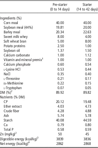
The basal feed was formulated in order to meet or exceed the nutritional requirements of pigs according to National Research Council (1998) but for Zn. Supplemental Zn sources were added to the basal diet in order to provide the amount of Zn necessary for each experimental diet. Feed and water were provided ad libitum and piglets were fed the experimental diets for 42 days. At day 14, the diet was switched to meet the nutritional requirements of the pigs in the subsequent period (Table 1). The health status of animals was monitored throughout the study and any culling or mortality was registered.
Composition and analyzed nutrient content of basal diets (as-fed basis)

DM=dry matter.
1 Providing per kilogram of premix: vitamin A, 1 500 000 UI; vitamin D3, 170 000 UI; vitamin E, 4000 mg; vitamin B1, 200 mg; vitamin B2, 500 mg; vitamin B6, 250 mg; vitamin B12, 4 mg; vitamin H, 15 mg; vitamin K, 250 mg; vitamin PP, 3000 mg; d-pantothenic acid, 1500 mg; choline cloride, 38 500 mg; folic acid, 100 mg; Mn, 6100 mg from manganese oxide; Fe, 15 000 mg from iron sulfate; Cu, 7600 mg from copper sulfate; Co, 50 mg; I, 150 mg from potassium iodide; Se, 30 mg from sodium selenite.
2 Analyzed total Zn in pre-starter diet was 2070 mg/kg, 165 mg/kg and 441 mg/kg for pZnO, mZnO-300 and mZnO-800 groups, respectively; Zn content in starter diet was 2330 mg/kg, 145 mg/kg and 355 mg/kg for pZnO, mZnO-300 and mZnO-800 groups.
3 According to the equation proposed by Whittemore (Reference Whittemore1987).
4 According to the equation proposed by Noblet et al. (Reference Noblet, Fortune, Shi and Dubois1994).
Piglets were individually weighed at the beginning of the study (day 0), after 14 days (day 14) and at the end of the study (day 42). Feed intake was measured per pen, by weighing refusals, during the same time intervals. Average daily gain (ADG), average daily feed intake (ADFI) and gain to feed ratio (G:F) were calculated for each interval (0 to 14 days, 14 to 42 days) and for the overall period (0 to 42 days). On days 7 and 14, feces were scored according to the following scale: 0, dry, hard, well formed feces; 1, soft but formed feces; 2, pasty feces green or brown in color; 3, viscous feces light in color; 4, fluid feces light in color; 5, watery feces, continuous (Peace et al., Reference Peace, Campbell, Polo, Crenshaw, Russell and Moeser2011). Fecal scores were conducted by personnel unaware of the experimental treatments.
At days 14 and 42 of the study, six pigs per treatment (one per six randomly selected pen per treatment) were euthanized by penetrating captive bolt followed by exsanguination to collect intestinal samples: the ileum was longitudinally cut 10 cm from the ileal–cecal valve to expose mucosa, washed with phosphate-buffered saline solution to remove mucus and digesta, then scraped gently with a glass slide, packed, immediately frozen in liquid N2 and stored at −80°C until gene expression analysis. Histological samples were instead collected and fixed in formalin until staining.
At the time of sacrifice, blood samples were also collected to perform plasma Zn analysis. Blood was collected by jugular venipuncture using 10cc heparinized (lithium heparin) disposable syringes (Becton Dickinson, Franklin Lakes, NJ, USA) and centrifuged at room temperature (RT) for 10 min at 3000×g; plasma was transferred with Pasteur pipette in a plastic test tube and immediately frozen at –80°C until analysis.
Chemical analysis
The dry matter, CP, ether extract, crude fiber, ash and starch contents of the feed were determined according to the AOAC (2000) methods. Zn in the feed was determined through incineration at 600°C overnight followed by microwave assisted acid digestion (EPA method 3052, 1996). Also Zn in plasma was hydrolyzed with HCl. Zn was then analyzed with Inductively Coupled Plasma Atomic Emission Spectrometry (EPA method 6010C, 2000) by using the Optima 2100 DV ICP/OES instrument (PerkinElmer Inc., Shelton, CT, USA). Standard solution of Zn analysis was certified reference material (Sigma-Aldrich, Milan, Italy).
Ileal histometry and immunohistochemistry
The analyses herein described were conducted according to Vitari et al. (Reference Vitari, Di Giancamillo, Deponti, Carollo and Domeneghini2012). Haematoxylin and eosin staining was carried out to assess the ileum micro-anatomical structure and perform histometry. For histometry, the following parameters were evaluated per section: villous height (10 villi measured per section), crypt depth (10 crypts measured per section), the villous height to crypt depth ratio (V:C). In other sections, the ileum mucin profile, and the respective histometry (number of mucous cells) were determined by staining sections with the Alcian Blue 8GX pH 2.5-periodic acid Schiff (AB-PAS) sequence, which reveals neutral (PAS-reactive, purple stained) and acid (AB-reactive, azure stained) glycoconjugates. The number of mucous cells was determined in five villi and five crypts of each section.
Other ileum sections were processed by immunohistochemistry to reveal proliferating epithelial cells (PCNA; 1:1000; clone PC10; Sigma-Aldrich). Proliferating cells were counted in six crypts of each section. For histology, histochemistry and immunohistochemistry, all the observations were made by a single investigator, blind to the pig groups, using an Olympus BX51 Microscope (Olympus Italia Srl, Milan, Italy) equipped with a digital camera and DP software for Computer-Assisted Image Acquirement and Management.
Inflammatory cytokines and TJ components gene expression analysis
Gene expression analysis was performed according to Herfel et al. (Reference Herfel, Jacobi, Lin, Fellner, Walker, Jouni and Odle2011). Ileal scraping samples obtained at day 14 of the study were disrupted by grinding in liquid N2 with mortar and pestle, then homogenized using TissueLyser (Qiagen, Hilden, Germany). Total RNA was isolated using RNeasy Mini Kit (Qiagen) according to the manufacturer’s instructions. Genomic DNA contamination was removed by treatment with deoxyribonuclease (RNase-Free DNase Set; Qiagen). RNA yield and quality were determined spectrophotometrically using A260 and A280 nm measurements. A total of 1 µg of RNA was reverse-transcribed with iScript cDNA Synthesis Kit (Bio-Rad Laboratories Inc., Hercules, CA, USA) according to the manufacturer’s instructions. Resulting complementary DNA (cDNA) was quantified with Quant-iT Oligreen ssDNA Assay (Life Technologies Italia, Monza, Italy) to normalize the quantity of cDNA template used for amplification by real-time PCR. Real-time PCR was performed using iCycler Thermal Cycler system and SybrGreen Supermix (Bio-Rad Laboratories Inc.). Thermocycling protocol included initial denaturation for 1 min and 30 s at 95°C, 40 cycles of denaturation at 95°C for 15 s followed by 30 s of annealing and extension at 60°C. After amplification, all samples were subjected to a melt curve analysis, with a slow heating from 55°C to 95°C with a rate of 0.5°C/s to validate absence of non-specific products.
Gene expression was normalized using two housekeeping genes (HKs), coding for portions of porcine ribosomal subunit 60 S, such as ribosomal protein L35 and ribosomal protein L4. Average threshold cycle (C T ) was determined for each gene of interest, and geometric average was calculated for HKs assuming C T as number of cycles needed to reach a fixed arbitrary threshold. Delta C T was calculated as C T (gene of interest)−C T (HKs), then a modification of the 2–ΔΔC T (Livak and Schmittgen, Reference Livak and Schmittgen2001) method was used to analyze the relative expression (fold changes), calculated relative to the control group.
The sequences, expected product length, accession number in the EMBL database/GenBank and references of porcine primers are provided in Supplementary Table S1. Primer oligonucleotides for tumor necrosis factor-α (TNF-α), and interferon-γ (IFN-γ), were designed using PrimerQuest software (IDT, Integrated DNA Technologies, www.idtdna.com). Primers were obtained from Life Technologies (Life Technologies Italia).
ELISA quantification of inflammatory cytokines
Inflammatory cytokines protein levels were analyzed using commercial ELISA kit specific for porcine cytokines (Quantikine ELISA, R&D Systems Inc., Minneapolis, MN, USA). Before analysis, ileal mucosa scrapings were disrupted by grinding in liquid N2 with mortar and pestle, added with lysis buffer (10 mM-2-amino-2-hydroxymethyl-propane-1,3-diol(Tris)-HCl, 1 mM-ethylenediaminetetraacetic acid (EDTA), 0.5%-Triton X100) and homogenized using TissueLyser (Qiagen). Analyses were performed according to manufacturer’s instructions. Results refer to picograms of cytokine per 100 mg of tissue (pg/100 mg).
TNF-α and TJ components Western blot quantification
TNF-α, occludin and zonula occludens-1 (ZO-1) protein levels were analyzed by Western blot. Briefly, mucosal scraping of ileum (∼1 g) from each animal were homogenized in a 2 ml solution containing ice-cold lysis buffer (50 mM-2-amino-2-hydroxymethyl-propane-1,3-diol (Tris)-HCl, 150 mM-NaCl, 0.1%-SDS, 0.5%-sodium deoxycholate, 1%-NP-40, pH 7.4) supplemented with a protease inhibitor cocktail (EuroClone, S.p.A., Milano, Italy). The homogenates were centrifuged at 10 000×g at 4°C for 5 min to remove all insoluble material. The supernatant was collected and the protein concentration of each sample was determined using a bicinchoninic acid protein assay kit (Pierce, Thermo Fisher Scientific Inc., Rockford, IL, USA). Total proteins (50 μg) were loaded either onto 10% SDS gels for occludin and TNF-α or onto 6% SDS gel for ZO-1, and subsequently electro-transferred onto nitrocellulose membranes. After blocking with 5% non-fat milk in Tris-buffered saline for 1 h at RT, membranes were incubated for 2 h at RT with the following primary antibodies: anti-occludin (1 : 500, Life Technologies Italia), anti-ZO-1 (1 : 500, Life Technologies Italia), anti-TNF-α (1 : 200, Abcam, Cambridge, UK) and anti-actin (1 : 2000, Sigma-Aldrich). Subsequently, they were washed and incubated for 1 h at RT with a horseradish peroxidase-conjugated secondary antibody (1 : 5000, Bio-Rad Laboratories Inc., Hercules, CA, USA). The blots were developed using the ECL system (GE Healthcare Europe, GmbH, Milan, Italy): immunoreactivities were detected by chemiluminescence autoradiography according to the manufacturer’s instructions. The optical densities of the protein bands of interest were determined densitometrically using the Scion Image software (Scion Corporation, Frederick, MD, USA) and the values were normalized to actin levels.
Statistical analysis
Animals were blocked in a completely randomized design and data were analyzed using one-way ANOVA with GLM procedure of SAS followed by Tukey post-hoc test to detect differences among treatments (SAS Institute Inc., release 9.2, Cary, NC, USA). The pen was the experimental unit for growth performance, whereas the pig was the experimental unit for histometry, immunohistochemical and inflammatory cytokines, and TJ components analyses. Differences were considered significant at P<0.05 and trends were defined at 0.05⩽P<0.1.
Results
Growth performance and plasma Zn
Animals maintained a good health status throughout the experiment and the overall mortality was 1.4% and was not affected by the treatments. Dead subjects were necropsied for gross examination and both clinical signs and lesions were consistent with respiratory problems associated with Streptococcus suis infection. Fecal score was not significantly different among the groups (average score 1.32).
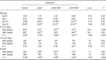
Growth performance is shown in Table 2. Compared with control, the pZnO group had greater ADG from 0 to 14 days, thus resulting in greater BW and G : F (P<0.05). Likewise, during the same weeks, the mZnO-800 group had an increased ADG compared with the control group; however, it did not affect G : F. The group mZnO-300 did not differ from the control group except for a lower feed intake (P<0.05).
Effect of feeding pharmacological ZnO or low doses of microencapsulated ZnO on growth performance in weaning piglets

ADG=average daily gain; ADFI=average daily feed intake; G : F=gain to feed ratio.
a,bValues within a row with different superscripts differ significantly at P<0.05.
A,BValues within a row with different superscripts differ significantly at P<0.01.
1 Treatments: control=basal diet providing Zn at 50 mg/kg; pZnO=basal diet+pharmacological ZnO providing ZnO at 3000 mg/kg; mZnO-300=basal diet+microencapsulated ZnO providing ZnO at 150 mg/kg; mZnO-800=basal diet+microencapsulated ZnO providing ZnO at 400 mg/kg (Zincoret-S, Vetagro SpA, Reggio Emilia, Italy).
From 14 to 42 days, although there were no significant differences among treatments, the ADG of all the groups receiving ZnO, regardless of the source and dose, tended to be greater than the control group (P=0.05). Between 0 to 42 days, pZnO and mZnO-800 pigs had higher ADG than the control pigs (P=0.02), whereas mZnO-300 did not differ from the control group. Nonetheless, at day 42, all of the groups weighed on average 1.6 to 2.2 kg more than control group and G : F was on average 10 points higher (P<0.05).
Zn source and amount significantly affected Zn plasma levels at both stages, as ZnO at 3000 mg/kg had the greatest values (P<0.01). In particular, plasma Zn concentration at day 14 was 15, 23, 14.5, and 18 µmol/l for control, pZnO, mZnO-300 and mZnO-800, respectively. At day 42, plasma Zn concentration was 10, 30, 10 and 12 µmol/l for control, pZnO, mZnO-300 and mZnO-800 groups, respectively.
Ileum histometry and immunohistochemistry
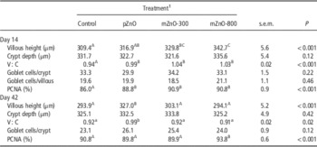
The histometrical results are summarized in Table 3. At day 14, the villi height was greater for mZnO-800 and mZnO-300 compared with control (P<0.05), whereas pZnO did not differ from control. mZnO-800 also had higher villi than pZnO (P<0.05), whereas mZnO-300 did not differ from pZnO. The crypt depth did not reveal significant differences among the groups, but the V : C ratio was greater in pZnO, mZnO-300 and mZnO-800 than in control piglets (P<0.01). Zinc oxide treatments did not influence the goblet cells numbers.
Effect of feeding pharmacological ZnO or low doses of microencapsulated ZnO on ileum histometry and immunohistochemistry in weaning piglets

V : C=villous height to crypt depth ratio; PCNA=proliferating cell nuclear antigen-positive cells.
a,bValues within a row with different superscripts differ significantly at P<0.05.
A,B,CValues within a row with different superscripts differ significantly at P<0.01.
1 Treatments: control=basal diet providing Zn at 50 mg/kg; pZnO=basal diet+pharmacological ZnO providing ZnO at 3000 mg/kg; mZnO-300=basal diet+microencapsulated ZnO providing ZnO at 150 mg/kg; mZnO-800=basal diet+microencapsulated ZnO providing ZnO at 400 mg/kg (Zincoret-S, Vetagro SpA, Reggio Emilia, Italy).
The percentage of PCNA-positive cells, which were counted in the ileum crypts, was higher in all ZnO supplemented groups compared with control group (P<0.01). At day 42, the villi length and V : C ratio were higher in the ileum of pZnO animals in comparison with the other groups (P<0.01). The crypt depth was not different among the groups. The percentage of proliferating cells was the highest in piglets of mZnO-800 group than in other groups (P<0.01). Goblet cell number was not affected by treatments.
Inflammatory cytokines profiling
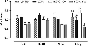
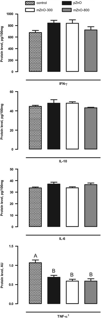
The mRNA and protein levels of inflammatory cytokines in ileum mucosa at day 14 are presented in Figures 1 and 2, respectively. Groups treated with microencapsulated ZnO tended to have reduced mRNA expression of interleukin-6 (IL-6), compared with both control and pharmacological ZnO groups (−25%, P=0.10). mZnO-800 group had the lowest expression of IFN-γ and significantly lower than mZnO-300 group (P=0.02). TNF-α mRNA was lower in both groups treated with microencapsulated ZnO compared with the others, although not significantly (P=0.18). No effects were observed in IL-10 gene expression.
Effect of feeding pharmacological ZnO or low doses of microencapsulated ZnO on gene expression of inflammatory cytokines in ileum mucosa of weaning piglets at day 14 post-weaning. Data are expressed as means (n=6) and s.e.m. represented by vertical bars. a,bValues with different superscripts differ significantly at P<0.05. Control=basal diet providing Zn at 50 mg/kg; pZnO=basal diet+pharmacological ZnO providing ZnO at 3000 mg/kg; mZnO-300=basal diet+microencapsulated ZnO providing ZnO at 150 mg/kg; mZnO-800=basal diet+microencapsulated ZnO providing ZnO at 400 mg/kg (Zincoret-S, Vetagro SpA, Reggio Emilia, Italy). A modification of the 2–ΔΔC T method was used to analyze the relative expression (fold changes), calculated relative to the control group (control; Livak and Schmittgen, Reference Livak and Schmittgen2001). IL-6=interleukin-6; IL-10=interleukin-10; TNF-α=tumor necrosis factor-α; IFN-γ=interferon-γ.

Effect of feeding pharmacological ZnO or low doses of microencapsulated ZnO on protein expression of inflammatory cytokines in ileal mucosa of weaning piglets at day 14 post-weaning. Data are expressed as means (n=6) and s.e.m. represented by vertical bars. A,BValues with different superscripts differ significantly at P<0.01. Control=basal diet providing Zn at 50 mg/kg; pZnO=basal diet+pharmacological ZnO providing ZnO at 3000 mg/kg; mZnO-300=basal diet+microencapsulated ZnO providing ZnO at 150 mg/kg; mZnO-800=basal diet+microencapsulated ZnO providing ZnO at 400 mg/kg (Zincoret-S, Vetagro SpA, Reggio Emilia, Italy). Data refer to picograms of cytokine per 100 mg of tissue (pg/100 mg) measured using ELISA method. 1 TNF-α expression was measured by Western blot and data refer to AU of protein expression. IFN-γ=interferon-γ; IL-10=interleukin-10; IL-6=interleukin-6; TNF-α=tumor necrosis factor-α; AU=arbitrary unit.

IFN-γ and IL-10 protein content tended to be higher in pZnO group and mZnO-300 group than in control and mZnO-800 groups (P=0.08). IL-6 protein content was not affected by the treatments. Both pharmacological ZnO and mZnO significantly reduced TNF-α expression compared with control group (P<0.0001).
TJ components profiling
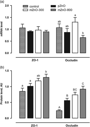
Figure 3 summarizes the results from gene and protein expression analysis of TJ components at day 14. Occludin gene expression was the lowest in mZnO-800 group and significantly lower than in mZnO-300 group (twofold lower; P=0.04). ZO-1 expression was not affected by the treatments.
Effect of feeding pharmacological ZnO or low doses of microencapsulated ZnO on gene and protein expression of tight junctions components in ileal mucosa of weaning piglets at day 14 post-weaning. (a) and (b) show gene and protein expression, respectively. Data are expressed as means (n=6) and s.e.m. represented by vertical bars. a,bValues with different superscripts differ significantly at P<0.05. A,B,CValues with different superscripts differ significantly at P<0.01. Control=basal diet providing Zn at 50 mg/kg; pZnO=basal diet+pharmacological ZnO providing ZnO at 3000 mg/kg; mZnO-300=basal diet+microencapsulated ZnO providing ZnO at 150 mg/kg; mZnO-800=basal diet+microencapsulated ZnO providing ZnO at 400 mg/kg (Zincoret-S, Vetagro SpA, Reggio Emilia, Italy). For gene expression, a modification of the 2–ΔΔC T method was used to analyze the relative expression (fold changes), calculated relative to the control group (control; Livak and Schmittgen, Reference Livak and Schmittgen2001). For protein expression, data refer to AU of protein expression measured using Western blot. ZO-1=zonula occludens-1; AU=arbitrary unit.

Both pharmacological ZnO and mZnO treatments had significantly higher occludin protein level than control group. Among the treatments, mZnO-800 group showed the highest level of occludin and was 1.6-fold higher than pZnO and 3.8-fold higher than control, respectively (P<0.001). ZO-1 protein level was significantly higher in mZnO-800 group compared with both control and pZnO groups (1.5 to 1.3-fold, respectively; P=0.01).
Discussion
Dietary inclusion of ZnO at 2000 to 3000 mg/kg is extensively used in worldwide pig production, and past research has consistently proved the beneficial effects during the post-weaning phase (Hill et al., Reference Hill, Mahan, Carter, Cromwell, Ewan, Harrold, Lewis, Miller, Shurson and Veum2001). However, increasing concerns on environmental pollution drove European legislators toward a more attentive policy on the excretion of heavy metals in manure (European Commission, 2003). With this perspective, one of the aim of this study was to investigate whether feeding low doses of ZnO in the microencapsulated form would allow to reduce the use of pharmacological Zn during the post-weaning phase.
Many mechanisms of action have been attributed to ZnO and numerous studies reported an improved feed intake as a possible way to growth enhancement (Hahn and Baker, Reference Hahn and Baker1993; Carlson et al., Reference Carlson, Hill and Link1999; Hill et al., Reference Hill, Mahan, Carter, Cromwell, Ewan, Harrold, Lewis, Miller, Shurson and Veum2001). The mechanism behind the increased feed intake has been recently explained by an increased IGF-I and IGF-I receptor expression in the intestinal mucosa, which in turn stimulates feed intake via ghrelin secretion from the stomach (Li et al., Reference Li, Yin, Li, Chen, Zang and Zhou2006; Yin et al., Reference Yin, Li, Li, Yue, Fang, Ni, Zhou and Wu2009). On the other hand, there is a consistent amount of research that has reported a growth improvement with little or no increase in voluntary feed intake in piglets (Case and Carlson, Reference Case and Carlson2002; Li et al., Reference Li, Yin, Li, Chen, Zang and Zhou2006; Ou et al., Reference Ou, Li, Cao, Li, Yin, Qiao and Wu2007). In our study, we measured a significant improvement of the ADG from days 0 to 14 due to the supplementation of ZnO at the two highest doses, which was not supported by an increased feed intake, and this would suggest the existence of an alternative mechanism promoting the growth. Carlson et al. (Reference Carlson, Hill and Link1999) suggested that a stimulation of metallothionein production in the small intestine would increase Zn retention in the enterocytes and consequently improve intestinal health status via higher protein turnover rate. Moreover, Zn has also been connected to the immune function due to its anti-oxidant and anti-inflammatory properties as Zn is critical to the development of the immune system. Sargeant et al. (Reference Sargeant, Miller and Shaw2011) recently demonstrated a modulation of the innate immune response of IPEC cells and an inhibition of nuclear factor-κ B following Escherichia coli infection mediated by Zn as ZnO. This decreased intestinal inflammatory response during challenging insults would eventually improve the intestinal integrity and barrier function, and, as a consequence, piglets would convert feed in a more efficient way and result in a better growth. Results from our study confirm previous findings on the anti-inflammatory properties of Zn: in fact, regardless of the source and dose, ZnO had a beneficial effect on the inflammatory status of the ileum. Inflammatory cytokines, and in particular TNF-α and IFN-γ, have a preponderant role in altering the intestinal permeability: they both decrease trans-epithelial electrical resistance and increase the para-cellular permeability by altering the number and depth of TJ strands and by causing pinocytosis of TJ proteins, respectively (Lallès, Reference Lallès2010). In particular, TNF-α activates apoptosis by activating the caspase protein superfamily and the apoptosis of adjacent epithelial cells would eventually contribute to the increase in permeability usually observed during inflammation (Al-Sadi et al., Reference Al-Sadi, Boivin and Ma2009). Zn, by inactivating caspase-3, inhibits apoptosis and counteracts the TNF-α effect (Perry et al., Reference Perry, Smyth, Stennicke, Salvesen, Duriez, Poirier and Hannun1997). These data agree well with the data on intestinal architecture: in fact the increased enterocyte proliferation rate of ZnO-treated animals, as reflected by PCNA staining, as well as the increased mucosa surface, as reflected by V : C ratio, clearly reflect the anti-apoptotic and enterotrophic effect of Zn.
IFN-γ has a pivotal role in controlling occludin, whose expression is correlated with enhanced barrier properties (Bruewer et al., Reference Bruewer, Utech, Ivanov, Hopkins, Parkos and Nusrat2005). In this study, we observed an inverse relationship between IFN-γ and occludin amount. In fact, IFN-γ mRNA was the lowest in pigs fed with ZnO at 400 mg/kg in the microencapsulated form (mZnO-800), whereas occludin amount was the highest despite the lowest mRNA levels. This would indicate a negative feedback mechanism of regulation of gene expression: the lower the level of IFN-γ, the higher the occludin amount and the less transcription of new mRNA is required. The role of the other cytokines in modulating the intestinal permeability and integrity is still not clear though it is known that IL-6, produced immediately after TNF-α and IL-1β in the inflammation cascade, contributes to the worsening of permeability and an increase in para-cellular flux, whereas anti-inflammatory IL-10 prevents disarrangement of TJ structure (Al-Sadi et al., Reference Al-Sadi, Boivin and Ma2009). In our study, IL-6 was only marginally affected by microencapsulated ZnO, whereas IL-10 was not affected by ZnO, regardless of the dose or source, and appeared to have a minor role compared with TNF-α and IFN-γ on the regulation of the TJ structure.
ZO is a family of proteins of the TJ, and in particular ZO-1, along with occludin, contributes to the maintenance of the epithelial integrity by connecting occludin to the β-actin filaments of the cell cytoskeleton (Fanning et al., Reference Fanning, Jameson, Jesaitis and Anderson1998). In this study, ZO-1 protein content was higher in pigs fed with the highest dose of mZnO compared with control and pZnO and this confirms the protective role of Zn from ZnO in preventing TNF-α and IFN-γ-mediated increase in para-cellular permeability and alteration of TJ functionality. Zang and Guo (2009) previously proposed the reduced intestinal permeability and the improved expression of TJ proteins as a mechanism behind the efficacy of pharmacological ZnO in piglets at weaning. In this study, we confirm the findings by Zang and Guo (2009) but we also explain the increased expression of TJ proteins as a consequence of reduced local inflammation. In fact, other authors suggest that Zn may halt the progression of gastrointestinal disease by halting the inflammatory process (Faa et al., Reference Faa, Nurchi, Ravarino, Fanni, Nemolato, Gerosa, Van Eyken and Geboes2008), but, despite the anti-inflammatory effect that we observed in this study could be generally attributable to Zn from ZnO, regardless of the source, the TJ protein structure was clearly modulated in a source-dependent way, as demonstrated by the relative abundance of both occludin and ZO-1, which were higher in mZnO-800 than in pZnO. Although reasons for this are not clear at the moment, some contribution might derive form the microencapsulation itself.
Plasma Zn was affected by pharmacological Zn at both stages as piglets receiving ZnO at 3000 mg/kg had plasma Zn in the range of 1.5 to 1.9 mg/l, which is lower than values reported by Hill et al. (Reference Hill, Mahan, Carter, Cromwell, Ewan, Harrold, Lewis, Miller, Shurson and Veum2001) but perfectly aligned with those reported by Carlson et al. (Reference Carlson, Hill and Link1999); piglets receiving microencapsulated ZnO, instead, did not differ from control and reported values comprised between 0.65 and 1.2 mg/l, which is in accordance with values for adequate Zn intake (Carlson et al., Reference Carlson, Hill and Link1999). However, according to Hahn and Baker (Reference Hahn and Baker1993), only Zn treatments that resulted in Zn plasma values of ∼1.5 mg/l were associated with improved gains that tended to be higher than those associated with Zn plasma values lower than 1.5 mg/l or higher than 3.0 mg/l. Interestingly enough, these data seem to be in contrast with what we observed in our study, as we described improved weight gains also at plasma Zn values below 1.5 mg/l. Nevertheless, Hahn and Baker (Reference Hahn and Baker1993) also reported that similar levels of supplementation from different Zn sources (i.e. ZnSO4 or Zn-methionine) were not as effective in improving weight gains despite higher Zn plasma values. This, in addition to the results we observed on the ileum mucosa, would further corroborate the hypothesis of a mechanism of action not directly correlated to Zn absorbability and Zn status and the possibility of a luminal effect rather than a systemic one.
In conclusion, 400 mg/kg of ZnO fed as microencapsulated were as effective as pharmacological ZnO at 3000 mg/kg in promoting and sustaining a healthy intestine in the first 2 weeks post-weaning, as revealed by histometry, inflammation mediators and TJ proteins. This resulted also in comparable performance in the overall 6 weeks post-weaning. In a context where policies on Zn and heavy metals usage in farm animal diets will rapidly evolve toward further reductions, we here propose microencapsulated ZnO as an effective alternative to pharmacological ZnO.
Acknowledgments
The authors gratefully acknowledge the Italian Fondazioni in rete per la Ricerca Agroalimentare, AGER Agricoltura e Ricerca, grant No. 2011-0280 and Vetagro SpA (Reggio Emilia, Italy) for providing microencapsulated ZnO. Andrea Piva serves as professor at the University of Bologna and is a member of the board of directors of Vetagro SpA.
Supplementary Material
To view Supplementary materials for this article, please visit http://dx.doi.org/10.1017/S1751731115001329