The amount of time children spend being physically active has been diminishing over the past several decades.Reference Williams, Hayman and Daniels 1 – Reference Strong 4 In addition, the rates of childhood overweight (body mass index greater than or equal to 85 percentile) and obesity (body mass index greater than or equal to 95 percentile) have increased between 1980 and 2004.Reference Ogden, Flegal, Carroll and Johnson 5 , Reference Hedley, Ogden, Johnson, Carrol, Curtin and Flegal 6 Although this prevalence of high body mass index for age has stabilized,Reference Ogden, Carroll and Flegal 7 in general, children and adolescents who are more physically active have greater cardiovascular fitness when compared with their less active peers.Reference Huang and Malina 8 , Reference Beunen, Malina, Renson, Simons, Ostyn and Lefevre 9 When taking into account body weight, there have been conflicting data on the effects of overweight on levels of cardiorespiratory fitness.Reference Stratton, Canoy and Boddy 10 – Reference Cooper, Poage, Barstow and Springer 12 However, a recent study found a decline in cardiorespiratory fitness not accounted for by an increase in body mass index.Reference Sandercock, Voss, McConnell and Rayner 13 Cardiorespiratory fitness is the most important factor in evaluating a child's overall fitness level because it takes into account the combination of multiple body systems and physiologies, including cardiovascular, respiratory, muscular, and metabolic.Reference Nassis and Sidossis 14
In our large paediatric exercise physiology laboratory, it has appeared that over the past decade children referred for exercise stress testing have demonstrated a declining level of cardiorespiratory fitness. Owing to the fact that many of our exercise stress test measurements of cardiorespiratory fitness are calculated using body weight, this apparent decrease could be due to increasing weight. We hypothesised that children were actually becoming less fit over time independent of an increase in overall body weight. The primary aim of this study was to evaluate exercise performance and body mass index in healthy children over an 8-year period to determine whether cardiorespiratory fitness has decreased independent of changes in body mass index in our patient population.
Materials and methods
Subjects
Inclusion criteria
After obtaining approval from the Institutional Review Board at The Children's Hospital of Philadelphia, subjects were included in our study if they were between the ages of 6 and 18 years and underwent maximal exercise stress testing at The Children's Hospital of Philadelphia between January, 1999 and December, 2006. These dates were chosen to enable comparison to the anthropometric data from the United States National Center for Health Statistics.Reference McDowell, Fryar, Hirsch and Ogden 15 , Reference McDowell, Fryar, Hirsch and Ogden 16 We divided the children into two groups based on the date of exercise stress test, with those in the Early Group performing exercise stress tests between 1999 and 2002 and those in the Later Group from 2003 to 2006.
Patients were identified by a search of the Exercise Physiology Laboratory database at The Children's Hospital of Philadelphia for all children with structurally and functionally normal hearts that performed a single maximal exercise stress test, defined as achieving a maximal respiratory exchange ratio greater than or equal to 1.10, using cycle ergometry during the above time period. Normal cardiac structure and function were determined by reviewing each patient's medical record for documentation by physical examination, electrocardiogram, and/or echocardiogram. Subjects were included if they met the above criteria and were referred for exercise stress test for any of the following reasons: chest pain, presyncope, syncope, palpitations, family history of congenital cardiac anomaly, or family history of sudden death.
Exclusion criteria
Children were excluded if they were under 6 years or over 18 years of age; had structural or functional heart disease, lung disease, or arrhythmia; were taking cardiovascular medication; performed more than one exercise stress test in our laboratory; or underwent treadmill exercise stress test. Children were also excluded if they did not perform a maximal exercise stress test.
Exercise protocol, cardiac monitoring, and metabolic measurements
Exercise protocol
Each subject's height and weight were measured before the exercise stress test. Height was measured to the nearest millimetre in bare stocking feet, with the subject standing upright against a wall-mounted stadiometer. Weight was measured to the nearest 10th of a kilogram, without shoes and in light clothes (t-shirt, underwear, shorts or pants, and socks), using a portable digital scale. Body mass index was calculated (kilogram per square metre) and the percentiles referred to the reference population of the 2000 Centers for Disease Control growth charts.Reference Kuczmarski, Ogden and Grummer-Strawn 17
Subjects exercised to maximal ability using an electronically braked cycle ergometer (SensorMedics, Yorba Linda, California, United States of America). The protocol consisted of 3 minutes of pedalling in an unloaded state (zero watts), followed by a ramp increase in work rate to maximal exercise capacity.Reference Cooper, Weiler-Ravell, Whipp and Wasserman 18 The steepness of the ramp protocol was designed to achieve predicted peak work rate in 10–12 minutes of cycling time. Pedalling cadence (revolutions per minute) was kept constant, with a goal of 80–90 revolutions per minute throughout the study. The children were able to view their cadence on a meter located in the centre of the bicycle handlebars.
Any exercise stress test was terminated if the patient exhibited signs of undue stress and/or requested termination of the exercise stress test. A fully trained clinical exercise physiologist and a staff paediatric cardiologist were present during the exercise stress testing.
Cardiac monitoring
A 12-lead electrocardiogram (Marquette Case-8000, Milwaukee, Wisconsin, United States of America) was obtained at rest, in the supine, sitting, and standing positions; during hyperventilation; during each minute of exercise; and during the 10 minutes of recovery. Cardiac rhythm was monitored continuously throughout the study. Significant ST segment depression was defined as 1 millimetre or more of flat or downsloping ST segment depression occurring greater than 60 milliseconds after the J-point.Reference Paridon 19 Blood pressure was measured in the right arm by auscultation at rest and every 3 minutes during exercise and recovery.
Metabolic measurements
Metabolic data were obtained throughout the exercise study and for the first 2 minutes of recovery on a breath-by-breath basis using a metabolic cart (SensorMedics V29, Yorba Linda, California, United States of America). Measured variables included: minute oxygen consumption and carbon dioxide production, physical working capacity (work rate), peak heart rate, and respiratory exchange ratio. The ventilatory anaerobic threshold was measured by the V-slope method.Reference Beaver, Wasserman and Whipp 20 Raw values were obtained and were indexed for ideal body weight in those patients whose weight exceeded 110% of ideal body weight at the time of exercise stress test.Reference Kuczmarski, Ogden and Grummer-Strawn 17
Statistical analysis
Descriptive statistics for all variables in the data set were computed using traditional measures of central tendency and variability. Several variables of exercise performance, including maximal minute oxygen consumption, ventilatory anaerobic threshold, maximal work rate before indexing for ideal body weight, and maximum heart rate were compared between the Early Group (exercise stress test 1999–2002) and the Late Group (exercise stress test 2003–2006) by two-tailed t-tests. Analysis of variance tests were performed to determine whether differences exist between various exercise and body composition variables divided by gender, age at exercise stress test (6–11 years and 12–18 years), year of exercise stress testing (Early and Late Groups), and reason for referral. Pearson correlation coefficient was used to evaluate the relationship between exercise performance variables and body weight. All analyses were computed using Statistical Analysis Software, version 9.1.
Results
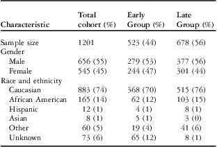
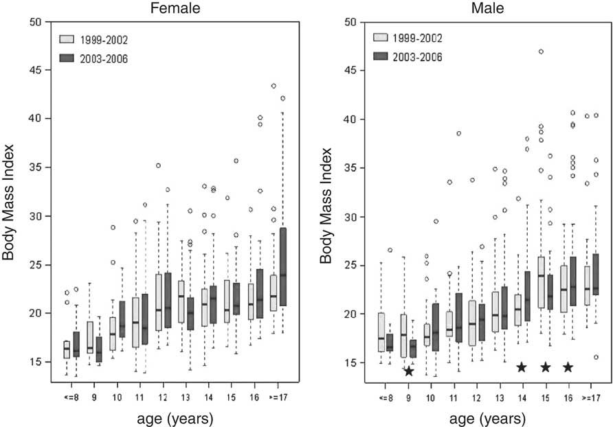
The study population consisted of 1201 children as shown in Table 1. There were slightly more male than female subjects in the cohort. The majority of the subjects were Caucasian, with African Americans making up the second largest cohort. Figure 1 is a box-plot showing the average body mass index between Early and Late Group, stratified by age and gender. Body mass index across the time periods 1999–2002 and 2003–2006 was not significantly different from the Centers for Disease Control national data for those two time periods (p-value equals 0.79).
Demographic information.

Early Group: 1999–2002; Late Group: 2003–2006
Average body mass index by gender and age, 1999–2002 (Early Group) and 2003–2006 (Late Group). ★ equals p-value less than 0.05.

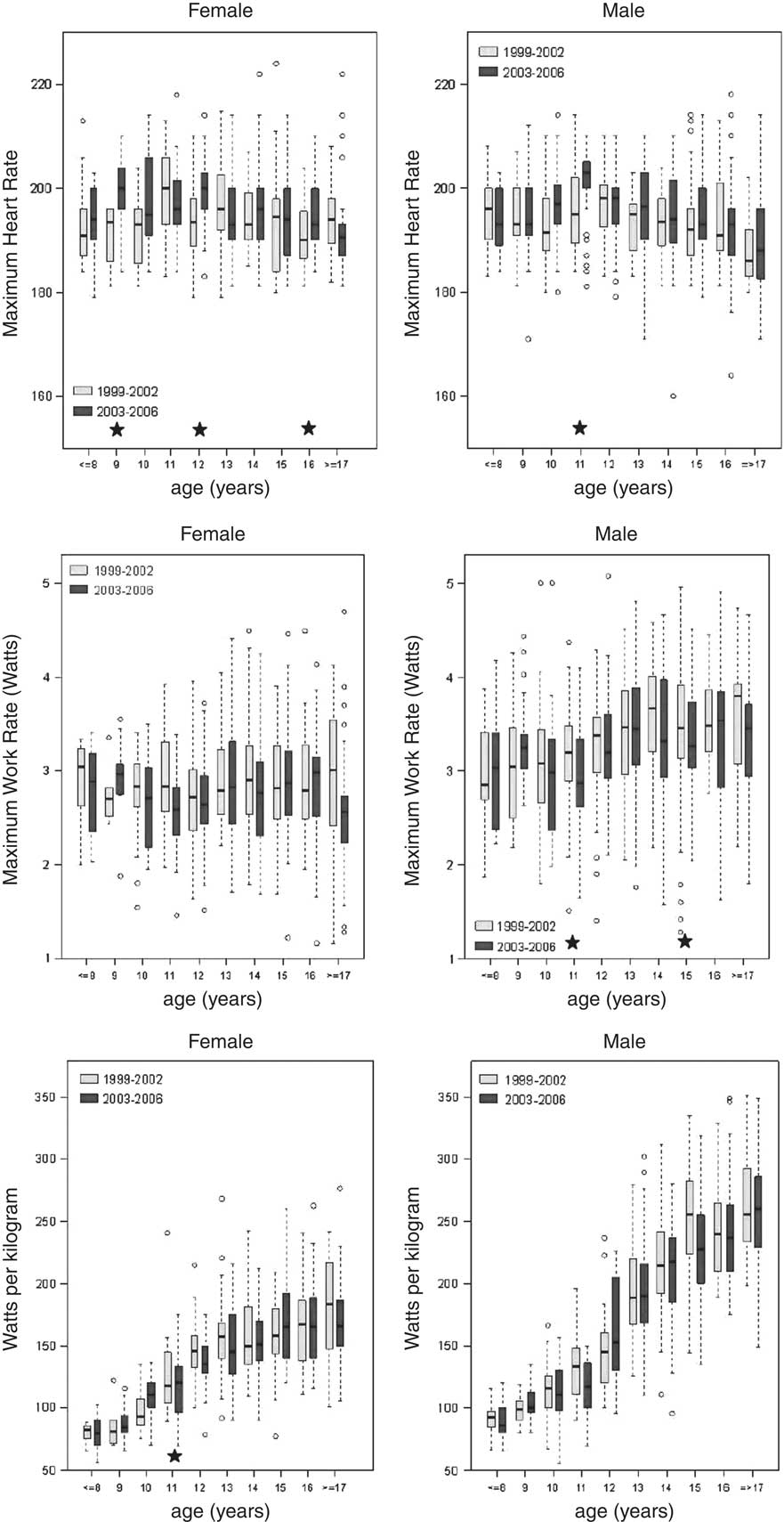
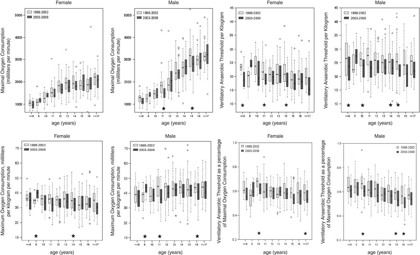
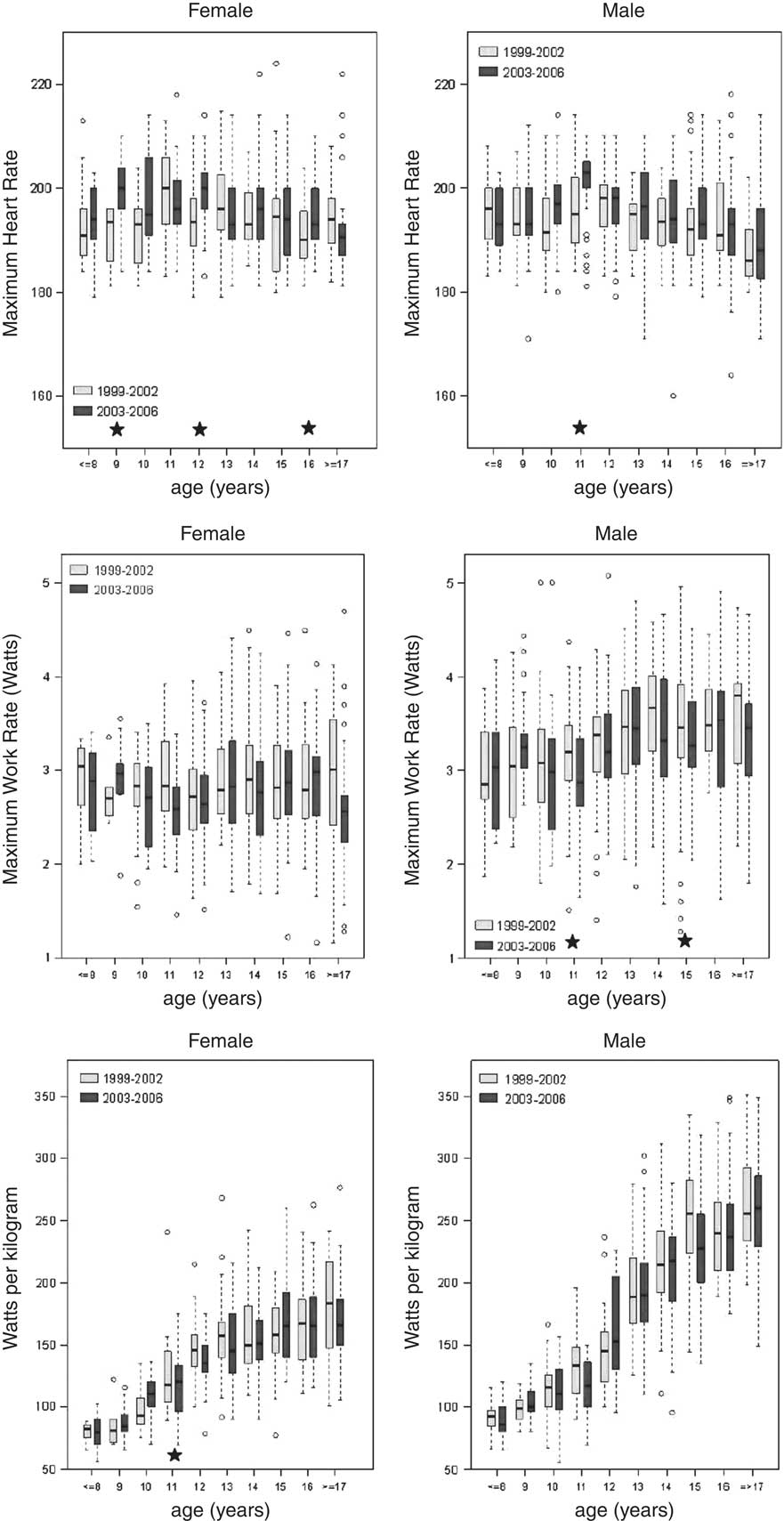
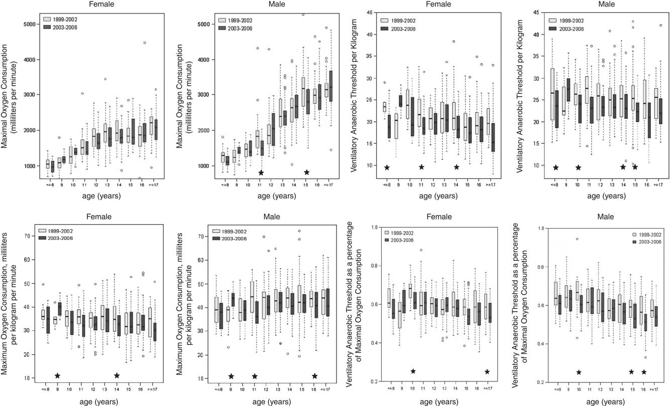
There was a negative trend (p-value greater than 0.15 and less than 0.20) among most indices of exercise performance between Early Group and Late Group. Figures 2 and 3 detail the cardiopulmonary exercise data between the two groups, stratified by age and gender. For the male subjects in the Late Group, the mean values at age 11 and 15 years for maximum work rate and oxygen consumption were significantly lower compared with the Early Group (p-value less than 0.05), but this did not remain significant when evaluated by body weight (watts per kilogram and oxygen consumption per kilogram). The youngest girls and those aged 11 and 14 years had lower ventilatory anaerobic threshold values in the Late Group compared with the Early Group (p-value less than 0.05); for boys, the 11, 15, and 16-year-olds had lower average ventilatory anaerobic threshold values in the Late compared with Early Group (p-value less than 0.05). There was significantly lower watts per kilogram among girls in the Late compared with Early Group 1 (2.7 plus or minus 0.1 versus 2.9 plus or minus 0.1, p-value less than 0.05) and lower ventilatory anaerobic threshold values among boys between Late compared with Early Group (23.4 plus or minus 1.6 versus 25.7 plus or minus 0.9, p-value less than 0.01).
Maximum heart rate and work rate by age and gender, 1999–2002 (Early Group) and 2003–2006 (Late Group). ★ equals p-value less than 0.05.
Maximal oxygen consumption and ventilatory anaerobic threshold by age and gender, 1999–2002 (Early Group) and 2003–2006 (Late Group). ★ equals p-value less than 0.05.
Discussion
This is the first study reporting changing trends in cardiorespiratory fitness and adiposity among children and adolescents in the United States. Despite an overall lack of association between changes in body mass index across these two time periods, a decline in many indices of cardiorespiratory fitness persisted among otherwise healthy children and adolescents. Our present findings support a recent study by Sandercock et alReference Sandercock, Voss, McConnell and Rayner 13 where they found a decline in cardiorespiratory fitness among 10-year-old English children, independent of changes in body mass index.
We examined the relationship of body mass index to cardiorespiratory fitness among otherwise healthy children and adolescents evaluated at our exercise laboratory between 1999 and 2006. Over this time period, in the United States, there has been an increase in childhood overweight and obesity, although those at the highest body mass index (greater than or equal to 97th percentile) have remained stable.Reference Kuczmarski, Ogden and Grummer-Strawn 17 Our study population overall did not show a statistically significant change in body mass index over this same time period; however, 9- and 15-year-old boys had a significant decrease in body mass index, whereas 14- and 16-year-old boys had a significant increase. Despite no significant change in body mass index, our population, on average, was above the 50 percentile for body mass index in both time periods with a majority at or above the 75 percentile.Reference Beaver, Wasserman and Whipp 20 This was seen in all age groups and in both genders. However, using body mass index as the sole indicator of overweight status may not differentiate between other factors, such as frame size and muscle mass. This is problematic, especially in athletic children, in that muscle weight is factored into the body mass index calculation; therefore, these children may be classified as overweight or obese despite having high levels of cardiorespiratory fitness.Reference Deurenberg, Weststrate and Seidell 21 Further prospective studies utilising measures such as bone densitometry scans could help distinguish between fat versus lean body tissue.
As a measure of overall health, cardiorespiratory fitness may be a better indication of cardiovascular health than body mass index alone, especially because it takes into account the cardiovascular, respiratory, muscular, and metabolic systems. There is strong evidence linking physical inactivity and poor cardiorespiratory fitness with greater risk of morbidity and mortality from all causes, including cardiovascular disease. 22 In one study, looking at adolescents and adults using data from the National Health and Nutrition Examination Survey from 1999 to 2002, the authors found that not only was low fitness in adolescents common (33.6 percent) but that it correlated with increased prevalence of risk factors for cardiovascular disease.Reference Carnethon, Gulai and Greenland 23 In addition, higher cardiorespiratory fitness appears to attenuate the morbidity and mortality associated with obesity, but the reverse does not apply. A recent study evaluated the prospective association between cardiorespiratory fitness, adiposity, and all-cause mortality in women and found that low cardiorespiratory fitness was an independent predictor of all-cause mortality. Physically fit women who were overweight had no greater risk of death compared with those of normal weight; however, women with low cardiorespiratory fitness had significantly higher risk of death regardless of body weight.Reference Farrell, Fitzgeral, McAuley and Barlow 24 Use of physical activity questionnaires or activity monitors in further studies may help correlate activity and cardiorespiratory fitness levels in children.
There were some potential limitations in the present study. This was a retrospective chart review and some patient data may have been unavailable. Subjects were enrolled after referral to a paediatric cardiologist. Although they were asymptomatic during exercise stress testing and had structurally normal hearts, the results may not be generalisable to the otherwise healthy paediatric population. Furthermore, assessments of physical activity level were not available.
In conclusion, we found that cardiorespiratory fitness in males and working capacity in females declined over time independent of body mass index. These data suggest that other measures, besides body mass index, may need to be evaluated in the assessment of cardiorespiratory health in the paediatric population. In addition, this may have implications for a need to adjust normative data for body habitus in exercise stress testing and to establish a robust set of cardiorespiratory fitness values in the United States for the present era.