Introduction
Sword-swallowing is an unusual vocation which derives its wonderment from the danger of the sword entering the neck, thorax and abdomen. This danger is substantial, as perforation of the digestive tract from the pharynx down to the stomach is possible. Hypopharyngeal perforation is an unusual complication. Most cases of such perforation are iatrogenic, being sustained during instrumentation of the pharynx and oesophagus. Hypopharyngeal perforation can lead to mediastinitis, sepsis and death.
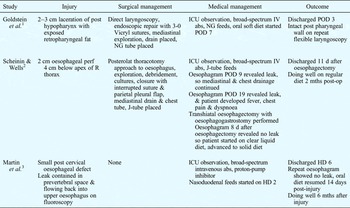
Three case reports of sword-swallowing injuries to the pharyngeal or oesophageal tissues have previously been published in the English language literature (Table I).Reference Goldstein, Yang and Brockenbrough1–Reference Martin, Steele, Mullenix, Long and Izenberg3
Reported cases of sword-swallower oesophageal and/or hypopharyngeal injuries

Post = posterior; NG = nasogastric; ICU = intensive care unit; iv = intravenous; abs = antibiotics; POD = post-operative day; perf = perforation; R = right; posterolat = posterolateral; d = day; mths = months; HD = hospital day
Case report
A 29-year-old male professional sword-swallower presented after a particularly difficult circus performance, during which he had been unable to fully pass a long metal sword. One hour after the performance, he had developed haemoptysis and severe throat pain, followed by shortness of breath on lying supine.
On presentation, his vital signs were stable and he was afebrile.
Examination revealed extensive subcutaneous emphysema of the bilateral anterior neck, with associated mild oedema and no erythema.
Flexible nasolaryngoscopy revealed a 1.5 cm laceration slightly to the right of the posterior hypopharyngeal wall midline, at the level of the arytenoids. No purulence or active bleeding was noted from the site, but surrounding mucosal oedema was present. The remaining hypopharyngeal and laryngeal structures were intact, with bilateral vocal fold mobility.
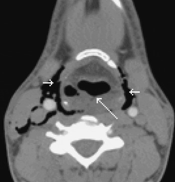
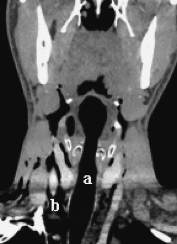
Plain X-ray films of the neck and chest revealed a large amount of air in the bilateral neck, tracking into the superior mediastinum. Computed tomography of the neck and chest revealed extensive subcutaneous emphysema and oedema of the parapharyngeal, carotid, retropharyngeal and anterior mediastinal spaces (Figures 1 and 2). Haematological studies revealed a white blood cell count of 12 500 cells per mm3.
Axial, contrast-enhanced computed tomography scan with soft tissue window, at the level of the hyoid bone, revealing posterior hypopharyngeal laceration (long arrow) and free air in the neck soft tissue bilaterally (short arrows).

Coronal, contrast-enhanced computed tomography scan with soft tissue window, at the level of the laryngeal and tracheal airways (a), revealing extensive free air (b) within the soft tissues of the neck extending into the superior mediastinum.

The patient was admitted to the intensive care unit and kept nil by mouth. An infectious disease consultation was obtained, and the patient was placed on prophylactic intravenous vancomycin and piperacillin-tazobactam, while being observed closely for mediastinitis and abscess formation.
The patient continued on a stable, afebrile course. His airway showed no signs of obstruction.
Flexible nasolaryngoscopy and a GastrografinTM (Bristol-Myers Squibb, Princeton, New Jersey, USA) swallow study were performed on hospital day six, revealing a healing hypopharyngeal laceration and no evidence of contrast leakage, respectively.
The patient was progressively advanced to a regular diet, and was discharged home on hospital day eight in a stable condition.
Discussion
The sword-swallowing performance requires the swallower to roll the tongue backwards to align the oropharynx with the upper oesophagus, to hyperextend the atlanto-occipital joint, and to hyperflex the cervical spine to vertically align the pharynx and oesophagus.Reference Devgan, Gross and McCloy4 The artist must desensitise the gag reflex, control retching, and learn to control the horizontal fibres of the cricopharyngeus and the lower oesophageal sphincter, which are not normally under voluntary control.
A survey of members of the Sword Swallowers' Association International (which recognises those who are able to swallow a non-retractable, solid steel blade of at least 2 cm width and 38 cm length) revealed a higher complication rate when the swallower was distracted, used multiple or non-straight swords, or when a previous injury was present.Reference Witcombe and Meyer5 Six of the 46 sword swallowers responding reported perforation of the pharynx or oesophagus, three of whom had been treated surgically and three conservatively.
Of related interest, the first rigid oesophagoscopy was performed on a professional sword-swallower in 1868, with passage of a 47 cm tube and successful examination of the oesophagus and gastric fundus.
Hypopharyngeal perforation from sword-swallowing is an uncommon but potentially fatal clinical entity. Pain is the most common symptom; the patient may also demonstrate dysphagia, haematemesis, dyspnoea, fever and shivering. Subcutaneous cervical emphysema is frequently seen on examination and imaging. A contrast swallow study with water-soluble contrast is the preferred test if the perforation is uncertain. The clinical presentation and successful care of the reported cases suggest that current upper digestive tract perforation treatment protocols will yield similar results for other causes of perforation. The overall mortality rate for oesophageal perforations has been reported as 6–44 per cent. In patients with cervical oesophageal perforation, a mortality rate of 6 per cent has been reported; the mortality rate for hypopharyngeal perforations is probably similar.Reference Goldstein, Yang and Brockenbrough1 Patients presenting for care less than 24 hours after injury have been found to have a significantly lower mortality rate than those presenting after 24 hours, due to earlier perforation repair and medical prevention of mediastinitis.Reference Goldstein, Yang and Brockenbrough1
• Sword-swallowing is an unusual vocation which may lead to substantial injury to the pharynx or oesophagus
• Surgical treatment may include endoscopic or open repair and drain placement
• A subset of patients may be treated successfully with conservative management, including intravenous antibiotics, nil by mouth status, and close observation in a monitored setting
The standard treatment for hypopharyngeal or cervical oesophageal perforation is open cervical exploration of the perforation, with external oversewing and drain placement. Endoscopic repair has been described for posterior pharyngeal injuries superior to the cricoid cartilage; the authors recommended open repair for lacerations inferior to the cricoid, or for large, complex injuries.Reference Goldstein, Yang and Brockenbrough1
Non-operative management has been recommended for a subset of patients with oesophageal perforation, and these indications have been expanded to patients with hypopharyngeal perforation.Reference Cameron6 This subset of patients includes those with contrast leakage that is well contained and localised, mild symptoms, and minimal evidence of sepsis. Nonsurgical management consists of stopping oral intake, intravenous administration of broad-spectrum antibiotics, judicious use of nasogastric decompression, and occasionally parenteral hyperalimentation.Reference Scheinin and Wells2 If the conservatively managed patient fails to improve, surgery is indicated.
Acknowledgement
The authors wish to thank Mrs Rebecca Colson, Administrative Assistant, for her help in the preparation of this manuscript.