Implications
Nowadays, prenatal vitamins supply (in ovo feeding (IOF) of exogenous vitamins) has been indicated to improve the hatchability and growth performance, antioxidation and immune function of broiler chickens. The present study showed that IOF of vitamin C could improve the antioxidant activity and immune function in plasma as well as reduce expression of pro-inflammatory cytokines in spleen. These findings provide a basis for future work on the use of vitamin C to improve immunity of broiler chickens.
Introduction
At the molecular level, nutrients can modulate gene expression by altering the epigenome and induce phenotypic changes by nutri-epigenetics (Burdge and Lillycrop, Reference Burdge and Lillycrop2010; Vickers, Reference Vickers2014). The gametogenesis and embryogenesis are two critical stages of epigenetic reprogramming in the process of animal development (Reik et al., Reference Reik, Dean and Walter2001). Simultaneously, these two stages are easily affected by the external environment, including nutrition. Unlike mammals, avian species, whose embryonic development is separated from the matrix, can only absorb nutrients from the fertilized egg itself to support embryonic and neonatal growth. Although it is possible to supply the specific nutrient for the matrix to accumulate it in the eggs, it is not for all of the nutrients. Deposition efficiency tends to be low. However, IOF technology provides the possibility of nutrient supply during embryogenesis in poultry (Zhai et al., Reference Zhai, Neuman, Latour and Hester2008; Kornasio et al., Reference Kornasio, Halevy, Kedar and Uni2011) and makes it possible to study the effect of specific nutrient supplied at embryonic age on the performance of broilers.
Hypoimmunity and numerous stresses are two major challenges in broiler industry. Hypoimmunity can lead higher susceptibility of broilers to various diseases and numerous stresses thereby accelerating the development of the disease, together increasing the risk of infections and death in broilers (Mujahid et al., Reference Mujahid, Pumford, Bottje, Nakagawa, Miyazawa, Akiba and Toyomizu2007; He et al., Reference He, He, Farrar, Ji, Liu and Ma2017). Vitamin C is a water-soluble vitamin with anti-stress (Khan et al., Reference Khan, Naz, Nikousefat, Selvaggi, Laudadio and Tufarelli2012), anti-oxidant (Abhilash et al., Reference Abhilash, Harikrishnan and Indira2012) and immune modulating function (Jena et al., Reference Jena, Panda, Patra, Mishra, Behura and Panigrahi2013). Therefore, vitamin C is used for increasing the immune and antioxidant capacity of broilers in the production process. In addition, epigenetic processes can be mediated by DNA methyltransferases, and DNA demethylase and studies have shown that vitamin C is involved in active DNA demethylation, histone demethylation and epigenome regulation (Young et al., Reference Young, Zuchner and Wang2015) as cofactors of related enzymes. The application of vitamin C treatment by IOF technology may intervene the epigenome reprogramming process of chicken embryos, regulate embryonic development and improve the performance of broiler chickens.
In the present study, we tested the hypothesis that IOF of vitamin C at embryonic age 15th day (E15) will improve the growth performance, antioxidation and immune function of broilers. Simultaneously, in order to explore the potential epigenetic mechanisms underlying such effects, we measured the expression of genes encoding DNA methyltransferases and demethylases. The information obtained from this study can provide theoretical basis for understanding the role of vitamin C on growth performance, antioxidation and immunoregulation in broiler chickens.
Material and methods
This experiment was approved by the Institutional Animal Care and Use Committee of Northwest A&F University (Yangling, China) and all procedures were performed in accordance with ethical guidelines for research in animal science.
Animals and sampling
Incubation
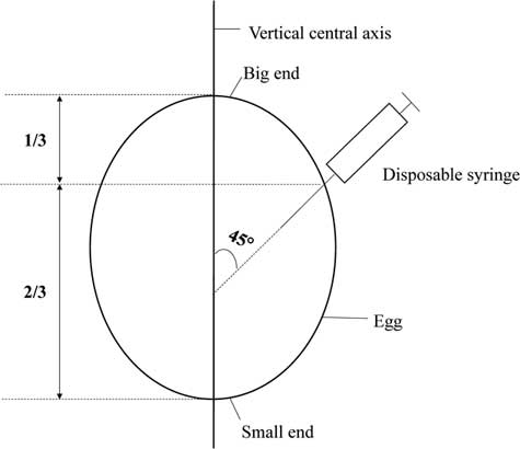
A total of 240 fertile eggs (Arbor Acres) with an average weight of 63±2.5 g were purchased from the Xianyang Dacheng Poultry Industry Co. Ltd (Xianyang, China). After disinfection, all eggs were randomly divided into normal saline (NS) and vitamin C (VC) group and incubated in an automatic incubator (9TV-2A; Beijing LanTianJiao Electronic Technology Co. Ltd). The hatchery program was as follows: the temperature was controlled at 38.0°C from E1 to 6, 37.8°C from E7 to 12, 37.6°C from E13 to 18 and 37.2°C from E19 to 21; the humidity was maintained at 60% from E1 to 6, 55% from E7 to 12, 60% from E13 to 18 and 70% from E19 to 21; an egg turning was executed for every 2 h until E19. On E15, eggs were candled, after which those unfertilized or with dead embryos were removed. After candling, every fertile egg was injected 0.1 ml of normal saline (0.9% aqueous sodium chloride solution) and 3 mg vitamin C (dissolved in 0.1 ml of normal saline) (A5960-25G; Sigma-Aldrich Inc., MO, USA) in NS and VC group, respectively. The big end of the egg was facing up. At a site of eggshell about one-third vertical height of egg from the big end, the needle entered a depth of about 2 cm into the egg at 45° to the vertical central axis (Figure 1). A 1-ml disposable syringe and a 22-gauge needle were used for injections into the eggs, and the injection site was the yolk sac.
Diagram showing the site and angle of broiler’s egg injection in side section. The disposable syringe is 1 ml with a 22-gauge needle. 1/3 stands one-third of the distance from the big end to the small end in the direction of the vertical central axis of the egg. 2/3 stands two-thirds of the distance from the big end to the small end in the direction of the vertical central axis of the egg.

Feeding
After hatching, 60 healthy new-hatched chicks with consistent BW were selected from each group and randomly allocated to six replicates with 10 birds per replicate. The experimental diet was commercial diet purchased from the Huaqin Agriculture and Animal Husbandry Technology Co. Ltd (Yangling, China) and the composition was listed in Supplementary Table S1. All birds were placed in three-layer wired battery cages and raised under a standard commercial broiler rearing environment. Rearing temperature was 32°C during the 1st week and then reduced by 2°C to 3°C/week until to 24°C. The relative humidity was set at 50% throughout the study. Birds had free access to water and commercial feed at all the time under 23 h lighting. The trial lasted for 42 days.
Sampling
On day 1 (D1), one bird was selected from each replicate with its weight close to average weight after the grouping was completed. On D21 and D42, one bird was selected from each replicate, but after fasting for 12 h. Blood samples were taken from the jugular vein on D1 and the wing vein on D21 and D42. The plasma samples were obtained after centrifuging at 3000 rpm for 5 min at 4°C and then frozen at −20°C for further analysis. After blood sampling, birds were euthanized by cervical dislocation and the spleen was collected. After that, the spleen samples were snap-frozen in liquid N2 and stored at −80°C for further analysis.
Hatchability and post-hatch performance parameters
At the end of the 21st day of incubation, the number of hatchling squabs was recorded. Body weight was recorded for each replicate on D1, D21 and D42. At the same time, feed intake was recorded for each group. After that, average daily feed intake (ADFI), average daily gain (ADG) and feed conversion ratio (FCR) were calculated. The formulae employed were as follows: the hatchability of fertilized eggs (%) = 100×(number of hatchling squabs/the number of fertilized eggs). Average daily feed intake (g feed/broiler per day) = feed intake/(the number of broilers × the number of days). Average daily gain (g BW/broiler per day) = (final weight−beginning weight)/(the number of broilers×the number of days). Feed conversion ratio = feed intake/(final weight−beginning weight).
Plasma parameters
The vitamin C content, the lysozyme activity, the concentration of immunoglobulin A (IgA), IgG and IgM, and total antioxidant capacity (T-AOC), glutathione peroxidase (GSH-Px), superoxide dismutase (SOD) and malondialdehyde (MDA) in plasma were detected using commercially available kits (A009, A050-1, E027, E026, E025, A015-1, A005, A001-1-1 and A003-1 respectively; Nanjing Jiancheng Bioengineering Institute, Nanjing, China) according to the manufacturer’s instructions. Sample quality control and testing parameter settings were strictly in accordance with the product specification.
On D35, one bird was selected from each replicate with its weight close to average weight and blood samples from the wing vein were collected into tubes equipped with heparin sodium. One millilitre of blood sample was mixed with an equal volume of precooling physiological saline. The total contents were slowly added into 2 ml of lymphocyte separation medium (P8740; Beijing Solarbio Science & Technology Co. Ltd) in 10-ml sterile centrifuge tube and centrifuged at 2000 rpm for 25 min. The lymphocyte-rich layer was removed and washed twice with phosphate buffer saline. The total contents were centrifuged at 2000 rpm for 15 min. Then, the washed lymphocytes were adjusted to a concentration of 5×10⁷ live lymphocytes/ml with cell culture medium (RPMI 1640). 80 μl of the diluted cell suspension per well was transferred to a 96-well microplate, and every nine wells were as a replicate. The control group, B lymphocyte proliferation group and T lymphocyte proliferation group were treated with 20 μl of RPMI 1640 medium, lipopolysaccharide (50 μg/ml) and concanavalin A (25 μg/ml), respectively. The 96-well microplate was incubated in incubator with 5% carbon dioxide concentration at 37°C for 44 h. After that, the proliferative efficiency of lymphocyte was determined by 3-(4,5-dimethyl-2-thiazolyl)-2,5-diphenyl-2H-tetrazolium bromide (MTT) assay (Mosmann, Reference Mosmann1983).
Therefore, 20 μl of MTT solution (5 mg/ml) per well was added and the 96-well microplate was further incubated at 37°C for 4 h. Then the plate was taken out and centrifuged at 1000 rpm for 10 min. The supernatant was removed and 100 μl of dimethylsulfoxide per well was added and then mixed thoroughly to dissolve the lymphocyte-rich layer for 5 min. The light absorption was detected at 570 nm using a microplate reader (Synergy HT; Biotek, VT, USA). The stimulation index (SI) was calculated by following formula: SI = OD570 (B or T lymphocyte proliferation group)/OD570 (the control group).
Real-time PCR for messenger RNA quantification in Spleen
RNA extraction from spleen tissue was executed according to TRIzol Reagent protocol (Invitrogen, Carlsbad, CA, USA). The concentration and purity of RNA was verified with a NanoDrop ND-1000 spectrophotometer (Nano-drop Technologies, DE, USA). Aliquots of RNA samples were subjected to electrophoresis with 1.4% agarose–formaldehyde gels stained with ethidium bromide to verify their integrity. After extraction, complementaryDNA (cDNA) was synthesized with a Primer Script RT Reagent Kit (TaKaRa, Dalian, China) following the manufacturer’s instructions and stored at −20°C. The messenger RNA (mRNA) levels of cytokines (interleukin (IL)-2, IL-4, IL-6, IL-1β, tumour necrosis factor-α (TNF-α) and interferon-γ (IFN-γ)), DNA methyltransferase (DNA methyltransferase 1 (DNMT1); DNMT3A; DNMT3B) and DNA demethylase (ten-eleven translocation protein 1 (Tet1); Tet2; Tet3; growth arrest and DNA-damage-inducible protein alpha (Gadd45α); Gadd45β; Thymine-DNA glycosylase (TDG); methyl-CpG-binding domain protein 4 (MBD4)) in spleen were determined with a SYBR® Premix Ex Taq Kit on the iCycler IQ5 (Bio-Rad Laboratories, Hercules, CA, USA). Melting curves and agarose gel electrophoresis were performed to insure a single specific PCR product for each target gene. Reaction system of 25 μl was as follows: 12.5 µl of SYBR® Premix Ex Taq II (2×), 1 µl of forward primer (10 µM/l), 1 µl of reverse primer (10 µM/l), 1 µl of cDNA and 9.5 µl of double-distilled water. Protocols were set as follows: 95°C for 10 min; 40 cycles of 95°C for 10 s, 60°C for 30 s, 72°C for 30 s; and 72°C for 5 min. The primers are listed in Supplementary Table S2. All samples were run in triplicate. After normalization to β-actin, the average cycle threshold (Ct) values were used for quantification by the 2−ΔΔCt method (Livak, Reference Livak and Schmittgen2001).
Statistical analysis
The hatchability was analysed using χ 2 test with the SPSS 20.0 (SPSS Inc., Chicago, IL, USA) and all other data were analysed by independent sample t test with the SPSS 20.0. A probability value of P < 0.05 was considered to be statistically significant.
Results
Hatchability and growth performance
The hatchability of VC and NS groups were 77.4% and 64.3%, respectively, and the χ 2 value and P-value of χ 2 test were 4.219 and 0.040, respectively. Compared with NS group, the hatchability was higher (P < 0.05) in VC group. As shown in Table 1, there was no significant difference on BW on D1, D21 and D42, as well as on ADFI, ADG and FCR from D1 to D21, from D21 to D42 and from D1 to D42 between NS group and VC group (P > 0.05).
Effect of in ovo feeding of vitamin C at E15 on BW and growth performance of broilers

E15 = embryonic age 15th day; NS = the normal saline group; VC = the vitamin C group; P-value = P value of independent sample t test; ADFI = average daily feed intake; ADG = average daily gain; FCR = feed conversion ratio.
Antioxidation in the plasma
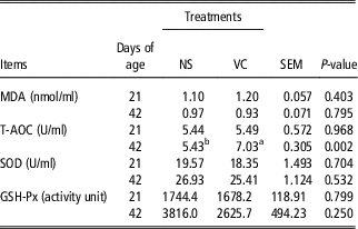
Compared with the NS group, T-AOC activity was significantly increased in broilers on D42 in the VC group (P < 0.05), but T-AOC activity on D21, GSH-Px and SOD activities and MDA contents on D21 and D42 did not differ (P > 0.05, Table 2).
Effect of in ovo feeding of vitamin C at E15 on antioxidation in plasma of broilers

E15 = embryonic age 15th day; NS = the normal saline group; VC = the vitamin C group; P-value = P value of independent sample t test; MDA = malondialdehyde; T-AOC = total antioxidant capacity; SOD = superoxide dismutase; GSH-Px = glutathione peroxidase.
a, bMeans within a row with different superscript letters are different at P < 0.05.
B/T lymphocyte stimulation index, lysozyme activity, and the concentration of immunoglobulin A, immunoglobulin G and immunoglobulin M in the plasma
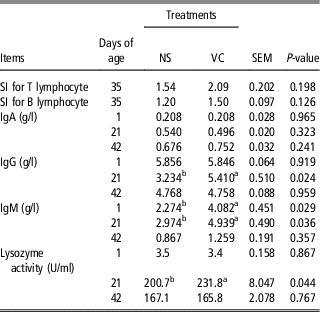
As shown in Table 3, there was no significant difference on SI for T or B lymphocytes between NS and VC broilers on D35 (P > 0.05). Compared with the NS group, lysozyme activity significantly increased on D21 in VC group (P < 0.05), but did not change on D1 and D42 (P > 0.05). In plasma, IOF of vitamin C significantly increased IgG on D21 and IgM on D1 and D21 (P < 0.05), but had no effect on the IgA contents on D1, D21 and D42, on the IgG contents on D1 and D42 and on the IgM contents on D42 (P > 0.05).
Effect of in ovo feeding of vitamin C at E15 on immune function in plasma of broilers

E15 = embryonic age 15th day; NS = the normal saline group; VC = the vitamin C group; P-value = P value of independent sample t test; SI = stimulation index; IgA = immunoglobulin A; IgG = immunoglobulin G; IgM = immunoglobulin M.
a,bMeans within a row with different superscript letters are different at P < 0.05.
Expression of cytokines in the spleen
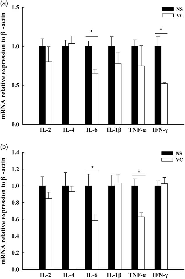
On D21, IOF of vitamin C significantly decreased the expression of IL-6 and IFN-γ (P < 0.05), but had no influence on the expression of IL-2, IL-4, IL-1β and TNF-α (P > 0.05, Figure 2a). On D42, IOF of vitamin C significantly decreased the expression of IL-6 and TNF-α (P < 0.05), but had no influence on the expression of IL-2, IL-4, IL-1β and IFN-γ (P > 0.05, Figure 2b).
Effect of in ovo feeding of vitamin C on the messenger RNA (mRNA) expression of interleukin (IL)-2, IL-4, IL-6, IL-1β, tumour necrosis factor-α (TNF-α) and interferon-γ (IFN-γ) on D21 (a) and D42 (b) in spleen of broilers. NS stands for 0.1 ml of normal saline injected into the yolk of fertile eggs on 15th day of embryonic age. VC stands for 3 mg vitamin C which was dissolved in 0.1 ml of normal saline injected into the yolk of fertile eggs on 15th day of embryonic age. Values are means±SEM from two different experiments in which each treatment was performed in triplicate with in experiment. Mean values with ‘*’ are different (P < 0.05, n = 6).

Expression of genes encoding enzymes related DNA methylation and demethylation in the spleen
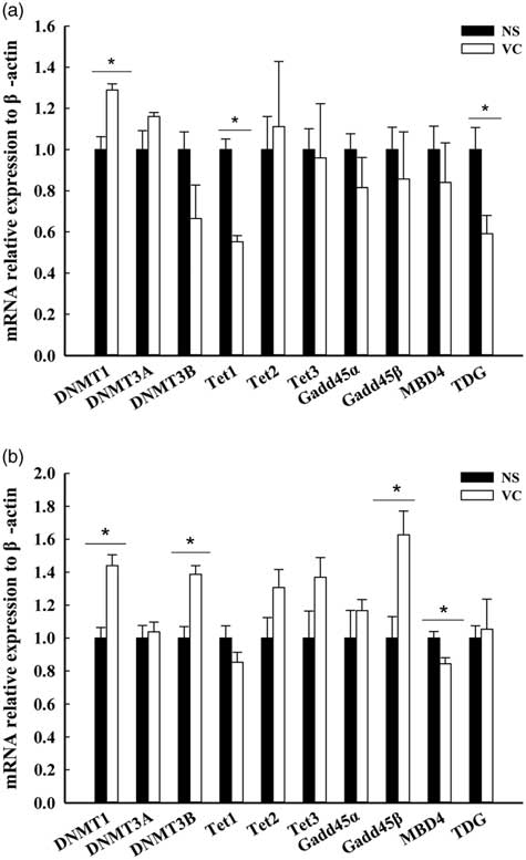
As shown in Figure 3a and b, IOF of vitamin C significantly increased the expression of DNMT1 on D21 and DNMT1 and DNMT3B on D42 (P < 0.05) and significantly decreased the expression of Tet1 and TDG on D21 and MBD4 on D42 (P < 0.05). On the contrary, vitamin C injection significantly increased the expression of Gadd45β on D42 (P < 0.05), but had no influence on the expression of other genes on the 2 days (P > 0.05).
Effect of in ovo feeding of vitamin C on the messenger RNA (mRNA) expression of genes related DNA methylation (DNMT1, DNMT3A and DNMT3B) and genes related DNA demethylation (Tet1, Tet2, Tet3, Gadd45α, Gadd45β, TDG, MBD4) on D21 (a) and D42 (b) in spleen of broilers. NS stands for 0.1 ml of normal saline injected into the yolk of fertile eggs on 15th day of embryonic age. VC stands for 3 mg vitamin C which was dissolved in 0.1 ml of normal saline injected into the yolk of fertile eggs on 15th day of embryonic age. Values are means±SEM from two different experiments in which each treatment was performed in triplicate with in experiment. Mean values with ‘*’ are different (P < 0.05, n = 6). DNMT1 = DNA methyltransferase 1; DNMT3A = DNA methyltransferase 3A; DNMT3B = DNA methyltransferase 3B; Tet1 = ten-eleven translocation protein 1; Tet2 = ten-eleven translocation protein 2; Tet3 = ten-eleven translocation protein 3; Gadd45α = growth arrest and DNA-damage-inducible protein alpha; Gadd45β = growth arrest and DNA-damage-inducible protein beta; TDG = Thymine-DNA glycosylase; MBD4 = methyl-CpG-binding domain protein 4.

Discussion
Vitamin C does not exist in newborn breeder eggs and can be biosynthesized by the developing embryo on the 3rd or 4th day of incubation. However, the synthetic quantities may not be sufficient during artificial incubation (Nowaczewski et al., Reference Nowaczewski, Kontecka and Krystianiak2012). In our study, IOF of vitamin C significantly increased the hatchability, which fell into line with the research results of other publications (Zakaria and Al-Anezi, Reference Zakaria and Al-Anezi1996; Elibol et al., Reference Elibol, Turkoglu, Akan and Erol2001; Nowaczewski et al., Reference Nowaczewski, Kontecka and Krystianiak2012). During late incubation, the generation of excessive metabolic heat from the developing embryos is the primary cause for dead embryos or culling of chicks, reducing the hatchability (Tullett, Reference Tullett1990). Vitamin C, as a cofactor of hydroxylase (Du et al., Reference Du, Cullen and Buettner2012), may regulate the production of glucocorticoides by 21-hydroxylase and β-hydroxylase, thereby promoting gluconeogenesis and enabling embryos due to higher nutrient content to adapt to the environment of incubation and pip of the eggshell. The increasing hatchability may benefit from this adaptation.
In our study, no significant impact of IOF of vitamin C on BW, ADFI, ADG and FCR could be found. Our previous research was conducted to explore the effect of IOF of folic acid at different times (E3, 7 and 11) on the growth performance of broilers. We found that IOF of folic acid at E11 was significantly increased ADG of broilers from D1 to D21 and D22 to D42, and BW on D1, D21 and D42, however, IOF of folic acid at E3 and 7 did not influence ADG and BW (Li SZ, Unpublished Data). Simultaneously, other unpublished data from our working group showed that IOF of vitamin C at E11 was beneficial for ADFI from D21 to D42 and D1 to D42, ADG from D1 to D21 and FCR from D1 to D21. Although the nutrients injected were different, the above results together clarified that the injection time directly affected the treatment effects of the nutrient. With regard to the BW, other studies had the same research results (Ipek et al., Reference Ipek, Sahan and Yilmaz2004; Bhanja et al., Reference Bhanja, Mandal, Agarwal, Majumdar and Bhattacharyya2007). The embryonic development process can be divided into three stages: establish germ (E1 to 7), embryo completion (E8 to 14) and preparation for emergence (E15 to 21) (Jr, Reference Jr2007). Embryonic developmental stages, nutritional needs and embryonic missions are different. Consequently, the injection time is a key factor for vitamin C treatment effects and the selection of injection time should be based on the biological function of nutrients and the nutritional needs during the embryonic development. Obviously, in terms of IOF of 3 mg/egg vitamin C, the E15 is not a good injection time for improving the growth performance of broilers.
As a non-specific immunity factor, lysozyme is produced by neutrophils and macrophages, important for bacteriolysis in peripheral blood (Möck and Peters, Reference Möck and Peters2010). In addition, vitamin C can accumulate in phagocytes (neutrophils and macrophages) and enhance phagocytosis of them (Carr and Maggini, Reference Carr and Maggini2017), which may be related to increased lysozyme activity. Like lysozyme, immunoglobulins also play a vital role in humoral immunity. Many studies have shown that vitamin C could enhance IgG and IgM content and lysozyme activities in serum (Peng et al., Reference Peng, Shi, Gao, Yin, Sun and Wang2013; Yao, Reference Yao2014). Although the role of vitamin C in lymphocytes is less clear, studies have shown that the gene regulatory functions of vitamin C may be involved in the immunomodulatory function of lymphocytes, affecting the expression of immunoglobulins (Carr and Maggini, Reference Carr and Maggini2017). In general, the results obtained from our research elucidated that IOF of vitamin C improves the immune status in plasma of broilers.
Antioxidant defense system of plasma, whose capacity is measured directly by T-AOC, GSH-Px, SOD, MDA and so on, is an important part of the body’s antioxidant defense system. In light of the results, we merely found that T-AOC in plasma on D42 was increased. Jena et al. (Reference Jena, Panda, Patra, Mishra, Behura and Panigrahi2013) found that vitamin C supplementation in dietary increased T-AOC of plasma. Studies have shown that older animals are more susceptible to the effects of oxidative stress because of the increasing production of reactive oxygen species (ROS) (Cui et al., Reference Cui, Kong and Zhang2012). Consequently, broiler chickens produced more ROS on D42 than on D21. It could be suggested that in the present study the higher T-AOC is accompanied with lower ROS production, especially in the VC broilers. Further investigations are necessary.
The spleen is the largest immune organ, playing an important role in cellular and humoral immunity in avain. In addition, birds are short of lymphatic vessels and lymph nodes, leading the spleen to a more prominent role in the function of immune system than other vertebrate’s. In the spleen of adult birds, filtration of blood (phagocytosis of damaged cells and antigens) and production, maturation and storage of lymphocytes are the two main ways in favour of immune defence (Smith and Hunt, Reference Smith and Hunt2004). Interleukin-2, IL-4, IL-6, IL-1β, TNF-α and IFN-γ are cytokines, which are important in response to infection and inflammation (Parkin and Cohen, Reference Parkin and Cohen2001). On the basis of their function, IL-2 and IL-4 are divided into anti-inflammatory cytokines and the others are pro-inflammatory cytokines (Carrillo-Vico et al., Reference Carrillo-Vico, Lardone, Naji, Fernández-Santos, Martín-Lacave, Guerrero and Calvo2005; You et al., Reference You, Luo, Zhang, Chen, He, Zhao, Zuo and Wu2011). Fluctuations in their mRNA levels can be used as indicators of the immune status in spleen (Alkhalifa, Reference Alkhalifa2015). Compared with NS group, we found that the expression of pro-inflammatory cytokines significantly decreased in VC group. As IOF of vitamin C improved the immunity and antioxidant capacity in plasma, which may reduce the amount of plasma antigens and oxygen free radicals, it could be suggested that the stimulus for the spleen to filtrate the blood is lowered and therefore the necessity for the expression of pro-inflammatory cytokines is reduced.
There is growing evidence that vitamin C plays an important role in epigenetics. It has been found that vitamin C not only participates in the reprogramming of somatic cells by the cofactor of DNA demethylase and histone demethylase (Young et al., Reference Young, Zuchner and Wang2015), but also promotes the whole-genome DNA demethylation of pluripotent stem cells in human which may be mediated by effects on activity of histone demethylase and (or) histone acetylase (Chung et al., Reference Chung, Brena, Kolle, Grimmond, Berman, Laird, Pera and Wolvetang2010). We detected the expression of enzymes related to DNA methylation (DNMT1, DNMT3A and DNMT3B) and DNA demethylation (Tet1, Tet2, Tet3, Gadd45α, Gadd45β, TDG and MBD4). We found that IOF of vitamin C promoted the expression of enzymes related to DNA methylation and reduced the expression of enzymes related to DNA demethylation (except Gadd45β) in spleen of broilers. The results suggested that the DNA methylation level may have an increased trend in spleen. However, it was contrary with the demethylation function of vitamin C (Chung et al., Reference Chung, Brena, Kolle, Grimmond, Berman, Laird, Pera and Wolvetang2010; Young et al., Reference Young, Zuchner and Wang2015). Methylation, occuring in gene regulatory elements, will suppress gene expression (Jones and Takai, Reference Jones and Takai2001). Vitamin C appears to regulate cytokines in a complex manner, likely due to its gene regulatory functions via regulation of epigenetic marks (Carr and Maggini, Reference Carr and Maggini2017). The changes in gene expression of methyltransferases and demethylases are consistent with the reduction in gene expression of pro-inflammatory cytokines. However, due to the lack of DNA methylation levels in gene regulatory elements of pro-inflammatory cytokines, whether DNA methylation variation is associated with the lower expression of pro-inflammatory cytokines in spleen remains to be further studied.
In summary, our results presented that IOF of vitamin C improved antioxidation and immune function in plasma, and this change may be related to the lower expression of pro-inflammatory cytokines in spleen. Changes in DNA methyltransferases and demethylases suggested an increased trend of the DNA methylation level in the spleen, however, whether DNA methylation variation is associated with the lower expression of pro-inflammatory cytokines in spleen remains to be further studied.
Acknowledgements
This work was funded by the National Key Research and Development Program of China (2017YFD0502200, 2017YFD0500500), the Program for Shaanxi Science & Technology (2017TSCXLNY-04-04, 2018ZDCXL-NY-0201, 2018ZDXM-NY-051) and the Program for Yangling Agricultural High-tech Industries Demonstration Zone (2018CXY-10).
Declaration of interest
The authors report no conflicts of interest. The authors alone are responsible for the content and writing of this article.
Ethics statement
The protocol applied to animal experiments was approved by the Institutional Animal Care and Use Committee of Northwest A&F University (Yangling, China).
Software and data repository resources
None of the data were deposited in an official repository.
Supplementary material
To view supplementary material for this article, please visit https://doi.org/10.1017/S1751731118003531