INTRODUCTION
Old World cutaneous leishmaniasis (CL) is transmitted by several different species of phlebotomine sandfly and is caused mainly by Leishmania major and Leishmania tropica parasites. Currently, it is one of the most important neglected tropical diseases and collectively affects up to 1–2 million people in over 60 countries, especially in the Middle East and Central Africa (Alvar et al. Reference Alvar, Velez, Bern, Herrero, Desjeux, Cano, Jannin, den Boer and Team2012). In 2008, approximately 100 000 new CL cases were reported in the Eastern Mediterranean Regional Office of the World Health Organisation region, with 12 countries reporting outbreaks (Postigo, Reference Postigo2010) (Alvar et al. Reference Alvar, Velez, Bern, Herrero, Desjeux, Cano, Jannin, den Boer and Team2012). The current number of Old World CL cases per year is predicted to be extremely high specially in countries at war or civil unrest due to human displacement and lack of control measures as a result of the Syrian civil war (Hotez, Reference Hotez2013). Old World CL also affects military troops deployed in the Middle East and Central Asia, with thousands of cases reported over the last 15 years.
CL is one of the most prevalent vector borne diseases throughout the Kingdom of Saudi Arabia (KSA). Despite the positive impact of programmes carefully designed to control and curtail CL cases in KSA, the disease remains a serious problem. This is mainly due to uncontrollable urbanization by expansion of cities toward leishmaniasis transmission areas, social factors related with Saudi culture and traditions, lack of leishmaniasis research, as well as lack of integration between governmental sectors. Anthroponotic CL (also known as wet leishmaniasis) is caused by L. tropica and is commonly found in Southwest provinces (al-Zaharani et al. Reference al-Zahrani, Peters, Evans, Smith and Ching Chin1989). By contrast, most CL endemic provinces have reported dry leishmaniasis, which is caused by L. major (Killick-Kendrick et al. Reference Killick-Kendrick, Bryceson, Peters, Evans, Leaney and Rioux1985; Elbihari et al. Reference Elbihari, Kawasmeh, Al-Naiem and Al-Atiya1987; El-Beshbishy et al. Reference El-Beshbishy, Al-Ali and El-Badry2013). In addition, Leishmania aethiopica, which so far has been only found in rock hyrax (Procavia capensis) in the Najran province, has also been suggested to be a new Leishmania species responsible for zoonotic CL in the Arabic Peninsula (Morsy et al. Reference Morsy, al Dakhil and el Bahrawy1997).
There is currently a great need to develop specific molecular and serological approaches for CL diagnosis in a clinical setting. In KSA, for instance, CL diagnosis relies exclusively on dermatological examination, which fails to detect the disease in a considerable number of patients. Furthermore, examination alone cannot identify the infective parasite species. Sensitive and reliable diagnostic methods are essential for patients receiving drug treatments and for active and passive surveillance in elimination settings.
Anti-α-galactosyl (anti-α-Gal) antibodies represent 1% of total human IgG (Galili, Reference Galili1993). In contrast to all other mammals, humans and Old World monkeys do not express terminal, non-reducing α-Gal epitopes freely on their cells. In humans, for instance, the α-Gal epitope is expressed only in a cryptic form in the blood group B glycolipid (Galα1-3[Fucα1-2]Galβ1-4Glc-ceramide). Therefore, terminal non-cryptic α-Gal epitopes are extremely immunogenic to humans, which produce high levels of natural anti-α-Gal antibodies directed to α-Gal-containing glycans expressed on lipopolysaccharides of Gram-negative bacteria of the gut microbiota (Galili, Reference Galili1993). In people infected with trypanosomatid parasites, high levels of anti-α-Gal have also been reported in both acute and chronic stages of Chagas disease (Avila et al. Reference Avila, Rojas and Garcia1988; Milani & Travassos, Reference Milani and Travassos1988; Almeida et al. Reference Almeida, Milani, Gorin and Travassos1991; Gazzinelli et al. Reference Gazzinelli, Pereira, Romanha, Gazzinelli and Brener1991). In Trypanosoma cruzi, the epitope is mainly expressed on O-linked glycans of surface mucin-like glycoproteins (Almeida et al. Reference Almeida, Ferguson, Schenkman and Travassos1994). Detection of anti-α-Gal levels is a biomarker for cure in drug-treated patients (Almeida et al. Reference Almeida, Krautz, Krettli and Travassos1993; de Andrade et al. Reference de Andrade, Zicker, de Oliveira, Almeida Silva, Luquetti, Travassos, Almeida, de Andrade, de Andrade and Martelli1996; Andrade et al. Reference Andrade, Martelli, Oliveira, Silva, Aires, Soussumi, Covas, Silva, Andrade, Travassos and Almeida2004; Almeida, Reference Almeida2014) and has also been exploited for diagnosis of Chagasic individuals in endemic (Almeida et al. Reference Almeida, Covas, Soussumi and Travassos1997) and non-endemic (Izquierdo et al. Reference Izquierdo, Marques, Gallego, Sanz, Tebar, Riera, Quinto, Aldasoro, Almeida and Gascon2013) settings. Moreover, in patients infected with New World Leishmania species (e.g. Leishmania mexicana spp., Leishmania braziliensis) and also patients suffering of visceral leishmaniasis from Venezuela, it has been found that the levels of anti-α-Gal antibodies are highly elevated (Avila et al. Reference Avila, Rojas and Garcia1988, Reference Avila, Rojas and Galili1989). Until now, no studies have reported on the levels of anti-α-Gal (Leish α-Gal) antibodies in Old World CL patients.
In this paper, we used a chemiluminescent enzyme-linked immunosorbent assay (CL-ELISA) to measure serum levels of α-Gal antibodies in individuals from different CL endemic regions of KSA. The CL-ELISA assay is highly sensitive and specific for CL diagnosis. We discuss the development and the possible implementation of this assay in a CL elimination setting such as KSA.
MATERIALS AND METHODS
Ethical approval
Ethical approval was obtained from both the Liverpool School of Tropical Medicine (12·06R) and the Saudi Ministry of Health (MoH) Ethical Committees. Written consent was obtained from patients in the ethical approval rubric.
Sample collection and sites
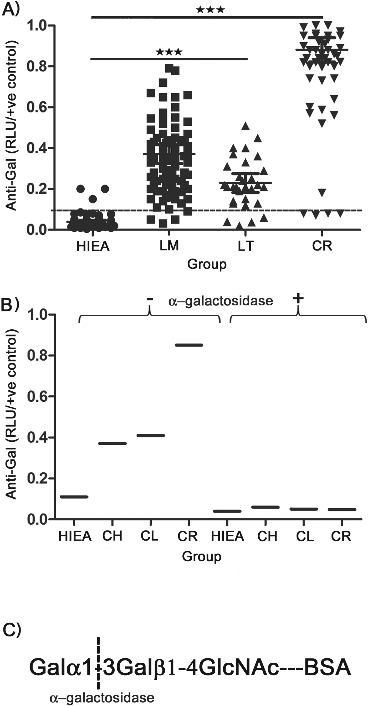
A total of 454 serum samples (119 females and 335 males, aged 18–60 years old) were obtained in two cohorts. In 2012, sera from 350 subjects were collected from five CL endemic regions from KSA: Al Qassim, Al Ahsa, Riyadh, Asir and Al Madinah. The cohort was divided into three groups of volunteers: healthy individuals from endemic areas (HIEA; 150); patients with active CL lesions (CL; 100); and cured CL patients (CR; 100), including both drug-treated (95) and self-cured (5). Patients were treated according to Saudi MoH guidelines, which recommend the application of topical azoles cream or azoles cream followed by 1–2 courses of sodium stibogluconate, depending on the patient's response. Cure or healing was defined by lesion size reduction, and induration or crusting. Samples were taken at either the leishmaniasis clinics or in the field with the help of the MoH leishmaniasis control team. HIEA volunteers were medically examined on their arms, feet, legs, hands, neck and face to discard any previous CL infection. Subsequently, in 2013, samples were collected from an additional 104 patients from the same endemic regions as above and used for skin biopsy, skin aspiration and serological assays. This latter cohort was characterized in detail in another study to be published elsewhere (Al-Salem WS et al. manuscript submitted). The pool of Chagasic sera used as positive control in Fig 1B was kindly provided by Dr Wendi Bailey (Liverpool School of Tropical Medicine).
High levels of anti-α-Gal IgG in CL patients from KSA. (A) CL-ELISA comparing the levels of anti-α-Gal in healthy individuals (HIEA), L. major-infected patients (LM); L. tropica-infected patients (LT) and cured individuals (CR). P<0·001 (***). (B) Pre-treatment of NGP Galα1-3Galβ1-4GlcNAc-BSA with CBAG abolishes recognition by anti-α-Gal antibodies in pool of sera from different groups. CH, Chagasic patients. (C) Cleavage specificity of CBAG on α-galactosylated NGP antigen.

Parasite collection and Leishmania spp. identification
Leishmania species were identified by polymerase chain reaction-restriction fragment length polymorphism (PCR-RFLP) analysis of the ribosomal Internal Transcribed Spacer 1 (IST1) as described elsewhere (el Tai et al. Reference el Tai, Osman, el Fari, Presber and Schonian2000). Detailed description about these analyses, including parasite isolation and patient's clinical descriptions, will be published elsewhere (Al-Salem WS et al. manuscript submitted).
Microscopy analysis
Samples for parasitological diagnosis were obtained from the edge of the lesion with a punch biopsy tool. The tissue sample was then transferred onto a clean microscope slide fixed in 10% buffered formaldehyde and processed for Giemsa staining. In addition, skin aspiration samples were taken and then cultured in M199 media containing Hanks medium (Invitrogen), 1% BME vitamins (Sigma-Aldrich), 20% heat-inactivated foetal bovine serum (HI-FBS) (PAA) and gentamycin sulphate (Sigma-Aldrich). Growing parasites were then isolated using DNeasy Blood and Tissue kit (Qiagen) and identified by PCR-RFLP (el Tai et al. Reference el Tai, Osman, el Fari, Presber and Schonian2000). Skin biopsies were collected for microscopy examination after Giemsa staining.
Chemiluminescent enzyme-linked immunosorbent assay
To determine levels of Leish anti-α-Gal IgG antibodies in human sera, 96-well ELISA microplates (NUNC) were coated overnight at 4 °C with 50 μL of 5 μg mL−1 neoglycoprotein (NGP) Galα1-3Galβ1-4GlcNAc-BSA (Galα1-3LacNAc-BSA) (V-Labs), diluted in carbonate–bicarbonate buffer (pH 9·6). After washing several times with 200 μL phosphate buffered saline (PBS)-0·05% Tween 20 (PBS-T), wells were blocked with PBS-1% BSA for 1 h at 37 °C. Human serum samples (diluted 1/400 in PBS-1% BSA) were then added and incubated at 37 °C for 1 h. After three washes with 200 μL PBS-T, 50 μL goat anti-human IgG (1 μg mL−1; Thermo Scientific), 50 μL donkey anti-goat IgG-biotin conjugate (1 μg mL−1; eBioscience) was added to each well. Streptavidin-horseradish peroxidase (1:2000; Zymed), diluted in PBS-BSA 1%, was added for 1 h at 37 °C. The reaction was developed by adding 50 μL Super-Signal (Pierce, Thermo) diluted 1:5 in 100 mm carbonate-bicarbonate buffer (pH 9·6). To determine the levels of anti-α-Gal IgM and IgA antibodies in human serum, samples were processed and analysed as indicated above except that 50 μL (1 μg mL−1) of biotin-conjugated goat anti-human IgM and 50 μL (1 μg mL−1) (ABD Serotec) of biotin-conjugated goat anti-human IgA (Abcam) were used as secondary antibodies. Plates were read in a Fluorostar Omega microplate reader (BMG Labtech) and values expressed as relative luminescent units (RLUs). To minimize interplate variation, values were normalized by dividing the mean value of the triplicate of each serum sample by the mean value of the positive control (CR) triplicate in each plate.
Coffee bean α-galactosidase (CBAG) treatment
For anti-α-Gal specificity (Fig 1B), 5 μg mL−1 of NGP Galα1-3LacNAc-BSA were treated overnight at 28 °C with 0·1 U per well of CBAG (Sigma). After incubation, the plates were washed several times with PBS-T and the CL-ELISA performed as described above. A pool of 10 sera (diluted 1/400 in PBS-1% BSA) from HIEA, CL, CR and Chagasic individuals was used as primary antibodies.
Statistical analysis
The Kruskal-Wallis one-way analysis of variance test was used to validate the significance among serum samples. GraphPad Prism 5 was employed for all data analyses.
RESULTS AND DISCUSSION
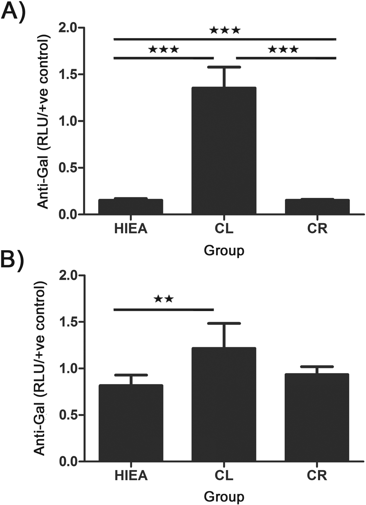
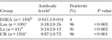
Serum, skin aspiration and biopsy samples were collected from five endemic regions of Saudi Arabia in two different cohorts. First, using a CL-ELISA with the NGP Galα1-3LacNAc-BSA as antigen, we determined the levels of anti-α-Gal IgG in patients infected with either L. major or L. tropica (Leish anti-α-Gal) and compared them to those from healthy individuals living in the same endemic areas (HIEA). We found that the mean Leish anti-α-Gal IgG titres of individuals infected with either L. major or L. tropica were significantly (P<0·005) higher than those of HIEA (Fig. 1A and Table 1). Patients infected with L. major showed a 12-fold increase in the antibody titres, while those infected with L. tropica had about an 8-fold increase in the mean Leish anti-α-Gal IgG titres when compared to HIEA. Strikingly, patients who responded to drug treatment and were considered cured (CR) (or self-healed) showed a 28-fold increase (P<0·001) in the mean titres of anti-α-Gal antibodies (Fig. 1A and Table 1). The CL-ELISA assay was specific for terminal αGal residues as treatment with CBAG abolished recognition of the resulting NGP (LacNAc-BSA antigen) by anti-α-Gal antibodies (Fig. 1B,C). Although the main immunoglobulin class produced was IgG, the anti-α-Gal IgM levels were found significantly higher (P<0·01) in patients with active CL compared to either HIEA or CR (Fig. 2A). Moreover, the levels of anti-α-Gal IgA antibodies were only slightly higher in CL-infected individuals compared to those from either HIEA or CR individuals (Fig. 2B). There appears to be variation in the Leish anti-α-Gal antibody levels depending on geographical region and age of the infection. For instance, patients from the Al Madinah villages (mainly infected with L. major) had the highest level of Leish anti-α-Gal antibodies compared to those from the Asir region, infected by L. tropica (data not shown). Furthermore, Leish anti-α-Gal antibodies were detectable as early as ~2 months post-infection and the titre tends to increase as the infection progresses (not shown), which explains why few infected individuals presented anti-α-Gal values below the cut-off line (Fig. 1A).
Levels of anti-α-Gal IgM (A) and IgA (B) in CL patients from KSA. HIEA, healthy individuals; CL, Leishmania-infected patients; CR, CL cured individuals. P<0·001 (***); P<0·01 (**).
Levels of anti-α-Gal antibodies in individuals from all CL-endemic areas
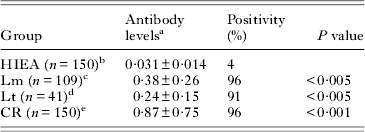
a As described in Fig 1A.
b Healthy individuals from a CL-endemic region (Al Ahsa).
c Leishmania major-infected patients.
d Leishmania tropica-infected patients.
e Cured individuals from Asir, Al Qassem, Al Madinah and Al Ahsa regions.
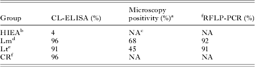
Identification of parasite species by RFLP-PCR analysis of the IST1 gene resulted in the detection of 92% and 91% of L. major and L. tropica cases, respectively. When compared to the results obtained by RFLP-PCR, we found that 96% and 91% of the L. major (95% CI; 94–98%) and L. tropica (95% CI; 86–98%) patients, respectively, had significantly high levels of Leish anti-α-Gal antibodies compared to HIEA group (Table 2). Moreover, the CL-ELISA test proved to be much more sensitive than microscopy analysis of biopsy samples taken from the same cohort of patients, which detected only 68 and 45% of the L. major and L. tropica infections, respectively (Table 1). Therefore, the CL-ELISA using Galα1-3LacNAc-BSA as antigen for detection of Leish anti-α-Gal IgG antibodies proved to be comparable to RFLP-PCR and more sensitive than microscopy analysis of biopsy samples. The CL-ELISA assay is cheaper than PCR-based techniques and its implementation would be easier at the level of district clinical laboratories. Furthermore, it has the potential to be adapted to alternative formats like lateral flow cassette, which will greatly reduce its costs and speed up diagnosis. Rapid diagnosis of CL is highly advantageous for screening populations at high risk such as displaced peoples during political unrest.
Comparison of CL-ELISA diagnostic assay with RFLP-PCR and microscopy
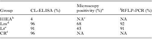
a As reported in Al-Salem WS et al. (manuscript submitted).
b Healthy individuals from a CL-endemic region (Al Ahsa).
c Not applicable.
d Leishmania major-infected patients.
e Leishmania tropica-infected patients.
f Cured individuals from Asir, Al Qassem, Al Madinah and Al Ahsa regions.
An early discrimination between a L. major and L. tropica infection is essential to administer the correct treatment regime, as infections with the latter tend to be more resistant to current anti-leishmanial drugs. One drawback of our CL-ELISA assay is its inability to discriminate between the aforementioned Leishmania spp., as the anti-α-Gal antibodies from all CL and cure patients are unable to discriminate the Galα1-3LacNAc-BSA antigen. This was surprising given the structure of the main surface glycoconjugates expressed by each of the parasites (see below). We are currently assessing the specificity of the Leish anti-α-Gal IgG antibodies from both types of infections against a non-commercial synthetic library of BSA-based NGPs that contain different terminal α-Gal epitopes (Ashmus et al. Reference Ashmus, Schocker, Cordero-Mendoza, Marques, Monroy, Pardo, Izquierdo, Gallego, Gascon, Almeida and Michael2013). Preliminary results suggest that sera from patients with either parasite species and cured individuals are able to recognize α-Gal epitopes on different oligosaccharidic structures (Alsalem W., Michael, K., Almeida, IC., and Acosta-Serrano, A., unpublished data).
Elevated levels of anti-α-Gal IgG levels have been previously detected in other kinetoplastid diseases, particularly in patients with Chagas disease (Avila et al. Reference Avila, Rojas and Garcia1988; Milani & Travassos, Reference Milani and Travassos1988; Almeida et al. Reference Almeida, Milani, Gorin and Travassos1991; Gazzinelli et al. Reference Gazzinelli, Pereira, Romanha, Gazzinelli and Brener1991), and either cutaneous or visceral leishmaniasis from Latin America (Avila et al., Reference Avila, Rojas and Garcia1988, Reference Avila, Rojas and Galili1989). Whereas the main target of the Chagasic anti α-Gal antibodies are a large family of surface mucin-like molecules (Almeida et al. Reference Almeida, Ferguson, Schenkman and Travassos1994), it is likely that the Leish anti-α-Gal antibodies are produced by the expression of glycoinositolphospholipids (GIPLs) containing terminal α-Gal residues (Avila et al. Reference Avila, Rojas and Acosta1991). This is the case of L. major GIPL2 and GIPL3, which contain oligosaccharides terminating in Galα1-3Galfβ- and Galα1-6Galα1-3Galfβ, respectively (McConville et al. Reference McConville, Homans, Thomas-Oates, Dell and Bacic1990). However, it is intriguing that although L. tropica patients show high levels of Leish anti-α-Gal antibodies, it has been reported that L. tropica promastigotes only express mannosylated (type-1) GIPLs, which contain no terminal α-Gal residues (McConville et al. Reference McConville, Schnur, Jaffe and Schneider1995). It is possible that this particular strain does not express α-galactosylated GIPLs or that this modification is stage-specific. Alternatively, terminal α-Gal epitopes can also be present on L. tropica surface glycoproteins.
While a high increase in the levels of anti-α-Gal antibodies appear to occur primarily in patients infected with kinetoplastid parasites, the use of anti-α-Gal antibodies for the detection of any form of leishmaniasis in the New World may not be specific because of the potential cross-reactivity with Chagasic anti-α-Gal, especially in areas were mixed infections can occur. This is clearly not the case for detecting patients with Old World CL as there is no Chagas transmission in areas affected by Old World CL, although it remains to be determined whether sleeping sickness patients also present an increase in the levels of anti-Gal antibodies.
It is intriguing why cured individuals showed higher levels of anti-α-Gal antibodies compared to patients with active CL, although it is possible that complete parasite clearance takes several years or is never accomplished. We could speculate that upon lysis of infected cells following chemotherapy, more α-Gal epitopes on intracellular amastigotes could be exposed to the host immune system, leading therefore to the activation of new or existing B-cell clones, resulting in a considerable increase in anti-α-Gal antibody titres. However, upon elimination of the glycan stimulus, the levels of anti-α-Gal antibodies should eventually considerably decrease in cured CL patients, as previously observed in patients with chronic Chagas disease subjected to chemotherapy (de Andrade et al. Reference de Andrade, Zicker, de Oliveira, Almeida Silva, Luquetti, Travassos, Almeida, de Andrade, de Andrade and Martelli1996; Andrade et al. Reference Andrade, Martelli, Oliveira, Silva, Aires, Soussumi, Covas, Silva, Andrade, Travassos and Almeida2004). To date, there is no obvious biomarker to differentiate individuals with an active Leishmania infection compared to cured ones and so an increase in the levels of anti α-Gal could be exploited as a potential biomarker for Old World CL cure.
ACKNOWLEDGEMENTS
We are indebted to the many Saudi volunteers who donated serum samples. We would like to thank the generous assistance of several colleagues from the Saudi MoH (Mr Saleh Alsenani (Al Madinah); Mr Abdulrhaman Alshahrani (Asir); Mr Khalid Alsuhaibani, Mr Abdulaah Alhogel, Mr Yahya Elwan and Mr Abdullah Alnosiri (Riyadh); Mr Saleh Alfahed, Mr Emad Albather, and Mr Omar Buhlaim (Al Ahsa)) for their contributions in sample collection. We thank Eng. Bader Al Malki (KACST) for logistical and technical support, Rabab Almalki and Lee R. Haines for artistic guidance, Emily Adams for useful suggestions, Deirdre P. Walsh for manuscript editing and Wendi Bailey for providing Chagasic sera.
FINANCIAL SUPPORT
This work was partially supported by the Saudi Arabian MoH and by a PhD studentship from the Saudi Cultural Bureau to WSA-S. ICA is supported by a grant (# 2G12MD007592) from the National Institute on Minority Health and Health Disparities (NIMHD), a component of the National Institutes of Health (NIH). DMF was supported by the Medical Research Council (UK) and São Paulo Research Foundation (Brazil).
 : The Ulcer, the Boil, the Sandfly, and the Conflict.