INTRODUCTION
Sea anemones of the family Phymanthidae Andres, Reference Andres1883 (Actiniaria: Actinoidea) are distinguished by verrucae on the distal column, no marginal sphincter muscle or a weak endodermal one, and two kinds of tentacles: marginal tentacles arranged in cycles that may have knoblike or branched protuberances, and discal tentacles arranged radially, typically very short, and vesicle-like (Carlgren, Reference Carlgren1949; Rodríguez et al., Reference Rodríguez, Daly, Fautin, Zhang and Shear2007).
Phymanthidae currently comprises two genera: Phymanthus Milne-Edwards & Haime, Reference Milne-Edwards and Haime1851 with eleven valid species; and Heteranthus Klunzinger, Reference Klunzinger1877 with two valid species (Fautin, Reference Fautin2013). These two genera are traditionally distinguished by the presence of lateral protuberances (papilliform or ramified) in the marginal tentacles and no marginal sphincter (or an indistinct one) in Phymanthus, whereas Heteranthus has smooth marginal tentacles without protuberances and a weak circumscribed marginal sphincter (Carlgren, Reference Carlgren1949).
Nevertheless, morphs with and without protuberances in the marginal tentacles (as well as intermediate morphs) have been reported in specimens of Phymanthus crucifer (Le Sueur, Reference Le Sueur1817) (Duerden, Reference Duerden1897, Reference Duerden1898, Reference Duerden1900, Reference Duerden1902; Stephenson, Reference Stephenson1922; Cairns et al., Reference Cairns, den Hartog, Arneson, Sterrer and Schoepfer-Sterrer1986). Verrill (Reference Verrill1900, Reference Verrill1905) suggested that morphs with and without protuberances in the marginal tentacles should be treated as separate species that could hybridize; however Duerden (Reference Duerden1897, Reference Duerden1900, Reference Duerden1902) argued that all forms should be treated as a single species based on the existence of forms with intermediate stages of tentacular protuberances. This morphological variability on marginal tentacles reported for P. crucifer challenges the value of this feature as a genus-level character within Phymanthidae.
Although the size of cnidae alone is not generally considered a specific taxonomic diagnostic character due to its variability within conspecific individuals (Fautin, Reference Fautin, Hessinger and Lenhoff1988, Reference Fautin2009; Williams, Reference Williams1996, Reference Williams1998, Reference Williams2000; Acuña et al., Reference Acuña, Excoffon, Zamponi and Ricci2003, Reference Acuña, Ricci, Excoffon and Zamponi2004; Ardelean & Fautin, Reference Ardelean and Fautin2004; Acuña & Garese, Reference Acuña and Garese2009), several studies have proposed quantitative analyses of the cnidae to help distinguish among colour morphs in some species (Allcock et al., Reference Allcock, Watts and Thorpe1998; Watts & Thorpe, Reference Watts and Thorpe1998; Manchenko et al., Reference Manchenko, Dautova and Latypov2000; Watts et al., Reference Watts, Allcock, Lynch and Thorpe2000). Watts & Thorpe (Reference Watts and Thorpe1998) found significant differences in the size of holotrichs in the acrorhagi of the upper-shore morphotype of Actinia equina (Linnaeus, Reference Linnaeus1758), suggesting that these could help distinguish between the mid- and lower-shore morphotypes of the species. Other attempts to distinguish between colour morphotypes using cnidae size alone found slight differences that do not support the use of this feature to separate species (Chintiroglou & Karalis, Reference Chintiroglou and Karalis2000).
In this study, we examined representatives of the three different marginal tentacular morphs of Phymanthus crucifer (with and without protuberances and intermediate forms) in order to identify morphological, cnidae and/or molecular distinctions that would enable separation of the morphs into different species or corroborate the broad phenotypic plasticity of P. crucifer. In addition, we tested the monophyly of Phymanthidae using three mitochondrial markers.
MATERIALS AND METHODS
Morphological and cnidae analyses
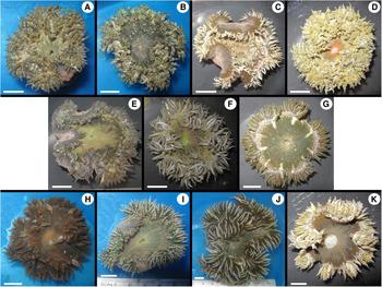
We catalogued the marginal tentacular morphotypes of Phymanthus crucifer as follows: morphotype 1 (M1), specimens with protuberances in all marginal tentacles; morphotype 2 (M2), specimens completely lacking protuberances in all marginal tentacles (i.e. smooth tentacles); and morphotype 3 (M3), specimens with some smooth marginal tentacles and some marginal tentacles with protuberances.
Twelve specimens (four per morphotype) were collected in La Gallega reef (19°13′13″N 96°07′37″W) of the Veracruz Reef System in the Gulf of Mexico in 2010; three additional specimens (one of each morphotype) were collected from Puerto Morelos reef (20°55′50.7″N 86°49′24″W) in the Mexican Caribbean (Figure 1). Collections were conducted by hand, snorkelling or SCUBA diving, and using a hammer and chisel. Collected specimens were transferred to the laboratory and maintained in an aquarium to register their colour while alive (Figure 2). Specimens were relaxed in a 5% MgSO4 seawater solution and fixed in 10% seawater–buffered formalin. Additionally, small samples of tissue were obtained from the pedal disc and preserved in 96% ethanol. Measurements of column height, as well as pedal and oral disc diameter were obtained from fixed specimens; fragments of selected specimens were dehydrated and embedded in paraffin. Histological sections 6–10 µm thick and stained with haematoxylin–eosin (Estrada-Flores et al., Reference Estrada-Flores, Peralta and Rivas1982) were prepared to examine internal anatomy.
Map of the southern Gulf of Mexico and Mexican Caribbean indicating the localities sampled in this study.

Images of specimens examined: (A–D) morphotype 1 (M1); (E–G) morphotype 2 (M2); (H–K) morphotype 3 (M3). Scale bars: 10 mm.

Data on cnidae were obtained from four representatives of each of the three morphotypes (a total of 12 individuals), all collected from La Gallega reef. Seven squash preparations were obtained from the main tissue types (~1 mm3) of each specimen. We analysed cnidae from the marginal tentacles tips (mtt), discal tentacles (dt), actinopharynx (ac), filaments (fi), column (co), vesicle-like marginal projections (vp), and protuberances on the marginal tentacles (pr/mt). For specimens of M2 (lacking protuberances), cnidae preparations of the marginal tentacles were obtained from regions where these protuberances regularly develop in morphotypes M1 and M3. From each of the seven squash preparations, the length and width of 40 undischarged capsules (replicates) of each type of cnidae were randomly measured using DIC microscopy 1000 × oil immersion (following Williams, Reference Williams1996, Reference Williams1998, Reference Williams2000).
Cnidae samples were ordered in a bi-dimensional space using principal component analysis (PCA). Differences in ordination given by morphotype, individual specimen and type of cnidae, as well as the interaction terms among these factors were analysed using a permutational MANOVA procedure (Anderson, Reference Anderson2001; McArdle & Anderson, Reference McArdle and Anderson2001). Differences among cnidae were analysed for each type of tissue separately. The PERMANOVA procedure was applied on resemblance matrices based on the Euclidian distance between samples. Although length and width of the capsules were in the same measurement scale, data were standardized and normalized prior to analyses. The statistical model used was given by:
 $$Y_{ijkl} = \alpha + M_i + I\lpar M\rpar _{\,j\lpar i\rpar } + T_k + MT_{ik} + I\lpar M\rpar T_{\,j\lpar i\rpar k} + \Sigma _{ijkl}$$
where Y is the response matrix with n samples (number of rows depending on tissue type; Table 2) * P = 2 variables (number of columns: length and width); M is a fixed factor representing morphotype (with three levels); α is the coefficient representing the intercept of the multivariate regression; I is a random factor representing individuals nested in M (with four levels); T is the fixed factor representing type of cnidae (with three or two levels, depending on tissue kind) and is orthogonal to M and I; MT and I(M)T are corresponding interactions terms; and Σ is the residual matrix. Permutation procedures were applied to obtain appropriate distributions for the pseudo-F statistic under the null hypothesis. All analyses were performed using permutations of residuals under the reduced model, resulting in a range from 909 to 999 unique permutations for each F-test. The experimental design was balanced in every case, and the partitioning of variation was achieved so that the test statistic (pseudo-F) represents the proportion of the variation in the bi-dimensional cloud that is explained by the source of variation being tested.
Specimens, as well as histological and cnidae preparations, were deposited in the Collection of Cnidarians of the Gulf of Mexico and Mexican Caribbean Sea (Registration code: YUC–CC–254–11) of the Unidad Multidisciplinaria de Docencia e Investigación en Sisal (UMDI-Sisal) at the Universidad Nacional Autónoma de México (UNAM).
Molecular analyses
Acquisition of molecular data followed the protocol detailed in Lauretta et al. (Reference Lauretta, Häussermann, Brugler and Rodríguez2013). We obtained DNA sequences of three mitochondrial (12S and 16S rDNA and cox3) regions for 14 specimens (11 from La Gallega reef and three from Puerto Morelos reef). Phymanthus crucifer haplotypes were compared to available GenBank sequence data for Phymanthus loligo (Hemprich & Ehrenberg in Ehrenberg, Reference Ehrenberg1834) and Heteranthus sp. (for GenBank accession numbers see Rodríguez et al. (Reference Rodríguez, Barbeitos, Brugler, Crowley, Grajales, Gusmão, Häussermann, Reft and Daly2014) and Crowther (Reference Crowther2013), respectively). Divergence estimates (based on the Kimura 2-parameter (K2P)) were obtained using Mega v.5.05 (Tamura et al., Reference Tamura, Stecher, Peterson, Filipski and Kumar2013).
Herein, we provide new sequences for Phymanthus crucifer which were added to the data matrix presented in Rodríguez et al. (Reference Rodríguez, Barbeitos, Brugler, Crowley, Grajales, Gusmão, Häussermann, Reft and Daly2014) after removing all hexacoral taxa not belonging to Actiniaria (except the antiphatharian Leiopathes Haime, Reference Haime1849, which was used as an outgroup) and adding Heteranthus sp.; for a complete account of taxa included in this study, we refer readers to Rodríguez et al. (Reference Rodríguez, Barbeitos, Brugler, Crowley, Grajales, Gusmão, Häussermann, Reft and Daly2014). New sequences have been deposited in GenBank (Table 1).
Voucher specimen location and GenBank accession numbers for new sequences provided in this study. See Rodríguez et al. (Reference Rodríguez, Barbeitos, Brugler, Crowley, Grajales, Gusmão, Häussermann, Reft and Daly2014) for a complete list of taxa and data included in the analysis and Crowther (Reference Crowther2013) for data regarding Heteranthus sp. UMDI-Sisal, Unidad Multidisciplinaria de Docencia e Investigación en Sisal, UNAM; AMNH, American Museum of Natural History.

Because all sequences were identical across 16S and cox3, only a single sequence for each gene was uploaded to GenBank (16S: KJ910345; cox3: KJ910346). Two haplotypes were recovered for 12S; thus a single sequence representing each haplotype was submitted to GenBank (haplotype 1: KJ910343; haplotype 2: KJ910344).
Morphological analysis of all three morphotypes; all measurements are in mm. pd, pedal disc diameter; ch, column height; od, oral disc diameter; nv, range of the number of verrucae per longitudinal row; sx, sex; (?), no gametogenic tissue present.

DNA sequences of each marker were separately aligned using MAFFT v.7 (online at http://mafft.cbrc.jp/alignment/server/; Katoh et al., Reference Katoh, Misawa, Kuma and Miyata2002, Reference Katoh, Kuma, Toh and Miyata2005; Katoh & Toh, Reference Katoh and Toh2008) using the following settings and parameters: Strategy, L-INS-i (recommended for <200 sequences with one conserved domain and long gaps); scoring matrix, 200PAM/k = 2; gap opening penalty, 1.53; offset value, 0.05; max. iterate, 1000; and retree, 1. We then concatenated the three mitochondrial markers to create a single dataset for 115 taxa and 2697 sites.
The Akaike information criterion (AIC) was implemented within jModelTest v.2.1.2 (Darriba et al., Reference Darriba, Taboada, Doallo and Posada2012) to determine the appropriate evolutionary model (TIM2 + I + G) and corresponding parameters (p-inv = 0.0470, gamma shape = 0.3360, freqA = 0.3034, freqC = 0.1821, freqG = 0.2212, freqT = 0.2933, (AC) = 1.3194, (AG) = 5.0386, (AT) = 1.3194, (CG) = 1.0000, (CT) = 8.7441, (GT) = 1.0000) (number of candidate models: 88; number of substitution schemes: 11; base tree likelihood calculations: BIONJ using PhyML v3.0 (Guindon et al., Reference Guindon, Dufayard, Lefort, Anisimova, Hordijk and Gascuel2010)).
We searched for optimal trees using maximum likelihood (ML) within PhyML v.3.0 (http://www.atgc-montpellier.fr/phyml/; Guindon & Gascuel, Reference Guindon and Gascuel2003). The following parameters were implemented within PhyML: substitution model = GTR + I + G (the online version of PhyML does not implement TIM2, and GTR had a ΔAIC of 2.3); substitution rate categories = 6; p-inv = 0.0470; gamma shape = 0.3360; starting tree = BIONJ; tree improvement = SPR & NNI; optimized tree topology and branch lengths; and bootstrap replicates = 350. We also conducted tree searches under maximum parsimony (results not shown) with TNT v.1.1 (random and consensus sectorial searches, tree drifting and 100 rounds of tree fusing; Goloboff et al., Reference Goloboff, Farris and Nixon2008). In all analyses, gaps (–) were treated as missing data. Trees of minimum length were found at least five times. The concatenated data set was subjected to 1000 rounds of bootstrap resampling to assess support for clades.
RESULTS AND DISCUSSION
Morphological analyses
All twelve specimens examined from La Gallega displayed external morphological diagnostic taxonomic features corresponding to Phymanthus crucifer, including verrucae in the distal column arranged in longitudinal rows, column coloration with flame-like staining pattern, discal tentacles arranged in radial rows from peristoma to margin, and marginal tentacles hexamerously arranged. The only external morphological difference among specimens, aside from coloration patterns, was the marginal tentacular protuberances. Internal anatomy was also similar in all the specimens (see González-Muñoz et al., Reference González-Muñoz, Simões, Sánchez-Rodríguez, Rodríguez and Segura-Puertas2012 for a complete description of the taxonomic diagnostic features of P. crucifer).
Size of specimens (pedal and oral disc diameter and column height) and number of verrucae per longitudinal row did not exhibit a consistent pattern that could be associated with any of the three marginal tentacular morphs (Table 2). The three morphotypes contained both relatively small and larger specimens, suggesting that the development of protuberances on marginal tentacles is not related to different growth stages of these organisms in the wild.
Colour patterns of the oral disc and tentacles varied among all the specimens examined but did not show a consistent pattern characterizing a particular morph (Figure 2A–K). The oral disc is mainly green, but presented a distinct tone, from olive green (Figure 2A, C–E, G) to dark green (Figure 2B, F); it could also be brown (Figure 2H, K), or with endocelic radial rows marking the arrangement of the discal tentacles (Figure 2I–J). The mouth was primarily the same colour as the oral disc or exceptionally bright green or bright orange in some specimens (Figure 2F, I and 2D, respectively). The peristoma often had a lighter tone than the rest of the oral disc (Figure 2B, G, H, K). Marginal tentacles without protuberances in representatives of morph M2 and some of M3 presented longitudinal rows of yellowish, brownish or white colorations (Figure 2E–G, I–J); and some marginal tentacles had purple shades at their tips (Figure 2I, K). Colour pattern is a controversial character to distinguish sea anemones; some species are distinguished by colour patterns while others have distinct colour morphs that are considered to be phenotypic plasticity due to local genetic adaptations (Stoletzki & Schierwater, Reference Stoletzki and Schierwater2005).
Phymanthus crucifer is dioecious and not thought to undergo asexual reproduction (Jennison, Reference Jennison1981). We found spermatic vesicles (males) in all three morphotypes (Table 2), but oocysts in only some specimens of morphs M2 and M3. Nevertheless, oocysts have been reported in specimens of morph M1 in previous studies (González-Muñoz et al., Reference González-Muñoz, Simões, Sánchez-Rodríguez, Rodríguez and Segura-Puertas2012). In most dioecious species of cnidarians, males and females are macroscopically indistinguishable (Fautin, Reference Fautin, Adiyodi and Rita1992), whilst sexual dimorphism has only been reported for a few hydrozoan and scyphozoan species (Fautin, Reference Fautin, Adiyodi and Rita1992), and for the actiniarian Entacmaea quadricolour (Leuckart in Rüppell & Leuckart, Reference Rüppell and Leuckart1828) (Scott & Harrison, Reference Scott and Harrison2009).
Crowther (Reference Crowther2013) suggested that the symbiotic relationship with zooxanthellae is likely associated with the formation of lateral protuberances in the tentacles as it occurs in other species such as Lebrunia coralligens (Wilson, Reference Wilson1890) and Lebrunia danae (Duchassaing & Michelotti, Reference Duchassaing and Michelotti1860). However, we found zooxanthellae in all specimens examined, including those without protuberances (M2). Quantitative comparisons of the densities of zooxanthellae within the different morphotypes may offer some insight about the feasibility of this hypothesis.
Cnidae analyses
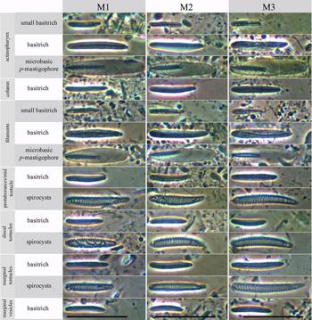
We found the same types of cnidae (cnidom) in all samples examined, regardless of morphotype (Figure 3). The cnidom of Phymanthus crucifer included basitrichs, microbasic p-mastigophores and spirocysts, as previously reported for the family and genus (Carlgren, Reference Carlgren1949). We did not find any additional types of cnidae in morphotypes M1 and M3 (those with protuberances in the marginal tentacles). It is unlikely that the protuberances on the marginal tentacles could be acting as structures for competition because agonistic behaviour in actiniarians is usually associated with the presence of holotrichs, a type of nematocyst in specialized structures such as acrorhagi and catch tentacles (Bigger, Reference Bigger, Hessinger and Lenhoff1988; Williams, Reference Williams1991) commonly found in some shallow water sea anemone species (Daly, Reference Daly2003; Fautin, Reference Fautin2009).
Cnida types and their distribution among tissues per morphotype (M1, M2, M3). Scale bars: 25 µm.

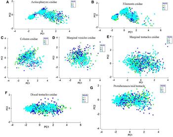
We measured 560 cnidae capsules per specimen, separated into 14 categories of cnidae (basitrichs, microbasic p-mastigophores and spirocysts) and tissue type; this added to a total of 6720 capsules measured (Figure 3). Our results showed no significant variation in the size of cnidae between morphotypes (Table 3), whereas cnidae varied in size within each morphotype depending on cnidae type and individual specimens (Figure 4A–G).
Probability associated with pseudo-F values obtained through restricted permutations of the residuals of MANOVA models applied to the similarity matrices (Euclidian distance) calculated from cnidae data sizes (length and width). ac, actinopharynx; co, column; fi, filaments; pr/mt, protuberances or middle part of the tentacle; dt, discal tentacle; mtt, marginal tentacle tip; vm, vesicle-like marginal projections.

The PCA ordination of samples from all tissue types showed that the first principal component explained from 60 to 94.5% of the variability of the cnidae size depending on the type of tissue being analysed (Table 3). Thus, the first principal component represents the variability in cnidae length. The percentage of variation explained by the second principal component was low (from 5.5 to 21.3%) in cnidae from ac, fi, pr/mt, dt and mtt, but relatively high in cnidae from co and vp (from 35.9 to 40.0%) (Table 3). This second principal component represents cnidae width.
In ac and fi the variation in cnidae width was higher for microbasic p-mastigophores than for basitrichs (Figure 4A–B). This was not the case in co, pr/mt, dt, mtt and vp tissues, in which cnidae width was similar among all types examined (Figure 4C–G).
Acuña et al. (Reference Acuña, Excoffon and Ricci2007, Reference Acuña, Ricci and Excoffon2011) only considered length when comparing cnidae sizes among specimens. Although our results confirm that length was the variable that explained most of the variation between samples (60–94.5%), we found slight differences in the width of some types of cnidae (e.g. microbasic p-mastigophores), a feature that should be considered in future studies.
The different morphotypes did not explain the variation of cnidae size in any of the tissues examined (Table 3: Morph). The ordination of samples from all types of tissue was similar regardless of the morphotype they came from (see Figure 4A–G). By contrast, differences in cnidae size among specimens within each morphotype were significant for all tissue types (Table 3: Ind(Morph) and Ind(Morph) × Type). Cnidae size (both length and width considered) also varied significantly depending on cnidae type (Table 3: Type), but differences in size between cnidae types were similar among all three morphotypes (Table 3: Morph × Type). Overall, these results suggest that individuals constitute the main source of variation when the size of cnidae are examined.
Principal component analyses of cnidae data (length/width) of all types of cnidae in each type of tissue; data from all specimens examined. Green dots, cnidae of M1; dark blue dots, cnidae of M2; light blue dots, cnidae of M3. Cnidae from: (A) actinopharynx; (B) filaments; (C) column; (D) marginal vesicles; (E) marginal tentacles; (F) discal tentacles; (G) protuberances midtentacle.

Edmands & Fautin (Reference Edmands and Fautin1991) noted that the size of nematocysts does not appear to correlate with animal size in Aulactinia veratra (Drayton in Dana, Reference Dana1846), and Acuña et al. (Reference Acuña, Excoffon and Ricci2007) suggest that there is no functional relationship between cnida length and body weight in Oulactis muscosa (Drayton in Dana, Reference Dana1846). Thus, although the diameter of the pedal disc is slightly variable between examined specimens of P. crucifer (Table 2), we found it unnecessary to include the pedal disc as a covariable in the analyses.
Molecular analyses
VARIATION WITHIN PHYMANTHUS CRUCIFER
Comparison of aligned sequences for cox3 (663 base pairs (bp) in length) and 16S (428 bp) did not reveal any variation among individuals or morphotypes from the Gulf of Mexico or Mexican Caribbean. However, mitochondrial 12S (824 bp) revealed two haplotypes that were distinguished by a single substitution (K2P distance = 0.1215%, see Table 4), but these haplotypes were not specific to any particular morphotype. While haplotype 1 (differentiated by a single adenine substitution) was specific to Gulf of Mexico specimens, it was shared by all three morphotypes. Haplotype 2 (differentiated by a single guanine substitution) was more broadly distributed, being shared between specimens in the Gulf of Mexico and Mexican Caribbean. Within the Gulf of Mexico, haplotype 2 was shared by M2 and M3, while in the Mexican Caribbean it was shared by all three morphotypes. Table 4 summarizes divergence estimates among sequences within morphotypes of Phymanthus crucifer and representatives of the family Phymanthidae (P. loligo and Heteranthus sp.).
Divergence estimates (K2P) based on sequence comparisons of the three mtDNA markers. Comparisons were made between Phymanthus crucifer and Phymanthus loligo, as well as between Phymanthus crucifer and Heteranthus sp. NA, not available.

12S, 792 base pairs (bp) compared; 16S, 428 bp compared; cox3, 513 bp compared.
Because mitochondrial DNA (mtDNA) exhibits low levels of sequence divergence within and among anthozoan species, finding no variation in sequences from conspecifics is not unexpected, even in those from potentially isolated populations that are geographically distant from each other (Shearer et al., Reference Shearer, Van Oppen, Romano and Wörheide2002; Hellberg Reference Hellberg2006; Brugler et al., Reference Brugler, France and Opresko2013). Sequence divergence based on 12S was 15–17 times higher between Phymanthus crucifer and P. loligo or Heteranthus sp. than between the two haplotypes obtained for P. crucifer. Thus, although anthozoan mtDNA is characterized by low levels of divergence, we would expect at least a similar degree of divergence among the morphotypes of P. crucifer if they were indeed distinct species. If all three P. crucifer morphotypes are indeed a single species, then mitochondrial 12S revealed, for the first time, intraspecific variation within sea anemones.
SYSTEMATICS AND TAXONOMIC STATUS OF PHYMANTHIDAE
A phylogenetic reconstruction based on the three concatenated mitochondrial genes recovered the two 12S-based Phymanthus crucifer haplotypes as sister taxa, and these as sister to P. loligo (Figure 5). However, Heteranthus sp. is recovered as sister to the actiniid genus Anemonia Risso, Reference Risso1826, thus rendering Phymanthidae polyphyletic. All studied members of Phymanthidae grouped within Actinoidea (Rodríguez et al., Reference Rodríguez, Barbeitos, Brugler, Crowley, Grajales, Gusmão, Häussermann, Reft and Daly2014), a superfamily of mainly shallow-water sea anemones (Rodríguez et al., Reference Rodríguez, Barbeitos, Daly, Gusmão and Häussermann2012, Reference Rodríguez, Barbeitos, Brugler, Crowley, Grajales, Gusmão, Häussermann, Reft and Daly2014). Our results concur with those of Crowther (Reference Crowther2013), who included a higher taxon sampling of the superfamily Actinoidea in her study of the families Thalassianthidae Milne-Edwards & Haime, Reference Milne-Edwards and Haime1851 and Aliciidae Duerden, Reference Duerden1895.
Phylogenetic reconstruction of the Actiniaria. Tree resulting from PhyML analysis of concatenated 12S, 16S and cox3. Grey boxes indicate superfamilies within the order; the name of each superfamily is inside or next to the coloured box. Species epithets are given only for genera represented by more than one species; for a complete list of taxa, see Rodríguez et al. (Reference Rodríguez, Barbeitos, Brugler, Crowley, Grajales, Gusmão, Häussermann, Reft and Daly2014). Numbers above the branches are bootstrap resampling values expressed as a percentage; values <50 not indicated; filled-in circles indicate nodes with support of 100%. Taxa in bold belong to Phymanthidae.

The presence of Phymanthus crucifer morphotypes without protuberances in the marginal tentacles renders Carlgren's (Reference Carlgren1949) major distinction between the two genera of Phymanthidae invalid. The marginal sphincter muscle, the other feature used by Carlgren (Reference Carlgren1949) to distinguish between these genera, is also problematic. Heteranthus is characterized by a weak but circumscribed marginal sphincter, whereas most species of Phymanthus lack a marginal sphincter (Carlgren, Reference Carlgren1949). However, Phymanthus muscosus (Haddon & Shackleton, Reference Haddon and Shackleton1893) has a very feeble sphincter muscle (Haddon, Reference Haddon1898). Carlgren (Reference Carlgren1900) initially placed Heteranthus within a different family, Heteranthidae Carlgren, Reference Carlgren1900, but he later placed it within Phymanthidae, based on similarities with Phymanthus (Carlgren, Reference Carlgren1943). We recovered Heteranthus as nested within Actiniidae (see Figure 5) suggesting that discal tentacles have evolved independently at least twice within Actinoidea. A comprehensive revision of the family Phymanthidae and a redefinition of its diagnostic characters are needed to establish its membership.
Based on external and internal morphological features, cnidae data, and mitochondrial DNA, we conclude that all morphotypes of Phymanthus crucifer represent a single species, despite differences in the presence or absence of protuberances in the marginal tentacles. The significance and function of the protuberances in the marginal tentacles remains unknown within P. crucifer, but might be related to specific adaptations to the surrounding environment.
ACKNOWLEDGEMENTS
Dr Judith Sánchez-Rodríguez (ICMyL) and B.S. Alejandro Córdova (FES-I) helped in the field; M.S. Maribel Badillo-Alemán (UMDI-Sisal) provided access and support to histological facilities; M.S. Gemma Martínez-Moreno and Dr Patricia Guadarrama-Chávez (UMDI-Sisal) helped with laboratory work and provided support in the microscopy laboratory; Dr Andrea Crowther (South Australian Museum) provided 12S and cox3 sequence data for Heteranthus sp. All specimens were collected under consent of Mexican law, collecting permit approved by Comisión Nacional de Acuacultura y Pesca (Number 07332.250810.4060). Comments of two anonymous referees improved this manuscript.
FINANCIAL SUPPORT
This work was partially supported by the Comisión Nacional de Ciencia y Tecnología (CONACyT) (R.G., grant number 35166/202677); CONACyT–SEMARNAT (N.S., grant number 108285); and DGAPA–PAPIME–UNAM (N.S., grant number PE207210).