Introduction
After the introduction of antibiotics, Streptococcus pneumoniae pericarditis cases decreased from 51% of all cases to 9% and reportedly mainly in adultsReference Dagan, Jacobs, Greenberg, Feigin, Cherry, Demmler and Kaplan 1 . The introduction of the 7-valent pneumococcal conjugate vaccine (PCV7) significantly reduced the incidence of invasive pneumococcal infection. Even though pneumococcal pericarditis is very rare in the current era, early detection of constrictive pericarditis as a potential complication is crucial to prevent mortality.
Case report
A 2-year-9-month old, previously well girl was referred to our centre for pericardiectomy due to suspected constrictive pericarditis. She had received all vaccinations except pneumococcal vaccine, which is not given routinely in Malaysia. Six weeks prior to this presentation, she was admitted to the referring hospital for persistent fever and cough for ten days, despite antibiotic from the outside clinic. She was noted to be in respiratory distress requiring Bilevel Positive Airway Pressure (BiPAP) support. She was diagnosed to have right lung empyema requiring chest drainage and complicated by necrotising pneumococcal pneumonia needing video-assisted thoracoscopic surgery for decortication and debridement of the affected right lung. Her pleural fluid and blood cultures were negative, but her urine for S. pneumoniae antigen was positive. She was covered with high-dose intravenous Crystalline-penicillin. Echocardiography which was performed for cardiomegaly demonstrated global pericardial effusion with no evidence of cardiac tamponade. She underwent pericardiocentesis, which drained 120 ml of pus. However, no organism was isolated from the pericardial fluid. Retroviral and immunodeficiency screening were also negative. Her antibiotic was upgraded to intravenous ceftriaxone when she did not respond much to the intravenous C-penicillin.
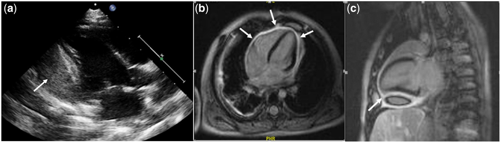
Her respiratory condition improved with ceftriaxone, but two weeks later, she was noted to have increasing peripheral oedema and abdominal distension related to ascites and hepatomegaly. Repeated echocardiography showed thickened and hyperechoic pericardium. There was a 12-mm pericardial effusion at the inferoposterior wall of the left ventricle (Fig 1a) with a preserved left ventricle function (ejection fraction of 67%). The presence of diastolic septal bounce and respiratory variation in ventricular filling suggested constrictive pericarditis. She was then referred to us for further management.
a: Parasternal long axis view on echocardiography depicting pericardial fluid collection posterior to the left ventricular free wall with thickening of the pericardium (arrow) b&c: CMRI images with pericardial late gadolinium enhancement (LGE) changes especially at the anterio-inferior aspect of the ventricles (arrows). There was also pericardial collection at the left inferolateral and inferior part of the heart that consisted of blood products.

On examination, she had periorbital oedema. She was mildly tachypneic on BiPAP support with a heart rate of 112 beats/minute, respiratory rate of 40 breaths/minute, blood pressure of 90/56 mmHg, and temperature of 37.2°C. Her breath sounds were reduced bilaterally with no added sounds. She had normal heart sounds with no murmur heard, and there was no pericardial rub. She had hepatomegaly of 4 cm with mild ascites.
Cardiac magnetic resonance imaging (CMRI) was conducted for suspected loculated pericardial effusion. It revealed global thickening of the pericardium with corresponding marked post-gadolinium enhancement suggesting ongoing inflammation. The pericardium measured up to 4.5 mm in thickness. There was also pericardial collection at the left inferolateral and inferior part of the heart that consisted of blood products (Fig 1b and c).
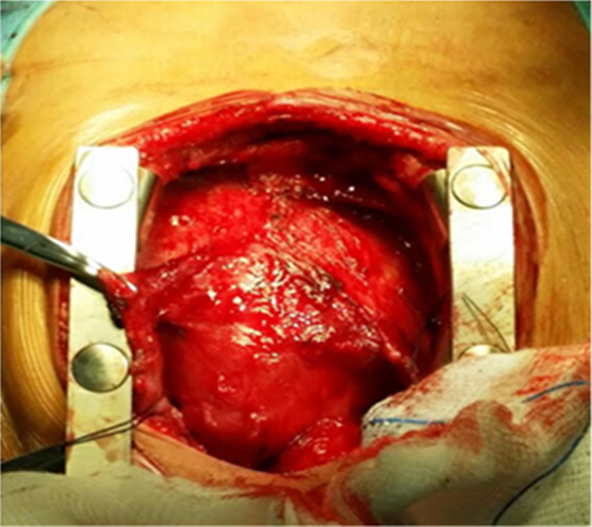
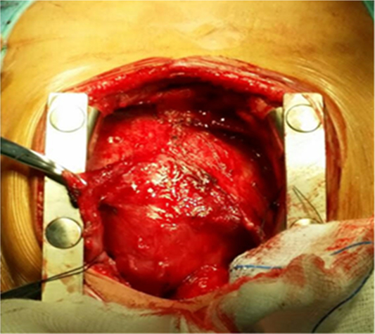
Total pericardiectomy was performed via median sternotomy. The parietal pericardium was thickened and adherent without calcification (Fig 2). The central venous pressure was immediately reduced by half after stripping the pericardium. The intra- and post-operative periods were uneventful with complete regression of heart failure. She was discharged well after completing intravenous antibiotics for six weeks. Her biopsy studies of the pericardial tissue reported as fibrotic pericardium with some hyaline degeneration. During the last follow-up appointment, she is asymptomatic, and echocardiogram revealed a normal shaped heart and function.
Intraoperative finding of thickened and adherent pericardium.
Discussion
Constrictive pericarditis is characterised by the appearance of signs and symptoms of right heart failure due to loss of pericardial compliance and restricted diastolic filling resulting from a chronic inflammatory process involving parietal and visceral pericardial layersReference Mariana, Ines and Rui 2 . Constrictive pericarditis is not common in children. Out of the 235 pericarditis patients treated at Toronto Hospital for Sick Children over approximately 30 years, only two developed pericardial thickening and constrictionReference Keith, Rowe and Vlad 3 . Common clinical findings include peripheral oedema, ascites, hepatomegaly, pleural effusion, and pericardial friction rub, and should be considered in the differential diagnosis of right heart failure. For our case, constrictive pericarditis was suspected early because of the evidence of right-sided heart failure following purulent pericarditis as a result of direct spread from lung empyema. Infections are the most common identifiable causes of pericarditis in children, particularly in tropical regions and in developing countries, even if, in a substantial proportion of cases, actual etiology cannot be determined.
Infectious aetiologies account for 5% of all cases of pericardial effusion, commonly, Staphylococcus aureus, S. pneumoniae, and Hemophilus influenzae Reference dos Santos, Moreira and Ribeiro 4 . Other reported pathogens include group A beta haemolytic streptococci, Mycoplasma pneumoniae and Chlamydia pneumoniae. Another important pathogen to consider in developing countries is Mycobacterium tuberculosis. In most cases, Streptococcus pneumoniae infection spreads from an intra-thoracic site, whereas the dissemination of Staphylococcus aureus is most often hematogenous. Majid and Omar showed that pneumococcal pericarditis is frequently associated with pneumonia and empyema, similar to our case where the patient developed purulent pericarditis following right lung empyemaReference Majid and Omar 5 .
Currently, pneumococcal pericarditis is uncommon due to the advent of penicillin therapy and the introduction of the pneumococcal vaccine. However, mortality remains high in cases of pneumococcal pericarditis associated with cardiac tamponade and constrictive pericarditisReference Tatli, Buyuklu and Altun 6 . No specific pneumococcal virulence factors have been shown crucial to cardiac involvement. However, Kan et al. found that in cases of cardiac infection with non-encapsulated S. pneumoniae, the bacteria had higher hydrophobicity and increased adherence to human epithelial cells than in infections of other tissuesReference Kan, Ries and Normark 7 .
Transthoracic echocardiography is usually the first diagnostic investigation performed for suspected constrictive pericarditis and can be very useful in the presence of the typical findings such as increased pericardial thickness and sometimes calcification, septal bounce, dynamic respiratory variation of cardiac inflow, and enhanced ventricular interactionReference Mariana, Ines and Rui 2 , Reference Keith, Rowe and Vlad 3 . However, echocardiography might not be confirmatory in all cases as some of these characteristic findings may not be present.
CMRI should be used when echocardiography is non-diagnostic or in suspected loculated, haemorrhagic pericardial effusion or pericardial thickeningReference Taylor, Dymarkowski and Verbeken 8 . Characteristic CMRI features include increased pericardial thickening and dilatation of the inferior caval vein, an indirect sign of impaired right ventricular diastolic filling. An advantage of CMRI over CT and echocardiography is its superior ability to characterise pericardial effusions and pericardial masses as well as the assessment of atrial/ventricular size and function, diastolic restraint, diastolic septal bounce, and myocardial tethering. Late gadolinium enhancement (LGE) inversion recovery images can evaluate both myocardial fibrosis and inflammation as well as pericardial inflammation; all possible findings in constrictive pericarditisReference Taylor, Dymarkowski and Verbeken 8 .
The management of pneumococcal pericarditis involves prolonged antimicrobial therapy of at least four weeks in conjunction with pericardiocentesis. With this combined treatment modalities, mortality can be reduced to less than 20%Reference Brian 9 . However, these combined therapies are often ineffective in draining thick, loculated fibrinous fluid. Pericardiotomy is the method recommended by the European Society of Cardiology guidelines, with its associated higher success rate and a lower incidence of constrictive pericarditis. Pericardiectomy is indicated in patients with dense adhesions, loculated effusion, recurrence of tamponade, and persistent infectionReference Mariana, Ines and Rui 2 , Reference Brian 9 , Reference Thompson, Burkhart and Dearani 10 .
In conclusion, despite advances in preventive, diagnostic, and treatment modalities, purulent pericarditis leading to constrictive pericarditis continues to be a serious complication following invasive pneumococcal infection. Therefore, constrictive pericarditis as a result of pneumococcal pericarditis should be considered and investigated early in a patient with pneumococcal pneumonia complicated with right-sided heart failure so that appropriate surgical intervention can be performed to prevent the mortality.
Acknowledgements
The authors are grateful to Dato Hamdan who did the successful operation and Dr. Koh Ghee Kiong, the head of the Pediatric Cardiology Unit and all the staff in Serdang Hospital who had taken care of this patient.
Conflict of Interest
None.
Financial Support
This case report received no specific grant from any funding agency or commercial or not-for-profit sectors. Conflicts of Interest None.
Ethical Standards
This case report complied with the ethical standards and was accepted by the National Medical Ethics Committee.