




No CrossRef data available.
Article contents
- Abstract
- Adolescent Changes in Behavior and Psychiatric Symptoms
- Hormones and Development
- Hormones and Brain Function
- The Neurohormonal Course of Pubertal Development
- Maturational Status, Hormones, and Behavior
- Mechanisms in Adolescent Risk for Psychopathology: Discontinuities in Development
- Conclusions
- References
Pubertal neuromaturation, stress sensitivity, and psychopathology
Published online by Cambridge University Press: 01 December 2004
Article contents
- Abstract
- Adolescent Changes in Behavior and Psychiatric Symptoms
- Hormones and Development
- Hormones and Brain Function
- The Neurohormonal Course of Pubertal Development
- Maturational Status, Hormones, and Behavior
- Mechanisms in Adolescent Risk for Psychopathology: Discontinuities in Development
- Conclusions
- References
Abstract
Normal adolescent development is often accompanied by transient emotional and behavioral problems. For most individuals with postpubertal-onset adjustment problems, there is a resolution by early adulthood and relative stability through the adult life span. But for a minority, adjustment problems escalate during adolescence and portend the development of serious mental illness in adulthood. In this article, we explore adolescent behavioral changes and neurodevelopmental processes that might contribute to stress sensitivity and vulnerability for the emergence of the mental disorders. Of particular interest is the role that hormonal changes might play in the expression of genetic vulnerabilities for psychopathology. Drawing on recent findings from clinical research and behavioral neuroscience, we describe the ways in which postpubertal hormones might alter brain function and, thereby, behavior. It is concluded that there are both activational and organization effects of hormones on the adolescent brain, and these contribute to developmental discontinuities in behavioral adjustment. Implications for adult psychopathology and preventive intervention are discussed.
Information
- Type
- Research Article
- Information
- Copyright
- © 2004 Cambridge University Press
The behavioral changes associated with adolescence have been the subject of both general public concern and scientific interest. There are long-standing cultural assumptions that adolescent behavior is often erratic and oppositional. Indeed, researchers have documented that parents tend to describe the teen years as the most challenging, and they typically report a relatively stable course beginning in early adulthood (Arnett, 1999). Further, cross-sectional and longitudinal studies of child behavior generally confirm the assumption that certain adjustment problems increase after the onset of puberty; and the debate has now shifted to the origins of normative adolescent behavior changes, namely, what biological and environmental factors are involved?
Advances in neuroimaging technology have made it possible to conduct studies of human brain development in vivo, and the findings have shown that neurodevelopment extends through adolescence. Neural circuitry is reorganized, and there are changes in the volume of some brain regions, with increases in white matter and decreases in gray matter (Benes, 2003a, 2003b; Blumberg et al., 2003; Chambers, Taylor, & Potenza, 2003; Durston, Hulshoff, Casey, Giedd, Buitelaar, & van Engeland, 2001; Paus, Zijdenbos, Worsley, Collins, Blumenthal, Giedd, Rapoport, & Evans, 1999). These developmental processes are presumed to subserve the enhancement of higher level cognitive abilities, including inhibitory capacities, during adolescence (Chambers et al., 2003; Tamm, Menon, & Reiss, 2002). There are also changes in adult brain morphology; however, they appear to be more gradual, and may not involve the emergence of new circuitry (Saito, Toyama, Uemura, Ishii, Senda, & Uchiyama, 2001), although more longitudinal studies of human brain morphology are needed to clarify the nature of postadolescent brain development.
The burgeoning field of developmental neuroscience has stimulated renewed interest in adolescence among clinical investigators. In particular, researchers in the field of psychopathology have begun to focus greater attention on adolescence as a critical period for the onset of certain psychiatric disorders (Benes, 2003a, 2003b; Walker & Bollini, 2002). They are seeking to understand why so many major mental illnesses have their prodromal stage in adolescence, with a full clinical onset that typically occurs in the early 20s, around the transition into adulthood. To date, relatively few cross-sectional or longitudinal studies have encompassed both the adolescent and early adult periods. Thus, our knowledge of the transitional process is limited. But numerous investigations of the adolescent period have been launched in the past decade, and we can anticipate that, in the future, the findings will shed greater light on the adolescent/early adult trajectory. At this juncture, the research has yielded some promising leads on neurodevelopmental processes that subserve behavioral changes in adolescence.
This article is concerned with the factors that contribute to adolescent vulnerability for the emergence of the mental disorders. The chief focus is the biological level of analysis. We address the role of hormones in altering brain function and behavior after the onset of puberty. Of particular interest is the role that hormonal changes might play in the expression of psychopathology. We begin with brief overviews of the behavioral and hormonal changes that accompany pre- and postpubertal development. Drawing on recent findings from both behavioral and molecular neuroscience, we then explore ways that hormones might act to alter brain function and, thereby, behavior. In particular, we consider the role that hormones might play in creating significant developmental discontinuities in behavioral adjustment (Cicchetti & Rogosch, 2002).
Although the gonadal hormones have been emphasized in discussions of hormone–behavior relations in adolescence, in this article we highlight the adrenal hormones, especially the hypothalamic–pituitary–adrenal (HPA) axis and release of the “stress” hormone, cortisol. Recent research findings indicate that the HPA axis may serve to stimulate the onset of pubertal development and play a role in the triggering latent liabilities for psychiatric disorders (L'Allemand, Penhoat, Lebrethon, Ardevol, Baehr, Oelkers, & Saez, 1996; Mesce, 2002). It may, in fact, render adolescence a stress-sensitive developmental period, as there is accumulating evidence that the neural systems involved in the stress response are augmented during adolescence. We present new findings to support this notion, then conclude with a discussion of activational and organizational effects of hormones, and how they might trigger developmental discontinuities in the phenotypic expression of genetic liabilities.
Adolescent Changes in Behavior and Psychiatric Symptoms
During the adolescent years there are normative improvements in several domains of cognitive functioning, and memory appears to reach a plateau during the late teens (Schneider, 2002). Entry into high school typically marks an expansion in social networks. There is no doubt that some experience adolescence as a very smooth transition to adulthood.
On average, however, after the age of 12, both children and their parents notice an increased occurrence of some problematic behavior (for a review, see Arnett, 1999). For example, adolescence is accompanied by increases in risk-taking behavior, such as substance use, as well as self-reports and parent reports of negative mood. On average, through the course of adolescence, youth report significant increases in feelings of awkwardness, distress, and embarrassment, and more frequent social conflicts. Although the relative prevalence of these trends in nonwestern countries has not been as well established, they have been consistently documented in the United States (Arnett, 1999).
For most individuals who experience them, adolescent adjustment problems are transient. Although our knowledge of the developmental course bridging adolescence and early adulthood is sketchy, there is evidence that adjustment problems decline in early adulthood, and the majority of young adults show significant improvements in behavioral control, positive affect, and self-confidence (McGue, Bacon, & Lykken, 1993; Roberts, Caspi, & Moffitt, 2001). It appears that even diagnosable personality disorders often resolve with the entry into adulthood. Longitudinal studies of youth have shown that the proportion who meet criteria for a personality disorder declines from adolescence to young adulthood (Grilo, Becker, Edell, & McGlashan, 2001). Further, the longitudinal stability of personality dimensions increases with age, and seems to reach an asymptote by 30 years (McCrae et al., 2000).
On the other hand, although the presence of adjustment problems, or even clinical disorder, during adolescence does not inevitably portend adult disorder, the risk for later psychiatric disorder is significantly greater among youth who manifest behavioral problems. As mentioned, the modal pathway for these youth is a rise in adjustment problems following the onset of puberty that escalates to the level of clinical disorder. The clinical threshold for mood and psychotic disorders is typically not exceeded until early adulthood, but for some, diagnosable Axis I psychiatric disorders arise in adolescence. Following 12 years of age, there are significant increases in the risk for onset of clinical depression, especially in girls (Angold & Rutter, 1992; Cyranowski, Frank, Young, & Shear, 2000). Likewise, risk for psychotic disorders rises (Benes, 2003a, 2003b; Stevens, 2002). In fact, there is a linear increase in the likelihood of psychotic symptoms with each year in adolescence, and this developmental trend is especially pronounced for males (Galdos & van Os, 1995).
Further, among those who succumb to schizophrenia during the peak risk period in early adulthood, research indicates that the onset of adolescence marks the point at which their social behavior and cognitive performance begins to diverge from that of individuals with healthy adult outcomes. Preschizophrenic children manifest an escalating rate of social adjustment problems through the teenage years (Neumann, Grimes, Walker, & Baum, 1995). A similar developmental trend has been found in the cognitive domain. Van Oel, Sitskoorn, Cremer, and Kahn (2002), examined the academic records of monozygotic and dizygotic twin pairs who were discordant for schizophrenia. They discovered that divergence in school performance occurred about 7.5 years earlier in the discordant twins than it did in control twins. The divergence was first detectable at 12 years of age, preceding the clinical onset of psychosis by 10 years. Taken together, these findings suggest that the first prodromal signs of schizophrenia manifest themselves as behavioral and cognitive signs around the onset of puberty.
Several recent models of the etiology of depression and psychosis have suggested that the longer the duration of clinical symptoms the worse the long-term prognosis. In the case of depression, Post (2002) has proposed a sort of “kindling” effect, such that the experience of an episode of depression “sensitizes” the individual's brain circuitry to subsequent episodes. Along these same lines, it has been suggested that there are adverse effects of untreated psychosis on brain function (Yung, Phillips, Yuen, Francey, McFarlane, Hallgren, & McGorry, 2003). These theories have led to a renewed emphasis on the potential importance of adolescence as the setting stage for adult mental health. Thus, adolescence is viewed as a transitional gateway for mental health in young adulthood, and investigators are formulating strategies for intervention, with the goal of preventing the onset of adult clinical disorder.
Hormones and Development
To appreciate the extent of the psychobiological transition that takes place in adolescence, it is first necessary to consider the nature of hormones and the pervasive effects that they are capable of exerting on the central nervous system. In recent years, scientific conceptualizations of the role of hormones in behavior have undergone a dramatic change. It is now known that hormone receptors exist throughout the brain. Moreover, hormones affect the way neurons function; they modulate the response parameters of neurons to neurotransmitters. This has been referred to as “metamodulation,” and it appears to be important in adaptive brain function (Mesce, 2002). The accumulating knowledge about the pervasive effects of hormones is one factor contributing to a resurgence of scientific interest in puberty.
Steriod hormones
Steroid hormones are a class of chemical compounds that are derived from cholesterol, and are primarily secreted by the endocrine glands, the adrenal, and the gonads (testes and ovaries; Kawata, 1995; Keenan & Soleymani, 2001; Mesce, 2002; Rieder & Coupey, 1999). The steroid hormones have widespread effects because they are transported through the bloodstream and trigger activity in the cells of various organs and serve to regulate a range of physiological functions. The adrenal steroids are secreted by the adrenal gland, and the sex hormones are largely produced by the gonads. But it is relevant to note that the adrenal also secretes some sex hormones.
There are actually two adrenal systems—the adrenal medulla and the adrenal cortex. The two adrenal systems are developmentally and anatomically related, although the hormonal products differ, as do the correlates for males and females (Fadalti, Petraglia, Luisi, Bernardi, Casarosa, Ferrari, Luisi, Saggese, Genazzani, & Bernasconi, 1999; Kawata, 1995). The adrenal medulla produces neurotransmitters, namely, the catecholamines, epinephrine, and norepinephrine. The adrenal cortex, on the other hand, produces androgen in both sexes. The adrenal cortex also produces mineralocorticoids and the glucocorticoids. Mineralocorticoids, such as aldosterone, help maintain osmotic balance in the body. Glucocorticoids, most notably cortisol in primates, were thus named because they serve to elevate blood glucose concentrations. Glucocorticoids influence many metabolic processes, particularly the formation of glucose. Further, glucocorticoids modulate blood pressure, activate the antiinflammatory and immunosuppressive responses, and are highly sensitive to environmental factors.
Glucocorticoid release from the adrenals is controlled by the HPA axis, which is influenced by a range of physical and social events. The HPA axis is particularly sensitive to stress, and is activated in response to physical and psychological factors that threaten homeostasis (Charmandari, Kino, Souvatzoglou, & Chrousos, 2003; Dorn & Chrousos, 1997). Thus, perturbations affect the organism through actions on hypothalamic secretagogues, corticotropin releasing hormone, and arginine vasopressin. This, in turn, triggers the release of adrenocorticotropin (ACTH) from the anterior pituitary. ACTH then causes the release of glucocorticoids (again, primarily cortisol in humans) from the adrenal cortex and into circulation. The rapid rise in cortisol levels can be adaptive because it can increase the availability of energy substrates.
The actions of cortisol are mediated by two types of receptors: mineralocorticoid receptors (MRs) and glucocorticoid receptors (GRs), also referred to as Type I and Type II receptors, respectively. These receptors are present on many cells throughout the body, including the brain and pituitary. The HPA axis is a self-modulating system, in that glucocorticoids act to suppress their own release through activation of MRs and GRs, which initiates both fast- and slow-acting negative feedback systems that inhibit ACTH release. These feedback systems affect the hypothalamus and pituitary, although other regions, such as the frontal cortex and hippocampus, also play a role. Thus, effects on the central nervous system are widespread.
The hypothalamic–pituitary–gonadal (HPG) axis governs the release of sex hormones (Buchanan, Eccles, & Becker, 1992; Rieder & Coupey, 1999). Like adrenal hormones, the gonadal hormones have pervasive neurophysiological effects. The principal male sex hormone, or androgen, is testosterone. It is mainly produced by the testes, although, as mentioned, it is also produced at lower levels by the adrenal cortex. Among females, low levels of androgen are produced by the ovaries. Another important adrenal androgen is dehydroepiandrosterone (DHEA). Both of these androgens promote the development and maintenance of reproductive function, and stimulate the production of skeletal muscles, bone, and secondary sex characteristics in the male.
Estrogens and progestins are the two classes of female sex hormones. Estrogens are secreted by the ovaries, and at lower levels by the adrenal glands and, in males, by the testes. (The most biologically active estrogen is estradiol.) Estrogens promote the development of female sex characteristics; they also stimulate physical growth and maturation. The main progestin is progesterone, a key hormone for maintaining pregnancy. Unlike androgens, estrogens, and progestins are secreted cyclically. As a result, their effects on the brain wax and wane through the cycle (Schmidt, Nieman, Danaceau, Adams, & Rubinow, 1998).
Hormones and Brain Function
Steroid hormones play such a major role in brain function that they are sometimes referred to as neurosteroids or neuroactive steroids (Stoffel–Wagner, 2001). Although they derive largely from the endocrine glands, and are consequently detectable in blood, there are also sites for production of steroid hormones in various brain regions (cortex, hypothalamus, pituitary, and hippocampus). Further, there are receptors for neurosteroids throughout the brain, and activation of these receptors can alter many aspects of neuronal function.
The effects of hormones on brain function are broad, and extend across the life span, beginning in the fetal period (Kawata, 1995; Keenan & Soleymani, 2001). Their pervasive effects are a consequence of their chemical properties. Steroid hormones can cross the blood–brain barrier, and thus have a significant impact on neuronal development and organization. Unlike neurotransmitters, however, they do not communicate through synapses. Rather, hormones diffuse in the extracellular space. In doing so, hormones influence individual neurons, as well as the structure and activity of neuronal circuits.
Two general classes of hormonal effects have been described—activational and organizational (Arnold & Breedlove, 1985). Activational effects are conceptualized as transient inductions of time-limited, functional changes in neural circuitry. Hormones can have activational influences on sensory processes, autonomic nervous system activity, and enzyme systems (and thus, cellular permeability to electrolytes, water, or nutrients). Changes in the autonomic nervous system can produce alterations in basal metabolic rate, blood pressure, sympathetic/parasympathetic tone, heart rate, and water and sodium retention, all of which may influence behavior. While receptors for gonadal and adrenal steroid hormones are present throughout the brain, they are especially dense in regions thought to be important for emotion and perception/interpretation of sensory information, such as the hypothalamus, amygdala, septal nucleus, and hippocampus. Menstrual cycle variations in mood and behavior are an example of activational effects that are likely produced by changes in neural circuitry that governs emotion (Kirschbaum, Kudielka, Gaab, Schommer, & Hellhammer, 1999).
In contrast, organizational effects are those that result in changes in the way the brain is organized—its structural characteristics. These effects have been well documented during the fetal period, when sex hormones guide sexually dimorphic brain development (Buchanan et al., 1992). Until recently, it was generally assumed that activational effects occur during adulthood, whereas organizational effects are restricted to the fetal neurodevelopment. But it now appears that some organizational effects of hormones occur later in life, including adolescence (Arnold & Breedlove, 1985; Charmandari et al., 2003). The magnitude of these effects is suggested by accumulating longitudinal studies of human adolescents, which demonstrate that the brain undergoes significant organizational changes during this period.
The Neurohormonal Course of Pubertal Development
Given the pivotal role of steroid hormones in the structural and functional characteristics of the central nervous system, we now turn to the hormonal processes involved in the transition from childhood to sexual maturity. Research has documented numerous changes in hormone secretion during adolescence, and it is likely that more will be discovered in the future. A comprehensive review of peri- and postpubertal hormone development is beyond the scope of this article; however, some trends will be briefly reviewed here. These illustrate the extent to which the neurohormonal milieu of the brain is transformed during puberty, setting the stage for discontinuities in behavioral development.
Pubertal development results from a combination of two maturational processes: adrenarche and gonadarche (Rieder & Coupey, 1999). Both adrenarche and gonadarche involve a cascade of hormonal events that entail changes in the secretion of steroid hormones. Although the causal neural mechanisms in adrenarche and gonadarche are not known, some of the hormonal processes and their correlates have been identified. It is known that, for both adrenarche and gonadarche, body fat (body–mass index [BMI]) must exceed a threshold, and adequate nutrition is essential. There is also evidence to suggest that leptin, an adipose tissue-derived hormone, plays a role in both. In the following sections, some key aspects of adrenarche and gonadarche will be briefly described.
Adrenarche
Adrenarche is the developmental prelude to sexual maturation (Rieder & Coupey, 1999; Weber, Clark, Perry, Honour, & Savage, 1997). It is a maturational process that begins in childhood, at about 6–8 years of age in humans. It has its onset about the same time as the preadolescent rise in BMI. Adrenarche is marked by a physiological increase in androgen secretion from the adrenal cortex, which takes place several years before the onset of puberty. The main hormonal product of adrenarche is the androgen, DHEA, and its sulfate (DHEAS).
It has been proposed that the HPA axis may be involved in triggering adrenarche (Weber et al., 1997). In particular, it has been suggested that pituitary release of ACTH and/or adrenal release of cortisol might play a role. Consistent with this, Weber et al. (1997) found that individuals with familial glucocorticoid deficiency were more likely to manifest a lack of adrenarche. Conversely, children who have an early onset of adrenarche, as measured by the early (before age 8) appearance of Tanner stage II–III pubic hair, show signs of heightened HPA activity (Dorn, Hitt, & Rotenstein, 1999). When compared to age-matched controls, girls with premature adrenarche show significantly higher concentrations of cortisol, as well as estradiol, thyroid-stimulating hormone, and adrenal androgens.
There is also a gradual increase in plasma insulin associated with the onset of adrenarche, and an increase in insulin-like growth factor I (IGF-I) levels (Guercio, Rivarola, Chaler, Maceiras, & Belgorosky, 2003). IGF-I is a hormone that promotes neurogenesis and neuromaturation. It has also been proposed that IGF-I might be a trigger to the commencement of adrenarche, although it does not necessarily remain elevated after the onset. In particular, growth hormone (GH) and IGF-I may be involved in the mechanism of adrenarche during prepuberty in girls.
In recent years it has been discovered that adipose tissue synthesizes a hormone called leptin that has been implicated in body weight and fat regulation (Faloia, Camilloni, Giacchetti, & Mantero, 2000; Weise, Eisenhofer, & Merke, 2002). Thus, adipose tissue is not solely devoted to energy storage and release, but rather is now viewed as an endocrine organ, as well as a potential target for steroid hormone activity. Leptin has received a great deal of attention as a neuromaturational trigger. Leptin levels increase during childhood in both sexes, and the rise is most pronounced in association with adrenarche. Although the role of leptin in adrenarche is not fully understood, there is a positive correlation between serum leptin and BMI in children. Another recent and potentially relevant finding is that adipose tissue produces cortisol from its inactive precursor, cortisone.
Gonadarche and postpubertal development
Gonadarche, the onset of puberty, is marked by activation of the HPG axis, resulting in a very rapid escalation in testosterone in males and estradiol in females (Buchanan et al., 1992; Nottelmann, Susman, Dorn, Inoff–Germain, Loriaux, Cutler, & Chrousos, 1987). The magnitude of the rise in gonadal hormones is striking (Mann, Johnson, Gimpel, & Castracane, 2003; Veldhuis, 1996). From infancy to age 12, the level of gonadal hormones is relatively stable. Then, between 12 years and early adulthood, pulsatile secretion of luteinizing hormone (LH), the hormone that stimulates gonadal growth and gonadal hormone secretion, increases more than 30-fold in boys and 100-fold in girls (Veldhuis, 1996). In girls, estradiol doubles from about 25 pg/ml in the 10–12 year period, to 50 pg/ml between 13 and 15 years. Then it skyrockets to an average of about 200 pg/ml between 16 and 20. Beyond age 20, estradiol levels never return to prepubertal values, although there is a very gradual decline to around 100 pg/ml during menopause and 75 pg/ml following menopause. Thus perimenopausal hormone changes are much less abrupt and of smaller magnitude than those occurring during normal adolescence. In males, the rise in testosterone is even more dramatic. Across the normal life span, testosterone increases from about 25 ng/dl prior to 12 years of age to over 200 ng/dl from 13 to 15 years, to 600 ng/dl between 16 and 20. Although testosterone declines very gradually after this peak in late adolescence, it remains well above 200 for most of the rest of the male life span.
For both sexes, compared to other life-course developmental changes in hormone levels, hormone changes during adolescence are unparalleled with respect to both magnitude and speed. In addition to pulsatile surges in LH, and rapid increases in testosterone and estradiol, there are also progressive postpubertal increases in other hormones, including allopregnanolone (a steroid hormone thought to originate in the ovaries and adrenal cortex), progesterone, and DHEA levels, in both boys and girls (Fadalti et al., 1999). In the absence of disease, endocrine changes are relatively mild following the early 20s, and extending throughout the adult life span.
Several factors have been hypothesized to trigger gonadarche. Central among these are two hormones implicated in adrenarche; namely, leptin and IGF. There is some evidence that a marked rise in leptin levels occurs just before the onset of puberty, and then leptin decreases to baseline levels after the initiation of puberty (Faloia et al., 2000). This suggests a role for leptin in triggering the onset of puberty. Further evidence to support this notion comes from experimental studies of rodents, which indicate that exogenous leptin administration advances the onset of pubertal development.
There are data indicating that IGF also regulates pubertal onset (Guercio et al., 2003; L'Allemand et al., 1996). In particular, it appears that an increase in IGF-1 may be involved in the stimulation of gonadotropin-releasing hormone activity. Conversely, it also appears that rising sex hormones contribute to increased GH and IGF-1 in children with normal puberty, as well as precocious puberty. The pubertal increase in sex steroids may therefore induce the neuromaturational events that activate the systems that control GH and IGF-1 secretion.
Adrenal hormones also change during puberty. Developmental research has shown that levels of the adrenomedullary hormone, epinephrine, and its metabolite metanephrine, decrease with pubertal maturation, and are higher in males than females (Weise et al., 2002). Weise and colleagues also found that decreases in epinephrine were most strongly linked with pubertal stage and leptin, whereas metanephrine was best predicted by estradiol and leptin. Further, epinephrine and metanephrine were inversely correlated with DHEAS, estradiol, testosterone, leptin, and insulin. In contrast, plasma norepinephrine, which is mainly derived from sympathetic nerve terminals, increases with advancing puberty and rising testosterone levels among boys. These findings suggest that sympathoadrenal hormones may be implicated in sexual maturation. In particular, the adrenal medulla might have a significant role in linking leptin and body weight to the timing of sexual maturation.
Puberty and the HPA axis
There is now mounting evidence of a pubertal increase in activity of the HPA axis. Most cross-sectional and longitudinal studies of salivary and urinary cortisol find an increase with age during the adolescent years. Cross-sectional studies of normal children reveal a gradual rise in salivary and urinary cortisol during middle childhood, then a marked increase that begins around 13 years of age and continues through adolescence (Kenny, Gancayco, Heald, & Hung, 1966; Kenny, Preeyasombat, & Migeon, 1966; Kiess, Meidert, Dressendorfer, Scheiver, Kessler, & Konig, 1995; Lupien, Wilkinson, Briere, Menard, Ng, Kin, & Nair, 2002; Wingo, 2002). Recent longitudinal studies have also revealed increases in cortisol release during adolescence, with the most significant augmentation occurring at 13 years (Wajs–Kuto, De Beeck, Rooman, & Caju, 1999; Walker & Bollini, 2002). Studies that have examined pubertal stage indicate that the changes are strongly linked with sexual maturation (Kenny et al., 1966; Kiess et al., 1995; Tornhage, 2002).
It is of interest, however, that studies that measure cortisol in plasma do not always find an increase in cortisol levels during the course of adolescence (Fadalti et al., 1999; Knutsson, Dahlgren, Marcus, Rosberg, Bronnegard, Stierna, & Albertsson–Wiklund, 1997). For example, a recent longitudinal investigation of serum cortisol in 28 adolescents found no developmental change (Knutsson et al., 1997). These discrepancies may be due to differences in subjects' responses to blood versus saliva sampling, or may reflect differential subject attrition based on sampling procedure. It has been shown that research participants are more likely to decline blood sampling, and those with greater psychiatric symptoms show higher attrition (Davies, Collins, Gregory, & Clarke, 1996; Keks, Copolov, Mackie, Stuart, Singh, McGorry, & Coffey, 1991). Thus, blood sampling may alter HPA activity, and thereby obscure the relation between cortisol levels and other variables. Consistent with this assumption, it appears that group differences in cortisol levels, including those induced by ACTH administration, are more readily detected in salivary assays than in plasma (Kirschbaum et al., 1999). Finally, the discrepancy between the developmental course of plasma and salivary cortisol may be a consequence of the fact that salivary cortisol is an index of plasma-free cortisol, and thus may be a better measure of neuroactive cortisol.
Cross-sectional data from a recent study of adolescents conducted in our laboratory replicates the age-related increase in salivary cortisol documented in previous reports. Our research is intended to track the developmental course of youth with personality disorders to identify risk factors for conversion to Axis I disorders. Participants range in age from 12 to 17 years. One diagnostic group (n = 26) comprises adolescents who met DSM-IV diagnostic criteria for schizotypal personality disorder (SPD). Another group (n = 28) meets criteria for one or more other Axis II personality disorder (OPD). The participants in the “normal” comparison group (n = 26) do not meet criteria for any DSM-IV disorder.
During the first assessment of this sample, cortisol was measured in saliva. A total of five saliva samples were taken over the course of the assessment, and they were averaged for the present analyses. All subjects gave their first sample between 9:00 and 9:30 a.m. Thereafter, saliva samples were collected every hour, for a total of five samples.
The correlation of age with cortisol was positive and significant for all groups at r = .30, .42, and .46 for the normal, OPD, and SPD groups, respectively. Curve fitting tests revealed that the relation between age and mean cortisol had no significant nonlinear component. Figure 1 shows the linear trend for residualized cortisol values, by age, for the three groups of youth. (Residualized cortisol values were obtained with regression analysis to control for potential effects of psychotropic medications.) As can be seen, the developmental trends are similar for the two groups.

The linear relationship between age and residualized cortisol levels for normal and personality disordered youth.
We have found the same linear trends in cortisol for males and females. These findings indicate that the developmental increase in cortisol secretion is similar across sex and diagnostic group. Second-wave assessments are now being conducted on this sample. This will allow us to address the question of longitudinal change in cortisol secretion and its relation with indicators of adjustment.
Maturational Status, Hormones, and Behavior
Given the significant developmental changes that are observed in both hormones and behavior, we now turn to the question of the relation between them. Are hormones driving developmental changes in adolescent adjustment problems?
There has been relatively little research on the behavioral correlates of hormone levels during adrenarche, but age at onset of adrenarche appears to have implications for adjustment. Dorn et al. (1999) compared children with on-time and premature adrenarche, and found that children with premature adrenarche had more self-reported psychiatric symptoms, including depression, more parent-reported behavior problems measured on the Child Behavior Checklist, and lower full-scale IQ. By parent report, a large proportion (44%) met diagnostic criteria for psychological disorders (primarily anxiety disorders). As mentioned previously, the premature adrenarche group also had significantly higher concentrations of adrenal androgens, estradiol and cortisol.
There is an empirical data base that documents an association between maturational status and behavioral disorders in adolescence. For example, it has been shown that in females the risk for depression rises with each stage of pubertal status (Angold, Costello, & Worthman, 1998; Patton, Hibbert, Carlin, Shao, Rosier, Caust, & Bowes, 1997). Further, early onset of menarche is linked with heightened behavioral problems (Kim & Smith, 1998). Girls with earlier menarche manifest a higher degree of anxiety and internalizing symptoms in later childhood. In contrast, for boys the onset and progression of puberty is most strongly linked with an increase in conduct disordered and risk-taking behavior (Buchanan et al., 1992).
Despite the association between pubertal status and certain forms of maladjustment, the findings from research directly examining the relation between sex hormone levels and adolescent behavior have been mixed. For example, higher levels of the adrenal androgens are associated with adjustment problems, especially in boys (Nottelmann et al., 1987). Also among boys, there is evidence of an association between testosterone levels and disruptive behavior (Buchanan et al., 1992; Susman, Dorn, & Chrousos, 1991). But there is no consistent evidence of a link between female hormones and adolescent adjustment (Buchanan et al., 1992; Warren & Brooks–Gunn, 1989). Based on their comprehensive review of the literature on gonadal hormones and behavior, Buchanan et al. (1992) concluded that the inconsistent findings may indicate that hormone effects during adolescence are best examined simultaneously. Thus, rather than affecting behavior in isolation from each other, hormones may have interactive effects that involve both augmentation and suppression.
Studies of the adrenal hormone, cortisol, have yielded more consistent evidence of a relation with behavior. Heightened activity of the HPA axis has been found in a range of adult psychiatric disorders (Muller, Holsboer, & Keck, 2002; Walker & Diforio, 1997; Wolkowitz, Epel, & Reus, 2001), and recent research indicates that this may also hold for some childhood adjustment problems and psychiatric symptoms (Birmaher & Heydl, 2001; Goodyer, Park, Netherton, & Herbert, 2001). Cortisol is also related to child temperament. The predominant trends in this literature indicate that elevated levels of baseline cortisol are typically associated with shyness, fearfulness, and behavioral inhibition (Goldsmith & Lemery, 2000; Kagan, Reznick, & Snidman, 1988), whereas reduced basal concentrations of cortisol are linked with increased aggression, hostility, and symptoms of conduct disorder (McBurnett, Lahey, Rathouz, & Rolf, 2000).
Cross-sectional studies, however, do not address causality in the cortisol–behavior relation. Does heightened cortisol precede decrements in adjustment, or is it a consequence? The results of longitudinal studies suggest that increased cortisol secretion predicts subsequent symptom severity for a variety of disorders in adolescence, suggesting a causal role for HPA activity. Several reports indicate that cortisol levels are linked with the course of depressive symptoms. Among adolescents the onset and persistence of depression is linked with cortisol hypersecretion at the initial assessment (Goodyer, Herbert, Moor, & Altham, 1991; Goodyer, Herbert, Tamplin, & Altham, 2000; Goodyer, Park, & Herbert, 2001; Goodyer, Park, Netherton, & Herbert, 2001). Along these same lines, Susman, Dorn, Inoff–Germain, Nottelmann, and Chrousos (1997) assessed a community sample of preadolescents and adolescents three times over a 12-month period. Subjects who showed an increase in cortisol release over the year also manifested more adjustment problems and depression at the third assessment. Similar findings have been reported in studies of adolescents with anorexia (Steiner & Levine, 1988), schizotypal symptoms (Walker et al., 2001), and mixed adjustment disorders (Granger, Weisz, McCracken, Ikeda, & Douglas, 1996).
Taken together, the above results suggest that the HPA axis acts as a generalized moderating system that has the potential to augment the expression of vulnerability to a variety of disorders. Although the determinants of individual differences in the relation between cortisol secretion and behavior dysfunction have not been systematically studied, it is likely that environmental and constitutional factors determine the nature and extent of the behavioral disturbance (i.e., depression, withdrawal, anxiety, or psychotic symptoms) that is associated with heightened cortisol release. In fact, such interactional processes may be critical to hormone–behavior relationships in general.
In support of this assumption, there is evidence that the association between hormones and adolescent behavior can be moderated by genetic and environmental factors. For example, evidence for genetic moderation is suggested by the finding that elevated cortisol is more strongly associated with behavior problems in boys and girls with fragile X than in their unaffected siblings (Hessl, Glaser, Dyer–Friedman, Blasey, Hastie, Gunnar, & Reiss, 2002). Environmental moderation is indicated by a report that the strength of the relation between testosterone and aggressive behavior is greater in adolescents exposed to more conflictual parent–child relationships (Booth, Johnson, Granger, Crouter, & McHale, 2003). Such findings raise the more general question of whether maturational status moderates the relation between stress and behavior. More specifically, is adolescence a period of heightened sensitivity to stress?
Adolescent stress sensitivity: The behavioral and biological levels
As with adults, research on children and adolescents has shown that exposure to stressful events is associated with increased risk for psychopathology (Chen, Matthews, & Boyce, 2002; Pine, 2003). Further, this effect may be at least partially mediated by activity of the HPA axis. For example, family conflict predicts cortisol levels, as well as the number of days ill, in children and adolescents (Flinn & England, 1997). Experimental studies indicate that, in response to a laboratory stressor (Trier Social Stress Test for Children), adolescents show increases in heart rate and cortisol (Buske–Kirschbaum, Jobst, Wustmans, Kirschbaum, Rauh, & Hellhammer, 1997).
Many have assumed that the behavioral changes that occur during adolescence are partially a consequence of additional external stressors impinging on the child during this developmental transition. But is there empirical support for the notion that exposure to stress is greater during adolescence? There is indeed evidence that the rate of occurrence of stressful events increases from childhood through adolescence (Gest, Reed, & Masten, 1999). But the increase is specific to certain classes of events; namely those that are classified as “nonindependent.” Events that are independent of the individual (i.e., physical illness in the family, natural disasters, academic problems) are relatively stable from childhood through adolescence. In contrast, negative events that can be linked to the individual's behavior (e.g., conflicts with friends or family members, interpersonal problems in work or school) increase significantly through adolescence. Further, the increase in negative, nonindependent experiences is linked with both the child's maladjustment and parental adjustment problems. Thus, the stressful events that increase during adolescence may be a partial consequence of the individual's behavior.
Of course, the physical changes associated with puberty may be inherently stressful because they involve a modification in physical appearance. As mentioned, there is evidence that experiencing puberty earlier is a risk for subsequent adjustment problems, particularly in girls (Hayward, Killen, Wilson, Hammer, Litt, Kraemer, Haydel, Varady, & Taylor, 1997). Girls who mature earlier than their peers manifest more problem behaviors than those who mature at more typical ages. There is also evidence of “accentuation,” such that girls with preexisting behavioral problems who mature early showed an exacerbation of problems, whereas those who mature at the same time or later than their peers do not manifest worsening of their behavioral problems (Caspi & Moffitt, 1991). In terms of social experiences, being either early or late in pubertal maturation might have negative effects on the individual because they experience a deviation from normal developmental timing (Brooks–Gunn, 1988; Neugarten, 1979). Thus, early-onset puberty may act as a stressor. Alternatively, early onset of hormonal changes may trigger biological processes that render the child more vulnerable to stress, and thereby contribute to adjustment problems.
In addition to early onset pubertal development acting as a stressor, it has been suggested by some that the influence may be in the opposite direction, such that negative life events trigger an early onset of puberty. There is evidence that factors such as nutrition, exercise (Brooks–Gunn, 1988; Malina, 1983), and interpersonal stress (e.g., Graber & Brooks–Gunn, 1996; Graber, Brooks–Gunn, & Warren, 1995) are correlated with the age at onset of puberty. It is possible that stress-induced activation of the HPA axis affects the timing of puberty by altering the gonadal hormone system. Thus, stress exposure, by augmenting HPA activity, may hasten the rise in glucocorticoid release that normally occurs at puberty, and thus trigger premature release of gonadal hormones.
This brings us back to the question of stress sensitivity: are adolescents more sensitive to stress than younger children or adults? Despite the strong evidence that adolescents experience greater negative affect than either younger children or adults (Arnett, 1999), there have been relatively few studies of developmental changes in the biobehavioral impact of stressful experiences. The published data that are available, however, suggest that adolescence may be linked with an increased vulnerability to stressful events, which is an actual change in how stress is experienced. It has been shown in studies of normal youth that the strength of the relationship between negative life events and dysphoric mood increases with age during adolescence (Rice, Harold, & Thapar, 2003). An excellent study by Silberg, Pickles, Rutter, Hewitt, Simonoff, Maes, Carbonneau, Murrelle, Foley, and Eaves (1999) explored the influence of genetic factors and environmental stressors on adolescent depression in twins. They replicated the developmental increase in depression during adolescence, especially among girls. They also observed a significant adverse effect of negative life events which was, again, more pronounced in the postpubertal girls. Thus, it appeared that the stress–behavior link grew stronger with each year during adolescence, consistent with greater stress sensitivity. Further, the analysis for genetic effects indicated that the influence of genetic factors on life events increased through adolescence, suggesting a developmentally related increase in genetic variance for life events. The authors conclude that latent genetic liabilities are expressed after puberty, a developmental phenomena that will be revisited later in this article.
With regard to the biological substrate for adolescent stress sensitivity, the postpubertal increase in cortisol secretion, reviewed earlier, offers one explanation: the HPA axis may become more responsive to external events. In conjunction with this, other neural systems involved in the biological response to environmental challenges may be subject to maturational change. Animal models yield support for this assumption. Research with rodents has revealed that the neural systems that govern the stress response mature during the postpubertal period (Kellogg, Awatramani, & Piekut, 1998). For example, stress responsiveness of cortical receptors for GABA, an inhibitory neurotransmitter, emerges over adolescence. Also, rodents manifest postpubertal maturation in the amygdala, a structure that projects to many other brain regions (e.g., cortex, hippocampus) that show age-related changes and are linked with the mature stress response.
The amygdala plays a similar role in humans, regulating the “affective” learning that takes place in relation to emotional and behavioral responses to stressful conditions. Through postmortem research, Benes (2003a, 2003b) has shown that amygdala fibers continue to develop throughout childhood, well into adolescence. These changes increase the ability of the amygdala to regulate emotional and attentional responses in the cingulate cortex. Because the cingulate region is a limbic brain area implicated in the pathophysiology of mental illness, maturation of the amygdalocingulate pathway during late adolescent years may influence the development of psychiatric symptoms.
Other evidence of heightened biological stress responsivity in human adolescents has come from psychophysiological studies. One investigation measured skin conductance responses to neutral (loud noise) and stressful (person screaming) sensory stimuli (Miller & Shields, 1980). Both adolescents and adults exhibited more pronounced responses, and more trials to habituate, following the stressful stimulus. But, compared to adults, adolescents showed more persistent skin conductance responding, taking significantly more exposure trials to habituate. In another study using a laboratory stress paradigm, it was shown that adolescents responded to stress with greater β-adrenergic activation than did younger children (Allen & Matthews, 1997). It is also of interest to note that exposure to stress may enhance sensitivity to subsequent stressors; youth who experience a higher level of stress in their lives manifest higher diastolic blood pressure in response to acute laboratory stressors (Matthews, Gump, Block, & Allen, 1997). Thus, consistent with the findings on cortisol secretion, psychophysiological research indicates that the postpubertal period may be associated with a more pronounced response to acute stress.
Mechanisms in Adolescent Risk for Psychopathology: Discontinuities in Development
We have reviewed research findings that document postpubertal changes in mood, behavior, hormone systems, stress sensitivity, and brain structure and function. We now turn to the question of causal mechanisms. Although not always explicitly stated, past models of the relation between hormones and behavior during adolescence have tended to focus on the activational effects of steroid hormones (i.e., transient, receptor-mediated effects; Nottelmann et al., 1987; Susman, Nottelmann, Inoff–Germain, Dorn, & Chrousos, 1987; Susman, Nottelmann, Dorn, Inoff–Germain, & Chrousos, 1989; Udry, Billy, Morris, Groff, & Raj, 1985; Udry & Talbert, 1988). Thus, the role of hormones in adolescent behavior has generally been viewed as transitory. Such an assumption is consistent with the evidence that (a) most adolescent adjustment problems are time-limited perturbations and are resolved before the individual reaches early adulthood and (b) the maximum longitudinal stability in personality and psychiatric disorder is reached in early adulthood. From this perspective, postpubertal hormonal “surges” would contribute to normative discontinuities in behavior that are a reflection of changes in brain function.
In addition, however, recent reports of postpubertal maturational changes in brain structure have highlighted the potential for organizational effects of hormones during adolescence. Like activational effects, organizational effects of hormones would contribute to developmental discontinuities in behavior. But organizational effects of hormones, by definition, involve more sustained changes in brain structure. Therefore, the impact on behavior would persist beyond adolescence. In this sense, organizational effects of hormones can mark a turning point in development. Several authors have described postpubertal maturational changes in brain circuitry that are likely to be playing a role in cognitive development. For example, maturation of limbic circuitry may underlie the transition from the impulsive emotional and cognitive styles that characterize children, to mature adult affective and cognitive functions (Benes, 2003a, 2003b; Chambers et al., 2003).
It is now well established that the organizational effects of hormones are mediated by genes. In recent years, our understanding of the impact of hormones on gene expression has increased dramatically (for an overview, see Kawata, 1995). A review of this rapidly expanding area of investigation is beyond the scope of this article. Just a few key principals should be noted. First, neurons have both surface and intracellular (nuclear) receptors for hormones. Second, when activated, the intracellular hormone receptors can initiate the process of gene expression through messenger RNA. Third, genes control both the growth and the death (apoptosis) of neurons and glia. Fourth, it is known that the expression of genes varies with development, and some are “turned on” (or off) during certain maturational stages.
For example, in humans, the expression of genes that govern the production of lipids and lipid proteins differs in adolescence when compared to adulthood (Snieder, van Doornen, & Boomsma, 1997). Similarly, in rodents, there appear to be developmental changes in the expression of genes implicated in the regulation of the HPA axis (Schmidt, Enthoven, van der Mark, Levine, de Kloet, & Oitzl, 2003). In particular, the expression of MRs in the brain changes over time, and these changes vary by brain region.
In humans, maturational changes in gene expression appear to be reflected in developmental changes in heritability estimates for behavioral characteristics that are partially genetically determined. For example, it has been documented that the heritability estimates for cognitive performance (Petrill, 2003), increase with age, especially in adolescence. Such effects may be mediated by normal maturational refinements in brain circuitry that result from the postpubertal expression of genes. Along these same lines, heritability estimates for personality characteristics appear to peak around puberty, and there is no evidence of new genetic contributions to individual differences in personality after age 30 (Viken, Rose, Kaprio, & Koskenvuo, 1994).
It is also plausible that hormonal changes during puberty trigger the expression of genetic vulnerabilities for dysfunctional behavior. Several lines of investigation lend support to this assumption. For example, Klump, McGue, and Iacono (2003) found that the intraclass correlations for eating disorder symptoms in postpubertal 11-year-old and 17-year-old twins were significantly higher than in prepubertal 11-year-old twins. Genetic factors accounted for none (0%) of the variance in 11-year-old prepubertal twins but explained 54% of the variance in 11-year-old and 17-year-old postpubertal twins. This indicates that the genetic influence on eating disorders is activated by puberty.
Similar developmental effects on heritability have been demonstrated for antisocial personality disorder and depression. In a recent twin study, researchers found that the best-fitting causal model to account for antisocial behavior was one that allowed for genetic influences on adolescents and adults that were not measurable in childhood (Jacobson, Prescott, & Kendler, 2002). In striking contrast, shared environmental influences did not vary as a function of age. The same pattern was observed for both males and females. There was also evidence that the sex difference in timing of puberty was responsible for an earlier presence of genetic effects among females. Further, it appears that the heritability estimates for antisocial behavior stabilize during adulthood (Lyons, Ture, Eisen, Goldberg, Meyer, Faraone, Eaves, & Tsuang, 1995).
Another twin study showed that the heritability of depression increases with age. As mentioned above, Silberg and colleagues (1999) used a twin paradigm and found that the relation between stress and depression increased during adolescence. These investigators also showed that the heritability estimate for depression increased significantly in adolescence, rising dramatically after puberty.
Taken together, these findings suggest that pubertal hormones may be triggering the expression of genes that are involved in both normal and abnormal developmental processes. In particular, some of the most serious psychiatric disorders appear to have their roots in adolescent neuromaturation. If this is true, then adolescence should be viewed as a critical stage in the etiology of mood and psychotic disorders that usually have their clinical onset in late adolescence/early adulthood. It is possible that future research on adolescent neuromaturaton will yield new insights about opportunities for preventive intervention. Intervention efforts focused on adolescence may hold great potential for preventing serious mental illness.
Conclusions
It has long been known that the adolescent period is accompanied by a rise in gonadal hormone levels that far exceeds the magnitude of hormonal change that occurs in any other postnatal stage. But more recently, the potential implications of these changes have been brought into clearer focus as our scientific understanding of neurohormonal effects on brain function has burgeoned. We have also witnessed great strides in our understanding of adolescent neurodevelopment, as researchers have documented significant adrenal hormone and brain changes during puberty. These changes, especially augmented activity of the HPA axis, may contribute to increased stress sensitivity.
Both activational and organizational effects of hormonal changes on the central nervous system have been demonstrated in various species, and it is likely that both occur in human adolescent development. Longitudinal and cross-sectional data on behavioral trends provide support for this assumption. Figure 2 is intended to illustrate three potential pathways through adolescence and into adulthood. There are, of course, many other possibilities, but those shown in the figure are intended to represent three prototypes. One is characterized by relative continuity in adjustment, with increasing social and cognitive competence, and a relatively smooth transition into early adulthood. In this case, hormonal changes augment functioning. A second developmental pathway involves an increase in certain adjustment problems following puberty, a decline in problems during late adolescence, and then stabilization beginning in early adulthood. In this case, activational effects of hormones are reflected in transient adjustment problems following the onset of puberty, but organizational hormone effects are presumably resulting in structural brain changes that give rise to healthy adult functioning. Finally, a modal pathway to clinical disorder in early adulthood is illustrated by a decline in adjustment during adolescence that persists and worsens into adulthood. This latter pathway is consistent with evidence that heritability estimates for some behavioral disorders appear to rise following puberty and reach adult levels by late adolescence. Thus, postpubertal hormones may be exerting organizational effects that impact both normal and atypical developmental trajectories.
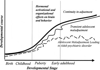
The developmental pathways through adolescence and into adulthood.
At the present time, these ideas must be considered speculative. It is obvious that researchers have not yet fully elucidated the range of hormonal processes that are associated with puberty. In particular, we do not yet understand the causal sequence of hormonal events that transpire during pubertal development. Nor do we know how this ongoing hormonal cascade influences gene expression and brain structure/function. Further, very few developmental studies have charted the transitional period from the onset of puberty into early adulthood. Thus, research has not established the growth curves for age-related changes in hormones, brain, and behavior during this broader period. But ongoing longitudinal studies are likely to yield valuable insights in the near future. These data will certainly have relevance for research on the genesis of psychopathology, as well as for studies of preventive intervention.
References
REFERENCES
Allen, M. T., &
Matthews, K. A.
(1997).
Hemodynamic responses to laboratory stressors in children and
adolescents: The influences of age, race, and gender.
Psychophysiology
34,
329–339.Google Scholar
Angold, A.,
Costello, E. J., &
Worthman, C. M.
(1998).
Puberty and depression: The roles of age, pubertal status and
pubertal timing.
Psychological Medicine
28,
51–61.Google Scholar
Angold, A., &
Rutter, M.
(1992).
Effects of age and pubertal status on depression in a large
clinical sample.
Development and Psychopathology
4,
5–28.Google Scholar
Arnett, J. J.
(1999).
Adolescent storm and stress, reconsidered.
American Psychologist
54,
317–326.Google Scholar
Arnold, A. P., &
Breedlove, S. M.
(1985).
Organizational and activational effects of sex steroids on brain
and behavior: A reanalysis.
Hormones & Behavior
19,
469–498.Google Scholar
Benes, F. M.
(2003a).
Schizophrenia, II: Amygdalar fiber alteration as etiology?
American Journal of Psychiatry,
160,
1053.Google Scholar
Benes, F. M.
(2003b).
Why does psychosis develop during adolescence and early adulthood?
Current Opinion in Psychiatry
16,
317–319.Google Scholar
Blumberg, H. P.,
Martin, A.,
Kaufman, J.,
Leung, H.,
Skudlarski, P.,
Lacadie, C.,
Fulbright, R. K.,
Gore, J. C.,
Charney, D. S.,
Krystal, J. H., &
Peterson, B. S.
(2003).
Frontostriatal abnormalities in adolescents with bipolar disorder:
Preliminary observations from functional MRI.
American Journal of Psychiatry
160,
1345–1347.Google Scholar
Birmaher, B., &
Heydl, P.
(2001).
Biological studies in depressed children and adolescents.
International Journal of Neuropsychopharmacology
4,
149–157.Google Scholar
Booth, A.,
Johnson, D. R.,
Granger, D. A.,
Crouter, A. C., &
McHale, S.
(2003).
Testosterone and child and adolescent adjustment: The moderating
role of parent–child relationships.
Developmental Psychology
39,
85–98.Google Scholar
Brooks–Gunn, J.
(1988).
Antecedents and consequences of variations in girls' maturational
timing.
Journal of Adolescent Health Care
9,
365–373.Google Scholar
Buchanan, C. M.,
Eccles, J. S., &
Becker, J. B.
(1992).
Are adolescents the victims of raging hormones? Evidence for
activational effects of hormones on moods and behavior at
adolescence.
Psychological Bulletin
111,
62–107.Google Scholar
Buske–Kirschbaum, A.,
Jobst, S.,
Wustmans, A.,
Kirschbaum, C.,
Rauh, W., &
Hellhammer, D.
(1997).
Attenuated free cortisol response to psychosocial stress in
children with atopic dermatitis.
Psychosomatic Medicine
59,
419–426.Google Scholar
Caspi, A., &
Moffitt, T. E.
(1991).
Individual differences are accentuated during periods of social
change: The sample case of girls at puberty.
Journal of Personality and Social Psychology
61,
157–168.Google Scholar
Chambers, R. A.,
Taylor, J. R., &
Potenza, M. N.
(2003).
Developmental neurocircuitry of motivation in adolescence: A
critical period of addiction vulnerability.
American Journal of Psychiatry
160,
1041–1052.Google Scholar
Charmandari, E.,
Kino, T.,
Souvatzoglou, E., &
Chrousos, G. P.
(2003).
Pediatric stress: Hormonal mediators and human development.
Hormone Research,
59,
161–179.Google Scholar
Chen, E.,
Matthews, K. A., &
Boyce, W. T.
(2002).
Socioeconomic differences in children's health: How and why do
these relationships change with age?
Psychological Bulletin
128,
295–329.Google Scholar
Cicchetti, D., &
Rogosch, F. A.
(2002).
A developmental psychopathology perspective on adolescence.
Journal of Consulting & Clinical Psychology
70,
6–20.Google Scholar
Cyranowski, J. M.,
Frank, E.,
Young, E., &
Shear, K.
(2000).
Adolescent onset of the gender difference in lifetime rates of
major depression.
Archives of General Psychiatry
57,
21–27.Google Scholar
Davies, P. S.,
Collins, D. L.,
Gregory, J. R., &
Clarke, P. C.
(1996).
Parents' and childrens' reactions to taking blood in a
nutrition study.
Archives of Disease in Childhood
75,
309–313.Google Scholar
Dorn, L. D., &
Chrousos, G. P.
(1997).
Neurobiology of stress: Understanding regulation of affect during
female biological transitions. Seminars
in Reproductive Endocrinology
15,
29–45.Google Scholar
Dorn, L. D.,
Hitt, S. F., &
Rotenstein, D.
(1999).
Biopsychological and cognitive differences in children with
premature vs. on-time adrenarche.
Archives of Pediatric & Adolescent Medicine
153,
137–146.Google Scholar
Durston, S.,
Hulshoff Pol, H. E.,
Casey, B. J.,
Giedd, J. N.,
Buitelaar, J. K., &
van Engeland, H.
(2001).
Anatomical MRI of the developing human brain: What have we learned?
Journal of the American Academy of Child & Adolescent
Psychiatry
40,
1012–1020.Google Scholar
Fadalti, M.,
Petraglia, F.,
Luisi, S.,
Bernardi, F.,
Casarosa, E.,
Ferrari, E.,
Luisi, M.,
Saggese, G.,
Genazzani, A. R., &
Bernasconi, S.
(1999).
Changes of serum allopregnanolone levels in the first 2 years of
life and during pubertal development.
Pediatric Research
46,
323–327.Google Scholar
Faloia, E.,
Camilloni, M. A.,
Giacchetti, G., &
Mantero, F.
(2000).
Adipose tissue as an endocrine organ? A review of some recent data.
Eating & Weight Disorders
5,
116–123.Google Scholar
Flinn, M. V., &
England, B. G.
(1997).
Social economics of childhood glucocorticoid stress response and
health.
American Journal of Physical Anthropology
102,
33–53.Google Scholar
Galdos, P., &
van Os, J.
(1995).
Gender, psychopathology, and development: From puberty to early
adulthood.
Schizophrenia Research
14,
105–112.Google Scholar
Gest, S. D.,
Reed, M. J., &
Masten, A. S.
(1999).
Measuring developmental changes in exposure to adversity: A life
chart and rating scale approach.
Development and Psychopathology
11,
171–192.Google Scholar
Goldsmith, H. H., &
Lemery, K. S.
(2000).
Linking temperamental fearfulness and anxiety symptoms: A
behavior–genetic perspective.
Biological Psychiatry
48,
1199–1209.Google Scholar
Goodyer, I.,
Herbert, J.,
Moor, S., &
Altham, P.
(1991).
Cortisol hypersecretion in depressed school-aged children and
adolescents.
Psychiatry Research
37,
237–244.Google Scholar
Goodyer, I. M.,
Herbert, J.,
Tamplin, A., &
Altham, P. M.
(2000).
Recent life events, cortisol, dehydroepiandrosterone and the onset
of major depression in high-risk adolescents.
British Journal of Psychiatry
177,
499–504.Google Scholar
Goodyer, I. M.,
Park, R. J., &
Herbert, J.
(2001).
Psychosocial and endocrine features of chronic first-episode major
depression in 8–16 year olds.
Biological Psychiatry
50,
351–357.Google Scholar
Goodyer, I. M.,
Park, R. J.,
Netherton, C. M., &
Herbert, J.
(2001).
Possible role of cortisol and dehydroepiandrosterone in human
development and psychopathology.
British Journal of Psychiatry
179,
243–249.Google Scholar
Graber, J. A., &
Brooks–Gunn, J.
(1996).
Transitions and turning points: Navigating the passage from
childhood through adolescence.
Developmental Psychology
32,
768–776.Google Scholar
Graber, J. A.,
Brooks–Gunn, J., &
Warren, M. P.
(1995).
The antecedents of menarcheal age: Heredity, family environment,
and stressful life events.
Child Development
66,
346–359.Google Scholar
Granger, D. A.,
Weisz, J. R.,
McCracken, J. T.,
Ikeda, S. C., &
Douglas, P.
(1996).
Reciprocal influences among adrenocortical activation, psychosocial
processes, and the behavioral adjustment of clinic-referred
children.
Child Development
67,
3250–3262.Google Scholar
Grilo, C. M.,
Becker, D. F.,
Edell, W. S., &
McGlashan, T. H.
(2001).
Stability and change of DSM-III-R personality disorder dimensions
in adolescents followed up 2 years after psychiatric
hospitalization.
Comprehensive Psychiatry
42,
364–368.Google Scholar
Guercio, G.,
Rivarola, M. A.,
Chaler, E.,
Maceiras, M., &
Belgorosky, A.
(2003).
Relationship between the growth hormone/insulin-like growth
factor-I axis, insulin sensitivity, and adrenal androgens in normal
prepubertal and pubertal girls.
Journal of Clinical Endocrinology & Metabolism
88,
1389–1393.Google Scholar
Hayward, C.,
Killen, J. D.,
Wilson, D. M.,
Hammer, L. D.,
Litt, I. F.,
Kraemer, H. C.,
Haydel, F.,
Varady, A., &
Taylor, C. B.
(1997).
Psychiatric risk associated with early puberty in adolescent girls.
Journal of the American Academy of Child & Adolescent
Psychiatry
36,
255–262.Google Scholar
Hessl, D.,
Glaser, B.,
Dyer–Friedman, J.,
Blasey, C.,
Hastie, T.,
Gunnar, M., &
Reiss, A. L.
(2002).
Cortisol and behavior in fragile X syndrome.
Psychoneuroendocrinology
27,
855–872.Google Scholar
Jacobson, K. C.,
Prescott, C. A., &
Kendler, K. S.
(2002).
Sex differences in the genetic and environmental influences on the
development of antisocial behavior.
Development and Psychopathology
14,
395–416.Google Scholar
Kagan, J.,
Reznick, J. S., &
Snidman, N.
(1988).
Biological Bases of childhood shyness.
Science
240,
167–171.Google Scholar
Kawata, M.
(1995).
Roles of steroid hormones and their receptors in structural
organization in the nervous system.
Neuroscience Research
24,
1–46.Google Scholar
Keenan, P. A., &
Soleymani, R. M.
(2001).
Gonadal steroids and cognition. In
R. Tarter &
M. Butters (Eds.),
Medical neuropsychology: The impact of disease on behavior.
Critical Issues in Neuropsychology (pp.
181–197).
New York:
Plenum Press.
Keks, N. A.,
Copolov, D. L.,
Mackie, B.,
Stuart, G. W.,
Singh, B. S.,
McGorry, P. D., &
Coffey, C.
(1991).
Comparison of participants and nonparticipants in a neuroendocrine
investigation of psychosis.
Acta Psychiatrica Scandinavica
83,
373–376.Google Scholar
Kellogg, C. K.,
Awatramani, G. B., &
Piekut, D. T.
(1998).
Adolescent development alters stressor-induced Fos immunoreactivity
in rat brain.
Neuroscience
83,
681–689.Google Scholar
Kenny, F. M.,
Gancayco, G. P.,
Heald, F. P., &
Hung, W.
(1966).
Cortisol production rate in adolescent males in different stages of
sexual maturation.
Journal of Clinical Endocrinology
26,
1232–1236.Google Scholar
Kenny, F. M.,
Preeyasombat, C., &
Migeon, C. J.
(1966).
Cortisol production rate II: Normal infants, children and adults.
Pediatrics
37,
34–42.Google Scholar
Kiess, W.,
Meidert, A.,
Dressendorfer, R. A.,
Scheiver, K.,
Kessler, U., &
Konig, A.
(1995).
Salivary cortisol levels throughout childhood and adolescence:
Relation with age, pubertal stage and weight.
Pediatric Research
37,
502–506.Google Scholar
Kim, K., &
Smith, P. K.
(1998).
Childhood stress, behavioral symptoms and mother–daughter
pubertal development.
Journal of Adolescence
21,
231–240.Google Scholar
Kirschbaum, C.,
Kudielka, B. M.,
Gaab, J.,
Schommer, N. C., &
Hellhammer, D. H.
(1999).
Impact of gender, menstrual cycle phase, and oral contraceptives on
the activity of the hypothalamus–pituitary–adrenal
axis.
Psychosomatic Medicine
61,
154–162.Google Scholar
Klump, K. L.,
McGue, M., &
Iacono, W. G.
(2003).
Differential heritability of eating attitudes and behaviors in
prepubertal versus pubertal twins.
International Journal of Eating Disorders
33,
287–292.Google Scholar
Knutsson, U.,
Dahlgren, J.,
Marcus, C.,
Rosberg, S.,
Bronnegard, M.,
Stierna, P., &
Albertsson–Wiklund, K.
(1997).
Circadian cortisol rhythms in healthy boys and girls: Relationship
with age, growth, body composition and pubertal development.
Journal of Clinical Endocrinology & Metabolism
82,
536–540.Google Scholar
L'Allemand, D.,
Penhoat, A.,
Lebrethon, M. C.,
Ardevol, R.,
Baehr, V.,
Oelkers, W., &
Saez, J. M.
(1996).
Insulin-like growth factors enhance steroidogenic enzyme and
corticotropin receptor messenger ribonucleic acid levels and
corticotropin steroidogenic responsiveness in cultured human
adrenocortical cells.
Journal of Clinical Endocrinology & Metabolism
81,
3892–3897.Google Scholar
Lupien, S. J.,
Wilkinson, C. W.,
Briere, S.,
Menard, C.,
Ng, Y.,
Kin, N. M., &
Nair, N. P.
(2002).
The modulatory effects of corticosteroids on cognition: Studies in
young human populations.
Psychoneuroendocrinology
27,
401–416.Google Scholar
Lyons, M. J.,
Ture, W. R.,
Eisen, S. A.,
Goldberg, J.,
Meyer, J.,
Faraone, S. V.,
Eaves, L. J., &
Tsuang, M. T.
(1995).
Differential heritability of adult and juvenile antisocial traits.
Archives of General Psychiatry
52,
906–915.Google Scholar
Malina, R. M.
(1983).
Menarche in athletes: A synthesis and hypothesis.
Annals of Human Biology
10,
1–24.Google Scholar
Mann, D. R.,
Johnson, A. O.,
Gimpel, T., &
Castracane, V. D.
(2003).
Changes in circulating leptin, leptin receptor, and gonadal
hormones from infancy until advanced age in humans.
Journal of Clinical Endocrinology & Metabolism
88,
3339–3345.Google Scholar
Matthews, K. A.,
Gump, B. B.,
Block, D. R., &
Allen, M. T.
(1997).
Does background stress heighten or dampen children's
cardiovascular responses to acute stress? Psychosomatic
Medicine
59,
488–496.Google Scholar
McBurnett, K.,
Lahey, B. B.,
Rathouz, P. J., &
Rolf, L.
(2000).
Low salivary cortisol and persistent aggression in boys referred
for disruptive behavior.
Archives of General Psychiatry
57,
38–43.Google Scholar
McCrae, R. R.,
Costa, P. T., Jr.,
Ostendorf, F.,
Angleitner, A.,
Hrebickova, M.,
Avia, M. D.,
Sanz, J.,
Sanchez–Bernardos, M. L.,
Kusdil, M. E.,
Woodfield, R.,
Saunders, P. R., &
Smith, P. B.
(2000).
Nature over nurture: Temperament, personality, and life span
development.
Journal of Personality & Social Psychology
78,
173–186.Google Scholar
McGue, M.,
Bacon, S., &
Lykken, D. T.
(1993).
Personality stability and change in early adulthood: A behavioral
genetic analysis.
Developmental Psychology
29,
96–109.Google Scholar
Mesce, K. A.
(2002).
Metamodulation of the biogenic amines: Second-order modulation by
steroid hormones and amine cocktails.
Brain, Behavior & Evolution
60,
339–349.Google Scholar
Miller, E. M., &
Shields, S. A.
(1980).
Skin conductance response as a measure of adolescents'
emotional reactivity.
Psychological Reports
46,
587–590.Google Scholar
Muller, M.,
Holsboer, F., &
Keck, M. E.
(2002).
Genetic modification of corticosteroid receptor signalling: Novel
insights into pathophysiology and treatment strategies of human
affective disorders.
Neuropeptides
36,
117–131.Google Scholar
Neugarten, B. L.
(1979).
Time, age, and the life cycle.
American Journal of Psychiatry
136,
887–894.Google Scholar
Neumann, C. S.,
Grimes, K.,
Walker, E. F., &
Baum, K.
(1995).
Developmental pathways to schizophrenia: Behavioral subtypes.
Journal of Abnormal Psychology
104,
558–566.Google Scholar
Nottelmann, E. D.,
Susman, E. J.,
Dorn, L. D.,
Inoff–Germain, G.,
Loriaux, D. L.,
Cutler, G. B., Jr., &
Chrousos, G. P.
(1987).
Developmental processes in early adolescence: Relations among
chronological age, pubertal stage, height, weight, and serum levels of
gonadotropins, sex steroids, and adrenal androgens.
Journal of Adolescent Health Care
8,
246–260.Google Scholar
Patton, G. C.,
Hibbert, M. E.,
Carlin, J.,
Shao, Q.,
Rosier, M.,
Caust, J., &
Bowes, G.
(1997).
Menarche and the onset of depression and anxiety in Victoria,
Australia.
Journal of Epidemiology & Community Health
50,
661–666.Google Scholar
Paus, T.,
Zijdenbos, A.,
Worsley, K.,
Collins, D. L.,
Blumenthal, J.,
Giedd, J. N.,
Rapoport, J. L., &
Evans, A. C.
(1999).
Structural maturation of neural pathways in children and
adolescents: In vivo study.
Science
283,
1908–1911.Google Scholar
Petrill, S. A.
(2003).
The development of intelligence: Behavioral genetic approaches. In
R. Sternberg &
J. Lautrey (Eds.),
Models of intelligence: International perspectives (pp.
81–89).
Washington, DC:
American Psychological Association.
Pine, D. S.
(2003).
Developmental psychobiology and response to threats: Relevance to
trauma in children and adolescents.
Biological Psychiatry
53,
796–808.Google Scholar
Post, R. M.
(2002).
Do the epilepsies, pain syndromes, and affective disorders share
common kindling-like mechanisms?
Epilepsy Research
50,
203–219.Google Scholar
Rice, F.,
Harold, G. T., &
Thapar, A.
(2003).
Negative life events as an account of age-related differences in
the genetic aetiology of depression in childhood and adolescence.
Journal of Child Psychology & Psychiatry & Allied
Disciplines
44,
977–987.Google Scholar
Rieder, J., &
Coupey, S. M.
(1999).
Update on pubertal development.
Current Opinion in Obstetrics & Gynecology
11,
457–462.Google Scholar
Roberts, B. W.,
Caspi, A., &
Moffitt, T. E.
(2001).
The kids are alright: Growth and stability in personality
development from adolescence to adulthood.
Journal of Personality & Social Psychology
81,
670–683.Google Scholar
Saito, R.,
Toyama, H.,
Uemura, K.,
Ishii, K.,
Senda, M., &
Uchiyama, A.
(2001).
Quantitative estimation of brain atrophy and function with PET and
MRI two-dimensional projection images.
Kaku Igaku–Japanese Journal of Nuclear Medicine
38,
201–209.Google Scholar
Schmidt, M.,
Enthoven, L.,
van der Mark, M.,
Levine, S.,
de Kloet, E. R., &
Oitzl, M. S.
(2003).
The postnatal development of the
hypothalamic–pituitary–adrenal axis in the mouse.
International Journal of Developmental Neuroscience
21,
125–132.Google Scholar
Schmidt, P. J.,
Nieman, L. K.,
Danaceau, M. A,
Adams, L. F., &
Rubinow, D. R.
(1998).
Differential behavioral effects of gonadal steroids in women with
and in those without premenstrual syndrome.
New England Journal of Medicine
338,
209–216.Google Scholar
Schneider, W.
(2002).
Memory development in childhood. In
U. Goswami (Ed).,
Blackwell handbook of childhood cognitive development (pp.
236–256).
Malden, MA:
Blackwell.
Silberg, J. L.,
Pickles, A.,
Rutter, M.,
Hewitt, J.,
Simonoff, E.,
Maes, H.,
Carbonneau, R.,
Murrelle, L.,
Foley, D., &
Eaves, L.
(1999).
The influence of genetic factors and life stress on depression
among adolescent girls.
Archives of General Psychiatry
56,
225–232.Google Scholar
Snieder, H.,
van Doornen, L. J., &
Boomsma, D. I.
(1997).
The age dependency of gene expression for plasma lipids,
lipoproteins, and apolipoproteins.
American Journal of Human Genetics
60,
638–650.Google Scholar
Steiner, H., &
Levine, S.
(1988).
Family environment of adolescents and coping in the hospital.
Psychoneuroendocrinology
13,
333–338.Google Scholar
Stevens, J. R.
(2002).
Schizophrenia: Reproductive hormones and the brain.
American Journal of Psychiatry
159,
713–719.Google Scholar
Stoffel–Wagner, B.
(2001).
Neurosteroid metabolism in the human brain.
European Journal of Endocrinology
145,
669–679.Google Scholar
Susman, E. J.,
Dorn, L. D., &
Chrousos, G. P.
(1991).
Negative affect and hormone levels in young adolescents: Concurrent
and predictive perspectives.
Journal of Youth and Adolescence
20,
167–190.Google Scholar
Susman, E. J.,
Dorn, L. D.,
Inoff–Germain, G.,
Nottelmann, E. D., &
Chrousos, G. P.
(1997).
Cortisol reactivity, distress behavior, and behavioral and
psychological problems in young adolescents: A longitudinal
perspective.
Journal of Research on Adolescence
7,
81–105.Google Scholar
Susman, E. J.,
Nottelmann, E. D.,
Dorn, L. D.,
Inoff–Germain, G., &
Chrousos, G. P.
(1989).
Physiological and behavioral aspects of stress in adolescence. In
G. Chrousos &
P. Gold (Eds.),
Mechanisms of physical and emotional stress (pp.
341–352).
New York:
Plenum Press.
Susman, E. J.,
Nottelmann, E. D.,
Inoff–Germain, G. E.,
Dorn, L. D., &
Chrousos, G. P.
(1987).
Hormonal influences on aspects of psychological development during
adolescence.
Journal of Adolescent Health Care
8,
492–504.Google Scholar
Tamm, L.,
Menon, V., &
Reiss, A. L.
(2002).
Maturation of brain function associated with response inhibition.
Journal of the American Academy of Child & Adolescent
Psychiatry
41,
1231–1238.Google Scholar
Tornhage, C. J.
(2002).
Reference values for morning salivary cortisol concentrations in
healthy school-aged children.
Journal of Pediatric Endocrinology & Metabolism
15,
197–204.Google Scholar
Udry, J. R.,
Billy, J. G.,
Morris, N. M.,
Groff, T. R., &
Raj, M. H.
(1985).
Serum androgenic hormone motivate sexual behavior in adolescent
boys.
Fertility & Sterility
43,
90–94.Google Scholar
Udry, J. R., &
Talbert, L. M.
(1988).
Sex hormone effects on personality at puberty.
Journal of Personality & Social Psychology
54,
291–295.Google Scholar
van Oel, C. J.,
Sitskoorn, M. M.,
Cremer, M. P., &
Kahn, R.
(2002).
School performance as a premorbid marker for schizophrenia: A twin
study.
Schizophrenia Bulletin
28,
401–414.Google Scholar
Veldhuis, J. D.
(1996).
Neuroendocrine mechanisms mediating awakening of the human
gonadotropic axis in puberty.
Pediatric Nephrology
10,
304–317.Google Scholar
Viken, R. J.,
Rose, R. J.,
Kaprio, J., &
Koskenvuo, M.
(1994).
A developmental genetic analysis of adult personality: Extraversion
and neuroticism from 18 to 59 years of age.
Journal of Personality & Social Psychology
66,
722–730.Google Scholar
Wajs–Kuto, E.,
De Beeck, L. O.,
Rooman, R. P., &
Caju, M. V.
(1999).
Hormonal changes during the first year of oestrogen treatment in
constitutionally tall girls.
European Journal of Endocrinology
141,
579–584.Google Scholar
Walker, E., &
Bollini, A. M.
(2002).
Pubertal neurodevelopment and the emergence of psychotic symptoms.
Schizophrenia Research
54,
17–23.Google Scholar
Walker, E. F., &
Diforio, D.
(1997).
Schizophrenia: A neural diathesis-stress model.
Psychological Review
104,
667–685.Google Scholar
Walker, E. F.,
Walder, D. J., &
Reynolds, F.
(2001).
Developmental changes in cortisol secretion in normal and at-risk
youth.
Development and Psychopathology
13,
721–732.Google Scholar
Warren, M., &
Brooks–Gunn, J.
(1989).
Mood and behavior at adolescence: Evidence for hormonal factors.
Journal of Clinical Endocrinology & Metabolism
69,
77–83.Google Scholar
Weber, A.,
Clark, A. J.,
Perry, L. A.,
Honour, J. W., &
Savage, M. O.
(1997).
Diminished adrenal androgen secretion in familial glucocorticoid
deficiency implicates a significant role for ACTH in the induction of
adrenarche.
Clinical Endocrinology
46,
431–437.Google Scholar
Weise, M.,
Eisenhofer, G., &
Merke, D. P.
(2002).
Pubertal and gender-related changes in the sympathoadrenal system
in healthy children.
Journal of Clinical Endocrinology & Metabolism
87,
5038–5043.Google Scholar
Wingo, M. K.
(2002).
The adolescent stress response to a naturalistic driving stressor.
Dissertation Abstracts International: Section B: The Sciences &
Engineering,
63,
1082
(Transaction Periodicals Consortium, Rutgers University).Google Scholar
Wolkowitz, O.,
Epel, E., &
Reus, V.
(2001).
Stress hormone-related psychopathology: Pathophysiological and
treatment implications.
World Journal of Biological Psychiatry
2,
115–143.Google Scholar
Yung, A. R.,
Phillips, L. J.,
Yuen, H. P.,
Francey, S. M.,
McFarlane, C. A.,
Hallgren, M., &
McGorry, P. D.
(2003).
Psychosis prediction: 12-month follow up of a high-risk
(“prodromal”) group.
Schizophrenia Research
60,
21–32.Google Scholar
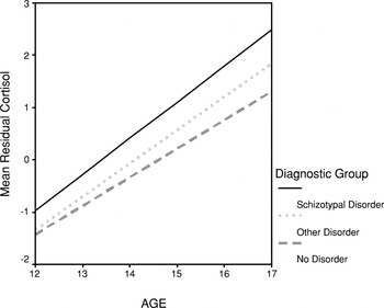
The linear relationship between age and residualized cortisol levels for normal and personality disordered youth.
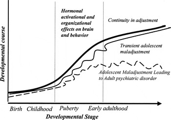
The developmental pathways through adolescence and into adulthood.
 You have
Access
- 115
- Cited by