Introduction
The importance of pre-operative assessment of the sphenoid sinus and neighbouring neurovascular structures has risen following the introduction of new endoscopic techniques. One of the rare but major complications of the growing number of endoscopic sphenoidotomy and endoscopic endonasal trans-sphenoidal approach procedures used to remove pituitary tumours is severe haemorrhage due to parasphenoidal carotid artery trauma.Reference Hudgins1 Therefore, it is essential pre-operatively to determine, as accurately as possible, the position of the internal carotid artery (ICA) in the lateral wall of the sphenoid sinus, in order to avoid intra-operative carotid injury.
It is challenging to evaluate the parasphenoidal structures with direct film X-rays and fluoroscopy. Currently, it has become routine to obtain a coronal computed tomography (CT) scan prior to endoscopic surgery. Anatomical variations of the sphenoid sinus and related anatomical structures have been demonstrated on gross anatomical and CT studies, with conflicting results.Reference Van Alyea2–Reference Unal, Bademci, Bilgili, Batay and Avci16 Magnetic resonance imaging (MRI) has advantages in pre-operative endoscopic planning, as it enables delineation of the extent of soft tissue tumours.Reference Som, Costantino and Silvers17 In dogs with a mass effect imaged by MRI, a diagnosis of neoplasia was significantly associated with vomer bone lysis, cribriform plate erosion, paranasal bone destruction, and sphenoid sinus and nasopharyngeal invasion by the mass; lack of a mass effect on MRI scans was significantly associated with inflammatory disease.Reference Miles, Dhaliwal, Moore and Reed18 Iatrogenic pseudoaneurysms of the ICA have been diagnosed on magnetic resonance angiography images.Reference Buerke, Tombach, Stoll, Heindel and Niederstadt19 Intra-operative MRI during neurosurgical procedures (including trans-sphenoidal procedures for pituitary tumour removal) can assist the surgeon in safely removing the tumour.Reference Anand, Schwartz, Hiltzik and Kacker20
Magnetic resonance imaging of the pituitary gland is undertaken pre-operatively for patients undergoing endoscopic trans-sphenoidal tumour removal. There is no radiation risk, and multiplanar imaging is possible. In this study, we sought to determine whether MRI scanning has a role in the evaluation of the sphenoid sinus and ICA. We also sought to establish reference measurements for the minimal distance between the internal carotid arteries, which we termed the ‘internal carotid interval’. To pursue our research objectives, we reviewed a series of 90 consecutive cranial MRI studies.
Materials and methods
A total of 167 patients undergoing cranial MRI were consecutively enrolled over a two-month period (June to July 2006). The scans were performed to investigate headache. Patients with a known pre-existing condition which might disturb the normal anatomy of the sphenoid and parasphenoidal structures, such as surgery or tumour with metastasis, were excluded from the study. Twenty-eight patients were thus excluded. In addition, 49 patients refused to participate in the study. Informed consent was obtained from all patients, following institutionally approved procedures, regulations and documentation. The study protocol was approved by our hospital's institutional review board. A total of 90 patients were included in the study, equating to a total of 180 sphenoid sinus sides.
The radiological anatomy of the sphenoid sinus and neighbouring structures was evaluated on cranial MRI studies. All examinations were performed on a 1.5 T, whole body MRI system (Excite; General Electric, Milwaukee, Wisconsin, USA) with a 33 mT/m maximum gradient capacity. The following images were obtained: spin echo, T1-weighted scans (TR, 500 msec; TE, 9.6 msec; slice thickness, 5 mm; interslice gap, 1.5 mm; FOV, 24 × 18 cm; matrix, 320 × 192; NEX, 2), and fast-recovery, fast spin echo, T2-weighted scans (TR, 4240 msec; TE, 98.1 msec; slice thickness, 5 mm; interslice gap, 1.5 mm; FOV, 24 × 18 cm; matrix, 352 × 224; NEX, 2).
Patients were classified according to their sphenoid sinus pneumatisation type (assessed on midplane, sagittal, T1-weighted MRI scans) as follows: conchal, presellar, sellar and occipitosphenoidal (Figure 1). The conchal type comprises a very small sinus which is separated from the sella turcica by a bony partition approximately 10 mm thick. The presellar type consists of a sinus which does not extend posterior to a line running perpendicular to the sphenoidal plane through the tuberculum sellae. The sellar type consists of a sinus which extends behind the aforementioned line. Finally, the occipitosphenoidal type comprises a sinus the pneumatisation of which extends across the occipital synchondrosis.
Sagittal magnetic resonance imaging scan showing the types of sphenoid sinus pneumatisation observed: (a) conchal; (b) presellar and (c) sellar. (A fourth type, occipitosphenoidal, was also looked for but not observed.)

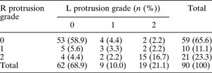
Protrusion of the internal carotid artery (ICA) into the sphenoid sinus was noted. The degree of such protrusion was classified according to the percentage of artery diameter exposed, as 30–50 per cent, termed grade one, or 50–100 per cent, termed grade two. The sphenoidal septum or crest may extend and attach to the wall of the ICA protrusion; any such variation was noted (Figure 2a). The distance between both ICAs was calculated (Figure 2b and 2c). Pterygoid process pneumatisation was deemed significant if it extended beyond a plane tangential to the most inferolateral aspect of the foramen rotundum and vidian canal in the axial and coronal planes. Anterior clinoid pneumatisation in the axial and coronal planes was also noted.
Axial magnetic resonance imaging scans showing (a) protrusion of the internal carotid arteries into the sphenoid sinus (arrows); a septum or crest (arrowhead) is attached on the right side. Also shown are the (b) shortest and (c) longest internal carotid intervals detected.

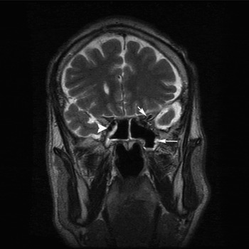
All these evaluations were conducted on T2-weighted images; however, in seven cases the findings were confirmed on pre- or post-contrast T1-weighted images. Mucosal disease was evaluated on T2-weighted images in all cases (Figure 3). All MRI images were assessed by three radiologists (HGH, AS, EY); any disagreements were discussed until consensus was achieved.
Coronal, T2-weighted magnetic resonance imaging scan showing pneumatisation of the left anterior clinoid process (short arrow) and left pterygoid plate (long arrow). There is also protrusion of the right internal carotid artery (arrowhead). Note the mucosal disease on both sides.

Statistical analysis
Data analysis was performed using the Statistical Package for the Social Sciences for Windows version 11.5 software. The Shapiro–Wilk test was used to determine whether the distributions of continuous variables were normal or not. Data were shown as mean ± standard deviation (SD) for continuous variables. Categorical variables were presented as percentages. Means were compared using Student's t-test when the number of independent groups was two. Differences between the means of more than two groups were evaluated using one-way analysis of variance. Nominal data were analysed using the chi-square test or Fisher's exact test, where applicable. The McNemar test was applied for the comparison of the two sides. Odds ratios and 95 per cent confidence intervals (CIs) were calculated. A p value of less than 0.05 was considered statistically significant.
Results
The study included 90 patients (34 males and 56 females; mean age ± SD, 42.92 ± 18.94 years; age range 12–87 years). The following types of sphenoid sinus pneumatisation were observed: conchal type in three patients (3.3 per cent); presellar type in 13 (14.4 per cent); sellar type in 74 (82.2 per cent); and occipitosphenoidal type in none (0 per cent).
Data on the incidence and degree of internal carotid artery (ICA) protrusion are summarised in Table I.
Incidence of internal carotid artery protrusion into sphenoid sinus
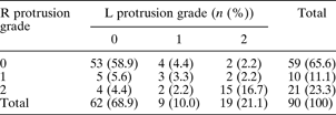
Data represent patients. R = right side; L = left side
We observed sphenoidal septum or crest attachment to a right-sided ICA protrusion in eight patients (8.9 per cent) and to a left-sided ICA protrusion in nine patients (10 per cent); such attachment occurred in 17 patients (18.9 per cent) in total. There was no statistically significant difference in the occurrence of sphenoidal septum or crest attachment to an ICA protrusion, comparing the right and left sides (p = 1.000).
The closest distance between the right and left internal carotid arteries in the parasphenoidal region varied between 9.04 and 24.26 mm. The mean ± SD for this parameter was 16.18 ± 3.20 mm for female patients (range, 9.40–23.82 mm) and 15.56 ± 3.70 mm for male patients (range, 9.04–24.26 mm). There were no statistically significant differences between the means of this parameter, comparing the two genders (p = 0.401).
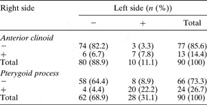
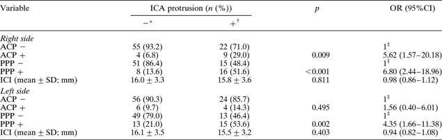
Anterior clinoid pneumatisation was observed in 16 patients (17.8 per cent) and pterygoid process pneumatisation in 32 patients (35.5 per cent) (Table II). The presence of anterior clinoid pneumatisation was found to be a predictive factor associated with ICA protrusion only on the right side (odds ratio, 5.62; 95 per cent CI, 1.57–20.18; p = 0.009). The presence of pterygoid process pneumatisation was found to be a predictive factor associated with both right-sided and left-sided ICA protrusion (for right side: odds ratio 6.80, 95 per cent CI 2.44–18.96, p < 0.001; for left side, odds ratio 4.35, 95 per cent CI 1.66–11.38, p = 0.002).
Incidence of sphenoid sinus pneumatisation

Data represent patients. −= pneumatisation absent; + = pneumatisation present
There was no statistically significant difference in the mean internal carotid interval, comparing patients with and without ICA protrusion (p = 0.548). Table III summarises these findings.
Internal carotid artery protrusion into sphenoid sinus, by anterior clinoid and pterygoid process pneumatisation and internal carotid interval
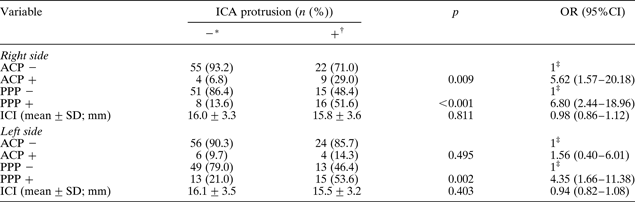
Data represent patients unless otherwise specified. *Right side, n = 59; left side, n = 62. †Right side, n = 31; left side, n = 28. ‡Reference category. ICA = internal carotid artery; OR = odds ratio; CI = confidence intervals; ACP = anterior clinoid pneumatisation; PPP = pterygoid plate pneumatisation; −= absent; + = present; ICI = closest distance between right and left ICAs in parasphenoidal region; SD = standard deviation
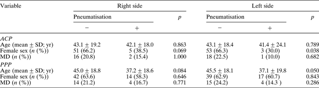
There was no statistically significant association between anterior clinoid pneumatisation and age (p = 0.758). However, patients with pterygoid process pneumatisation were found to be younger on average than those without (p = 0.009). There was no statistically significant association between pterygoid process pneumatisation and patient sex (p = 0.646). However, anterior clinoid pneumatisation was more frequent in males (p = 0.004). The presence of mucosal disease had no statistically significant association with either pterygoid process pneumatisation (p = 0.274) or anterior clinoid pneumatisation (p = 0.470). Please refer to Table IV for detailed analysis.
Anterior clinoid and pterygoid plate pneumatisation by patient age, sex and presence of mucosal disease
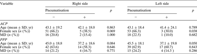
−= absent; += present; ACP = anterior clinoid pneumatisation; PPP = pterygoid plate pneumatisation; SD = standard deviation; yr = years; MD = mucosal disease
There was no statistically significant association between sphenoid sinus pneumatisation and either patients' sex (p = 0.986) or age (p = 0.650). There was also no statistically significant association between the internal carotid interval and the presence of sphenoid sinus pneumatisation (p = 0.538) (Table V).
Sphenoid sinus pneumatisation pattern by patient age, sex and internal carotid interval

* n = 3; †n = 74; ‡n = 13. SD = standard deviation; yr = years; ICI = closest distance between right and left internal carotid arteries in parasphenoidal region
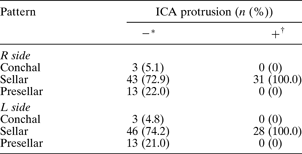
The incidence of sellar-type sphenoid sinus pneumatisation was higher in patients with ICA protrusion, compared with those without (p < 0.001). Presellar-type sphenoid sinus pneumatisation was found in patients without ICA protrusion, but did not occur in those with ICA protrusion (p < 0.001) (Table VI).
Internal carotid artery protrusion into sphenoid sinus, by sphenoid sinus pneumatisation pattern
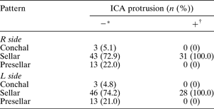
* Right side, n = 59; left side, n = 62. †Right side, n = 31; left side, n = 28. ICA = internal carotid artery; R = right; L = left
Discussion
The sphenoid sinus continues to pneumatise until adolescence.Reference Warwick and Williams21 In adulthood, the extent of sphenoidal pneumatisation is classified as conchal, presellar, sellar or occipitosphenoidal (Table VII).Reference Hammer and Radberg3, Reference Hamberger, Hammer, Norlen and Sjogren4 The current study assessed sphenoidal pneumatisation patterns on sagittal, T1-weighted MRI scans. In our patients, the extent of pneumatisation was not related to age or sex. Sellar pneumatisation was the most frequent type. As the degree of sphenoid sinus pneumatisation increases, the surrounding neurovascular structures project more into the sinus cavity, appearing as ridges. This may increase the risk of vessel and nerve injury during sphenoidotomy procedures.
Previously reported incidences of sphenoid sinus pneumatisation types

* Current study. – = not assessed
Using sagittal, T1-weighted MRI scanning, it is possible to evaluate the thickness of the posterior wall of the sphenoid sinus. In patients with a pituitary mass who may require a trans-sphenoidal surgical approach, the posterior extent of the main sphenoid sinus cavity should be carefully evaluated. If more than 1 or 2 mm of bone remains between the posterior margin of the pneumatised sinus and the anterior sellar wall (this occurs in less than 1 per cent of cases), surgeons should consider avoiding a transnasal, trans-sphenoidal hypophysectomy. A thick, bony posterior sinus wall may also be a relative contraindication.Reference Som, Shugar, Brandwein, Som and Curtin22 However, with the current, widespread use of high speed drilling, these operations can be performed much more safely.
The lateral wall of the sphenoid sinus is related to the orbital apex, optic canal, optic nerve, cavernous sinus and internal carotid artery.Reference Som, Shugar, Brandwein, Som and Curtin22 Surgeons should consider these important neighbouring neurovascular structures and take into consideration the possible anatomical variations, prior to any dissection in this region. Injuries of the internal carotid artery (ICA) may occur, not only due to anomalies and aneurysms of the artery, but also in cases of a missing bony covering of the artery in the sphenoid sinus when the sinus is not opened strictly paraseptally.Reference Johnson, Hopkins, Hanafee and Fisk9 In the latter case, tamponade compression or balloon occlusion of the ICA may be the only options. However, hemiplegia or death is the result in most such cases.Reference Hudgins1 On CT scans, the ICA, the optic nerve, and the more posterior cavernous sinus and its contents are only visualisable as indentations on the lateral walls of the sphenoid bone, depending on the degree of sphenoid sinus pneumatisation. However, on MRI, the ICA can be distinguished from the cavernous sinus surrounding it. Vascular anomalies and aneurysms can be diagnosed on MRI and MR angiography images.Reference Buerke, Tombach, Stoll, Heindel and Niederstadt19
The current study attempted to establish reference measurements for the minimal distance between the internal carotid arteries, termed the internal carotid interval. The closest distance between the internal carotid arteries in the parasphenoidal region varied between 9.04 and 24.26 mm. There was no statistically significant difference in this parameter between male and female patients. Furthermore, there was no statistically significant difference in the internal carotid interval between patients with and without ICA protrusion.
Various studies have reported ICA protrusion into the sphenoid sinus in between 5.2 and 72 per cent of cases.Reference Elwany, Elsaeid and Thabet8, Reference Johnson, Hopkins, Hanafee and Fisk9, Reference Teatini, Simonetti, Salvolini, Masala, Meloni and Rovasio11, Reference Arslan, Aydinlioglu, Bozkurt and Egeli13–Reference Unal, Bademci, Bilgili, Batay and Avci16 In our study, almost one-third (32.8 per cent) of our patients had ICA protrusion into the sphenoid sinus. The prevalence of sellar-type sphenoid pneumatisation was significantly greater in patients with ICA protrusion, and the prevalence of presellar-type pneumatisation was significantly greater in patients without ICA protrusion. Because of the high incidence of ICA protrusion into the sphenoid sinus, surgeons and radiologists should be aware of the degree of such protrusion prior to sphenoidotomy.
• Pre-operative assessment of the sphenoid sinus and neighbouring neurovascular structures has gained importance following the introduction of new endoscopic techniques
• Magnetic resonance imaging provides reliable information on anatomical variations of the sphenoid sinus
• This information could help avoid intra-operative carotid injury during endoscopic sphenoid sinus and trans-sphenoidal pituitary gland procedures
In our study, bony septum attachment to a protruding ICA was better evaluated on axial MRI scans. We observed this anatomical variant in 18.9 per cent of our patients; other authors have reported an incidence of 5.4 to 26.7 per cent.Reference Elwany, Elsaeid and Thabet8, Reference Sirikci, Bayazit, Bayram, Mumbuc, Gungor and Kanlikama14–Reference Unal, Bademci, Bilgili, Batay and Avci16 This variant is usually present at the site of fusion of several synchondroses of the sphenoid bone.Reference Elwany, Elsaeid and Thabet8 It may result in carotid avulsion during surgery.Reference Hudgins1
We found anterior clinoid pneumatisation and pterygoid process pneumatisation in 17.8 and 35.5 per cent of patients, respectively. Other authors have reported incidences of 6 to 24.1 per cent for anterior clinoid pneumatisation and of 29.3 to 39.7 per cent for pterygoid process pneumatisation.Reference Arslan, Aydinlioglu, Bozkurt and Egeli13–Reference Unal, Bademci, Bilgili, Batay and Avci16 We found the presence of pterygoid process pneumatisation to be a predictive factor associated with ICA protrusion. In contrast, other authors have found no statistically significant association between pterygoid process pneumatisation and ICA protrusion.Reference Sirikci, Bayazit, Bayram, Mumbuc, Gungor and Kanlikama14–Reference Unal, Bademci, Bilgili, Batay and Avci16
The literature gives conflicting accounts of the anatomy of the sphenoid sinus. Such variation may be the result of the number of patients studied, racial or population variability, or differences in methodology. On plain X-ray films, the sphenoid sinus cavity is best evaluated on the lateral, base and open-mouth Waters views. However, overlapping of structures may make assessment difficult. On CT views, neurovascular structures can be evaluated only as indentations of the cavernous sinus. On MRI views, however, it is possible to distinguish the carotid artery from the cavernous sinus surrounding it. The internal carotid arteries may be visualised along their whole length. The current study assessed patients who underwent MRI as an investigation for headaches. Therefore, paranasal sinus findings were incidental. Previously reported CT studies of sphenoid sinus anatomy used patients complaining of sinusitis; this may have created a bias. However, the current study also had several limitations. Computed tomography evaluates sinus disease and bony dehiscence more reliably.
Conclusion
Magnetic resonance imaging provides reliable information on variations of sphenoid sinus anatomy. In addition, MRI makes it possible to distinguish the internal carotid artery (ICA) from the cavernous sinus surrounding it. This information could help surgeons to avoid intra-operative ICA injury during endoscopic sphenoid sinus and trans-sphenoidal pituitary gland procedures. The distance between the ICAs, which we termed the internal carotid interval, can be as small as 9 mm in both genders. The internal carotid interval is not influenced by patients' sex or age, or by the pneumatisation pattern of the sphenoid sinus. There is no statistically significant association between the internal carotid interval and the degree of ICA protrusion into the sphenoid sinus. The sphenoid pneumatisation pattern, and the posterior sphenoid sinus wall, may be assessed quite accurately on sagittal, T1-weighted MRI scans. The incidence of sellar-type pneumatisation is greater in patients with ICA protrusion.