Bifidobacteria are common members of human gastrointestinal microflora and are known to contribute to health benefits to humans (Ventura et al. Reference Ventura, O'Connell-Motherway, Leahy, Moreno-Munoz, Fitzgerald and van Sinderen2007). Because they play an important role in the control of a balanced intestinal microflora, bifidobaceria are considered as probiotic and are used in preparation of functional foods as ingredients. From a functional food perspective, the presence of live bifidobacteria in products is particularly indispensable. As a general recommendation and in the absence of “dose response” data for many strains, it is suggested that probiotic products should contain at least 107 CFU/g (ml) (Ishibashi & Shimura, Reference Ishibashi and Shimamura1993). Probiotic yogurt is preferable carrier of bifidobacteria. We know it is easy to enumerate the amount of bifidobacteria in products by the plate counting when only bifidobacterial strains are added to products. However bifidobacterial strains are commonly used together with other probiotic strains or other lactic acid bacteria strains.
Culture-based methods, especially selective media are very useful in enumeration of bifidobacteria. It has previously been shown that a number of selective media, such as LP-MRS (Vinderola & Reinheimer, Reference Vinderola and Reinheimer1999), NPNL-MRS agar (Martin & Chou, Reference Martin and Chou1992), AMC (Payne et al. Reference Payne, Morris and Beers1999) and OG-MRS (Lim et al. Reference Lim, Huh, Baek and Kim1995) were suggested to perform a selective enumeration of bifidobacteria in various milk products. However these culture-based methods are labour intensive and time consuming. Triggered by these shortcomings, culture-independent methods such as real-time PCR (Masco et al. Reference Masco, Vanhoutte, Temmerman, Swings and Huys2007) and enzymatic-colorimetric assays (Bibiloni et al. Reference Bibiloni, Pérez and De Antoni2000) have been developed. In recent years, several molecular methods have been described: a PCR on the hsp60 gene for a rapid detection of bifidobacteria in raw milk and raw milk cheese samples (Delcenserie et al. Reference Delcenserie, Bechoux, China, Daube and Gavini2005); a PCR-denaturing gradient gel electrophoresis (DGGE) technique for the screening of the microbial composition of probiotic products (including bifidobacteria) (Fasoli et al. Reference Fasoli, Marzotto, Rizzotti, Rossi, Dellaglio and Torriani2003); the quantitative real-time PCR for quantification of Bifid. in human fecal samples (Gueimonde et al. Reference Gueimonde, Tölkkö, Korpimäki and Salminen2004), in probiotic products (Masco et al. Reference Masco, Vanhoutte, Temmerman, Swings and Huys2007) and in bacterial mixtures (Vitali et al. Reference Vitali, Candela, Matteuzzi and Brigidi2003); the pulsed-field gel electrophoresis (PFGE) method for identification of bifidobacterial strains in probiotic milk products (Grand et al. Reference Grand, Küffer and Baumgartner2003). Of all the advanced methods based on PCR techniques and newly-developed methods based on signal or probe enlargement, real-time PCR technology has been attracting attention as a reliable method (Vitali et al. Reference Vitali, Candela, Matteuzzi and Brigidi2003). However, conventional real-time PCR methods do not distinguish viable cells from dead cells.
The selective detection of viable but not dead bacteria is a major issue in nucleic acid-based detection (Rudi et al. Reference Rudi, Nogva, Moen, Nissen, Bredholt, Møretrø, Naterstad and Holck2002). Ethidium monoazide (EMA) is a DNA intercalating agent (Bolton & Kearns, Reference Bolton and Kearns1978) which is reported to selectively penetrate the membranes of dead cells, but is purportedly unable to penetrate the intact membranes of viable cells (Rudi et al. Reference Rudi, Nogva, Moen, Nissen, Bredholt, Møretrø, Naterstad and Holck2002; Nogva et al. Reference Nogva, Drømtorp, Nissen and Rudi2003). Nogva et al. (Reference Nogva, Drømtorp, Nissen and Rudi2003) reported that dead cells stained by EMA did not result in DNA amplification by real-time PCR. Rudi et al. (Reference Rudi, Nogva, Moen, Nissen, Bredholt, Møretrø, Naterstad and Holck2002), Nogva et al. (Reference Nogva, Drømtorp, Nissen and Rudi2003) and Wang et al. (Reference Wang, Li and Mustapha2009) used the EMA to discriminate between living and dead cells of Escherichia coli, Salmonella sp., Listeria monocytogenes and Campylobacter jejuni by PCR. However, it has been reported that DNA amplification from viable cells of certain bacteria, such as Enterobacter sakazakii (Cawthorn & Witthuhn, Reference Cawthorn and Witthuhn2008), List. monocytogenes (Nocker et al. Reference Nocker, Cheung and Camper2006; Flekna et al. Reference Flekna, Stefanic, Wagner, Smulders, Mozina and Hein2007; Pan & Breidt, Reference Pan and Breidt2007) and Camp. jejuni (Flekna et al. Reference Flekna, Stefanic, Wagner, Smulders, Mozina and Hein2007), can also be negatively influenced by EMA staining.
In the present work, the potential of EMA real-time PCR method for rapid detection of viable bifidobacteria present in probiotic yogurt was evaluated. For this purpose, a real-time PCR with molecular beacon assay was developed to determine Bifid. quantitatively in order to increase the sensitivity and specificity of assay. EMA treatment procedures were optimized for selectively amplifying a DNA target sequence via molecular beacon and real-time PCR from only viable cells of Bifid. in the presence of dead cells in probiotic yogurt. Furthermore, the EMA real-time PCR method was applied to enumerate viable bifidobacteria in commercial probiotic yogurt.
Materials and Methods
Bacterial strains, media and culture conditions
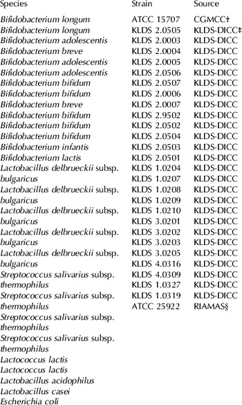
The strains used in this study are shown in Table 1. A total of 28 bacterial strains, including bifidobacterial (n=14) and non-bifidobacterial (n=14) strains, were used to evaluate the specificity of the molecular beacon probe and primers. Bifidobacterial strains were cultivated overnight in trypticase peptone yeast (TPY) broth under anaerobic conditions (80% N2, 10% H2, 10% CO2) at 37°C, and other lactic acid bacterial strains were cultivated overnight in Man Rogosa Sharpe (MRS) (Oxoid) broth under aerobic conditions at 37°C. Esch. coli was cultured aerobically in nutrition agar at 37°C. All cells were harvested from the early exponential growth phase. Colony forming units (CFU) of bifidobacterial cultures were determined by serial dilutions of cultures in maximum recovery diluents (MRD) (Oxoid) followed by pour plating with TPY. Plates were incubated at 37°C for 3 d under anaerobic conditions.
The bacterial strains used for determination of the specificity of the real-time PCR based on molecular beacon probes in this study

ATCC: American Type Culture Collection; KLDS: Key Laboratory of Dairy Science (Education Ministry of China)
† China General Microbiological Culture Collection Center (CGMCC)
‡ Dairy Industiral Microbiology Culture Collection Center (KLDS) (KLDS-DICC)
§ Research Institute of Applied Microbiology, Heilongjiang of Academy of Science
Preparation of probiotic yogurt samples containing bifidobacteria
The bifidobacterial cultures were prepared separately, and then added into the yogurt, in order to obtain high counts of bifidobacteria and avoid microbial competition with the starter cultures. Bifid. longum ATCC 15707 cultures were made by incubation overnight at 37°C with a 1% (v/v) inoculum in TPY under anaerobic conditions. Cells of culture were harvested by centrifugation at 2,500×g for 10 min and resuspended in 10% (w/v) sterilized reconstituted skim milk (RSM), the cell density of bifidobacteria in RSM was adjusted to 1011 CFU/ml approximately.
Full-fat milk powder (Nestlé) was reconstituted with water at 13% (w/v) total solids, and 7% (w/w) sucrose was added, then the total was pasteurized at 90°C for 15 min. After pasteurization, the milk mixture was cooled down to 45°C and commercial yogurt starter cultures contained Streptococcus salivarius subsp. thermophilus and Lactobacillus delbrueckii subsp. bulgaricus (YF-L902; Chr. Hansen, Denmark) was added. The milk mixture was stirred gently, and was brought to incubate at 43°C till pH 4·6 was obtained. This fermented milk is defined as yogurt samples. RSM suspension containing bifidobacteria (1011 CFU/ml) was added to above yogurt with a 1% (v/v) inoculum, and the probiotic yogurt samples containing bifidobacteria were obtained. Both yogurt samples and probiotic yogurt samples were taken and kept at 4°C overnight.
Total DNA extraction from probiotic yogurt samples
Total DNA of probiotic yogurt samples were extracted as previously described by Rademaker et al. (Reference Rademaker, Hoolwerf, Wagendorp and Giffel2006) with modifications. Probiotic yogurt samples of 25 ml were diluted in 225 ml of sterile PBS (phosphate-buffered saline, composed of 137 mm-NaCl, 2·7 mm-KCl, 10 mm-Na2HPO4, 2·0 mm-KH2PO4, pH 7·4). One milliliter of 10-fold dilution probiotic yogurt samples were mixed with 100 μl 18% (w/v) sodium citrate and 50 μl 1 m-NaOH at room temperature, then the mixture were centrifuged at 6600×g for 10 min in the Eppendorff tubes. The pellet was washed in PBS buffer. The washed pellet was resuspended in 100 μl MilliQ water, and was mixed with 100 μl 2% (w/v) Triton X-100 (Sigma). This mixture was heated at 100°C for 10 min, then immediately cooled in ice-water, and centrifuged at 6600×g for 10 min and the supernatant was stored at −20°C until its use.
Primers and molecular beacon probes design
The sequences of the genus-specific primers for genus Bifid. were designed on the basis of 16S rRNA and/or 16S rDNA sequences which were downloaded from the GenBank database (http://www.ncbi.nlm.nih.gov). The multiple alignments were prepared by the program Clustal W (http://www.ebi.ac.uk). The genus-specific primers for Bifid. were designed by primer design software (Primer 5.0) after comparison of unique bifidobacterial primer sequences with a large number of reference primer sequences (Kaufmann et al. Reference Kaufmann, Pfefferkom, Teuber and Meile1997; Venema et al. Reference Venema and Maathuis2003; Vitali et al. Reference Vitali, Candela, Matteuzzi and Brigidi2003; Matsuki et al. Reference Matsuki, Watanabe, Fujimoto, Kado, Takada, Matsumoto and Tanaka2004). Secondary structures and dimers formation were controlled by the Oligo Analyzer 6.0 Software (http://www.bioon.com/Soft/Class1/Index.html). The sequences of the primers were as follows, Bif-F: 5′-TCTGGCTCMGGATGAACGC-3′; Bif-R: 5′-CACCGTTACACCGGGAATTC-3′. These primers have a length of about 20–25 bases, a G/C content of over 50%, and a Tm of about 58°C. Primers used in this study were synthesized by TaKaRa (TaKaRa Biotechnology [Dalian] Co. Ltd.). Primers were resuspended in TE buffer (1 mm-EDTA-10 mm-Tris-HCl buffer, pH 8·0) and stored at −20°C. Aliquots (10 μm) of each primer were prepared and used for PCR.
Comparative analysis of the primary nucleotide sequences of the 16S rRNA of Bifid. longum ATCC 15707 (accession number M58739) amplicon generated by conventional PCR was used for the design of a molecular beacon probe. Fluorescein (6-FAM) was chosen as the chromophore (excitation at 488 nm and mission at 519 nm) and DABCYL was used as the quencher. Fluorescein and DABCYL were coupled to the 5' and 3' ends of the beacon probe respectively. The sequence of the probe (occupying position 161–180 of the 16S rDNA of Bifid. longum ATCC 15707) with fluorophore and quencher moieties was: 5′-FAM-CCAGGCATCCGGCATTACCACCCGTCCTGG-3′-DABCYL. Molecular beacon probes used in this study were synthesized by Sangon Company (Shanghai, China).
Molecular beacon real-time PCR
Real-time PCR amplification reactions were performed with the 7500 Real Time PCR System (Applied Biosystems, USA) using above primers and molecular beacon probes. Thermal cycling conditions were specified as follows: 1 cycle of 94°C for 3 min; 40 cycles of 94°C for 30 s, 40°C for 35 s, 59°C for 30 s and 72°C for 1 min. Fluorescent measurements were recorded during each first annealing step (40°C for 35 s). Data were analyzed using the Sequence Detection System software (version 1.9.1) (Applied Biosystems). For each PCR, 5 μl of template DNA was added to 45 μl PCR master (5 μl of 10×PCR buffer [TIANWEI, Beijing, China], 2·5 mm-MgCl2, 0·8 μm-each primer, 0·4 μm-beacons, 200 μm-each of the four deoxynucleoside triphosphates [TIANWEI, Beijing, China], 1·25 U of Taq DNA polymerase [TIANWEI, Beijing, China], and 16·5 μl water). The PCR buffer contained ROX as the reference dye for normalization of the reactions. Any possible fluctuations in ROX signal were used to correct the sample signal. Non-template controls were included in all PCR runs and tested negative.
Genomic DNA of bifidobacterial culture was extracted as previously described by Matsuki et al. (Reference Matsuki, Watanabe, Fujimoto, Kado, Takada, Matsumoto and Tanaka2004), and genomic DNA of non-bifidobacterial culture was extracted by using TIANamp Bacteria DNA Kit (TIANGEN BIOTECH [Beijing] Co., LTD). The genomic DNA of 14 bifidobacterial strains and 14 non-bifidobacterial strains was amplified to assess the ability of the real-time PCR assay to discriminate Bifid. from other bacterial strains as described above. All detections were performed in triplicate.
Influence of EMA concentration on differentiation between viable and heat-killed Bifid. longum ATCC 15707 in probiotic yogurt samples containing bifidobacteria
Ethidium monoazide bromide (Sigma) was dissolved in dimethylformamide (DMF) (Sigma) to yield a stock EMA solution containing 1 mg/ml, which was stored at −20°C in the dark. This EMA stock solution of different volumes (0·5 μl, 1·0 μl, 1·5 μl, 2·0 μl, 2·5 μl, 5 μl) were added respectively into 6 microcentrifuge tubes containing 1·0 ml of 10-fold dilution probiotic yogurt samples with 109 CFU/ml heat-treated dead bifidobacteria at 100°C for 5 min and 6 microcentrifuge tubes containing 1·0 ml of 10-fold dilution probiotic yogurt samples with 109 CFU/ml viable bifidobacteria. These sample tubes were then placed in the dark at room temperature for 5 min, and subsequently placed on ice and exposed to light for 2 min at a distance of 20 cm from the light source (OSRAM with a 650-W halogen light bulb, Osram, Germany) to activate and photolyse the EMA. The EMA was handled as a carcinogen. The DNA was extracted from the EMA-treated samples, and real-time PCR was performed to detect the amplification reactions of DNA as described above. All real-time PCR assays were performed at least three independent times with the mean Ct values and standard deviations reported.
Standard curve
The standard curve used to make correlation between viable counts determined by real-time PCR coupled with EMA and plate counts using the selective agar media was obtained from viable cells of Bifid. longum ATCC 15707 in yogurt. Bifid. longum ATCC 15707 culture was serially diluted with sterilized water to obtain a range of CFU equivalent to approximately 2×10⁸ CFU/ml, 2×10⁷ CFU/ml, 2×10⁶ CFU/ml, 2×10⁵ CFU/ml, 2×10⁴ CFU/ml, and then centrifuged at 2, 500×g for 10 min respectively. These bacterial cell pellets were resuspended in the yogurt which contained viable Strep. salivarius subsp. thermophilus and Lb. delbrueckii subsp. bulgaricus. EMA treatment of probiotic yogurt and DNA isolation were carried out as described above, then amplified by molecular beacon probe real-time PCR. Bifid. longum ATCC 15707 counts in different suspension were determined by LP-MRS agar (Vinderola & Reinheimer, Reference Vinderola and Reinheimer1999). By plotting the log CFU/ml against the Ct value, a linear relationship was formed.
Comparison of the EMA molecular beacon real-time PCR assay and plate counts for quantitative detection of bifidobacteria in commercial probiotic yogurt
Probiotic yogurts were purchased from Carrefour supermarket (Harbin, China). The total bifidobacteria of probiotic yogurts were detected by real-time PCR, and the viable bifidobacteria of probiotic yogurts were detected by EMA real-time PCR method established in this study and plate counting respectively. The results obtained by these methods were compared to elucidate the accuracy of EMA real-time PCR method. LP-MRS agar was used for enumerating bifidobacteria in probiotic yogurt (Vinderola & Reinheimer, Reference Vinderola and Reinheimer1999). All detections were performed in triplicate.
Statistical analysis
All cell amounts obtained by EMA real-time PCR and CFU counts were converted to log counts prior to statistical analysis. Paired Student's tests were performed using Microsoft Excel software (Windows XP) and SAS statistics software 8.0.
Results
Specificity of the real-time PCR based on molecular beacon probe
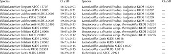
Genomic DNA extracted from bifidobacterial (n=14) and non-bifidobacterial strains (n=14) was amplified to assess the ability of the real-time PCR assay to discriminate bifidobacteria from non-bifidobacteria. Real-time PCR studies showed that all of the bifidobacterial strains were positively detected, and the non-bifidobacterial strains were not detected by the real-time PCR assay (Table 2).
Representative real-time PCR amplification results obtained from 14 bifidobacterial strains and 14 non-bifidobacterial strains

The values are means of triplicates±standard deviation (SD)
“—” means no fluorescence signal and no Ct value
Influence of EMA concentration on differentiation between viable and heat-killed Bifid. longum ATCC 15707 in probiotic yogurt containing bifidobacteria
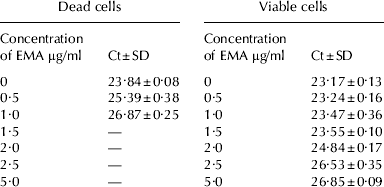
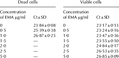
The amplification of DNA derived from dead cells still existed when EMA concentration was less than 1·5 μg/ml, otherwise there was no detectable difference in the PCR signals from dead bifidobacteria when EMA concentrations was 1·5 μg/ml or higher (Table 3). It was showed that the amplification of target DNA derived from dead cells of bifidobacteria in the real-time PCR was completely inhibited when EMA concentrations were 1·5 μg/ml or higher.
Real-time PCR amplification results of DNA derived from dead and viable cells of Bifidobacterium in yogurt treated with EMA of different concentrations
The values are means of triplicates±standard deviation (SD)
“—” means no fluorescence signal and no Ct value
When the EMA concentration was 2·0 μg/ml or less, little or no inhibition of the amplification of the target DNA derived from viable cells occurred in the real-time PCR procedure (Table 3). However there was significant inhibition of amplification when the EMA concentration was 2·5 or 5·0 μg/ml (P<0·05), but it also had the Ct values (Table 3). The Ct values obtained from EMA treatment (1·5 μg/ml) for viable bifidobacteria were not significantly different (P>0·05) from the Ct value of the control. Therefore, it was concluded that the minimum concentration of EMA was 1·5 μg/ml by this method.
Standard curve
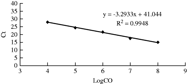
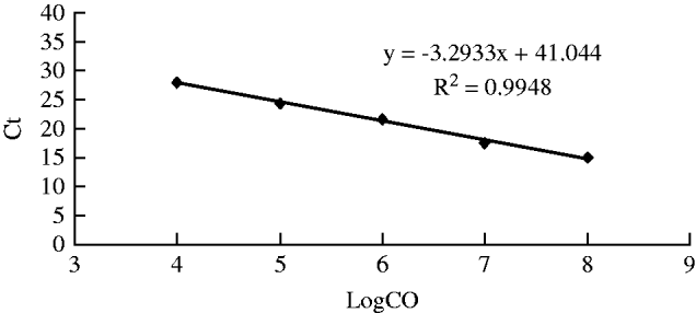
A linear relation (y=−3·2933x+41·044, x representative log CFU/ml, y representative Ct values, R2=0·9948) between bacterial concentrations and Ct values was obtained in the range of 104–108 CFU/ml for probiotic yogurt containing bifidobacteria using real-time PCR based on the molecular beacon probe (Fig. 1).
Standard curve for quantifying viable Bifidobacterium in probiotic yogurt containing bifidobacteria by real-time PCR combined with ethidium monoazide based on the molecular beacon probe.
Comparison of the EMA molecular beacon real-time PCR assay and plate counting for quantitative detection of bifidobacteria in commercial probiotic yogurt
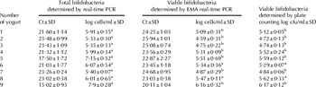
Comparison of the viable bifidobacterial cells obtained by culture and EMA real-time PCR showed that there was good correlation (Table 4). The number of viable bifidobacteria in probiotic yogurts determined by EMA real-time PCR was somewhat lower than that detected by plate counting, however there was no significant difference between them (P>0·05). The number of total bifidobacteria for some samples determined by real-time PCR was higher than that of the viable bifidobacteria determined by EMA real-time PCR and plate counting significantly (P<0·05). Table 4 illustrated EMA real-time PCR assay established in this investigation was a good method for direct determination of viable bifidobacteria in probiotic yogurts, and the detection can be carried out within 4 h.
The detection results of total and viable bifidobacteria in commercial probiotic yogurt determined by real-time PCR, EMA real-time PCR assay and plate counting (n=3)

a,b mean in row with a common superscript do not differ (P>0·05)
Discussion
Binding dyes and hybridization probes are all used to detect DNA amplification in real-time PCR experiments. For the former, the primers have to be highly specific to avoid synthesis of undesirable PCR products. The later uses a set of primers for amplification of a DNA fragment, and in addition, a highly specific probe labelled with a fluorogenic substance that is able to pair with the nascent DNA fragment emitting a signal upon interaction. The overall specificity of the PCR reaction is thus substantially enhanced. A real-time quantitative PCR method, based on the use of probes labelled with a stable fluorescent lanthanide chelate, is developed for the quantification of different human faecal bifidobacterial populations (Gueimonde et al. Reference Gueimonde, Debor, Tölkkö, Jokisalo and Salminen2007). Furthermore, molecular beacons, due to their stable stem-and-loop structure, have been demonstrated to be significantly more specific than dyes such as SYBR green I and other types of probes (Gubala & Proll, Reference Gubala and Proll2006).
In the present study, we designed the primer set of Bif-F/Bif-R which is genus-specific for Bifid. The real-time PCR method presented here is highly specific. The specificity of the primers and probes was tested both by identity searches in a nucleotide database (GenBank) and by the screening of 14 representative bifidobacterial and 14 non-bifidobacterial strains. The specificity of the probes ensures that no signal is generated by non-target amplicons. An important aspect of a microbiological detection assay is the ability to detect only viable bacteria in the samples and cultivation techniques have been relied on as a measure of cell viability. EMA treatment was selected because it has been shown that EMA can prevent amplification of DNA from dead bacteria in the PCR assay (Nogva et al. Reference Nogva, Drømtorp, Nissen and Rudi2003; Wang et al. Reference Wang, Li and Mustapha2009).
Varying EMA concentrations contribute a significant difference in Ct values for either viable or killed cells samples. EMA with final concentration of 100 μg/ml are used to treat Esch. coli O157:H7, Sal. enterica serovar Typhimurium cultures (Nocker & Camper, Reference Nocker and Camper2006) and Camp. jejuni (Rudi et al. Reference Rudi, Moen, Drømtorp and Holck2005a). But the optimum final concentration of EMA was 1·5 μg/ml when probiotic yogurt containing bifidobacteria was treated by EMA in this research.
Comparison of this direct quantitative method with standard culture technique for bifidobacterial counts in probiotic yogurt revealed that the assay described herein has the potential to be applied on a routine basis to probiotic yogurt for the detection of bifidobacteria. In agreement with previous publications introducing the application of EMA in combination with quantitative PCR to differentiate between viable and dead bacterial cells (Nogva et al. Reference Nogva, Drømtorp, Nissen and Rudi2003; Rudi et al. Reference Rudi, Naterstad, Drømtorp and Holo2005b), the efficacy of this DNA-intercalating dye in selecting against DNA from dead cells was confirmed (Nocker & Camper, Reference Nocker and Camper2006).
Of course, recent studies found that propidium monoazide (PMA) may exert less influence on DNA amplification from certain viable cells than EMA. PMA has been used to detect viable Ent. sakazakii, Esch. coli O157:H7, List. monocytogenes and Camp. jejuni with success (Nocker et al. Reference Nocker, Cheung and Camper2006; Flekna et al. Reference Flekna, Stefanic, Wagner, Smulders, Mozina and Hein2007; Pan & Breidt, Reference Pan and Breidt2007; Cawthorn & Witthuhn, Reference Cawthorn and Witthuhn2008). Continuation of this work using PMA instead of EMA would be an interesting next step.
In conclusion, the quantitative EMA molecular beacon-based real-time PCR assay described in this research are reliable and rapid methods for the quantification of viable bifidobacteria in probiotic yogurt. This culture-independent approach is promising for the direct and rapid detection of viable bifidobacteria in probiotic yogurt. Using this protocol, it was possible to detect bifidobacteria in probiotic yogurt within 4 h. The detection limit for quantitative real-time PCR analysis of bifidobacteria is about 104 cell/ml (g). The protocol has the potential for use in monitoring level of viable bifidobacteria in probiotic yogurt during manufacture or storage. This method is faster but requires expensive reagents and real-time PCR system.
This research was supported by grant ZJN03-3 from the Heilongjiang Province Natural Sciences Fund, China and National 863 Program (2008AA10Z335) of People's Republic of China.