Unlike pulmonary arteriovenous malformation, right pulmonary artery to left atrium fistula does not involve direct communication between artery and vein, as first described by Freidrich et al in 1950.Reference Friedlich, Bing and Blount 1 Clinical diagnosis is difficult due to normal cardiovascular examination, and varied age of presentation. The age of diagnosis ranges from infancy to adulthood, sometimes as late as 60 years. The diagnosis usually is established by suspicion of flow from right pulmonary artery to left atrium and subsequent bubble contrast echocardiography and/or CT scan or cardiac catheterization confirms the diagnosis. Treatment is often surgical patch closure. Recently, transcatheter closure by various devices like atrial septal defect devices, duct occluder, vascular plugs, or coils are being offered to close the fistula. We report a successful closure of a right pulmonary artery to left atrium fistula in an infant by a transcatheter transseptal approach using a duct occluder in an infant along with description of possible closure techniques.
Case report
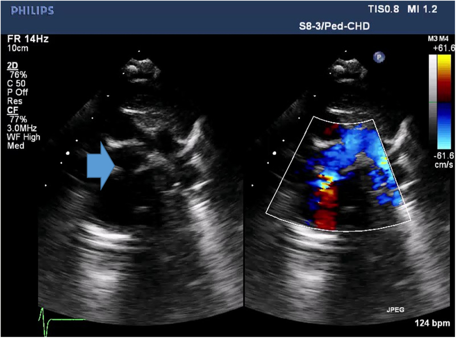
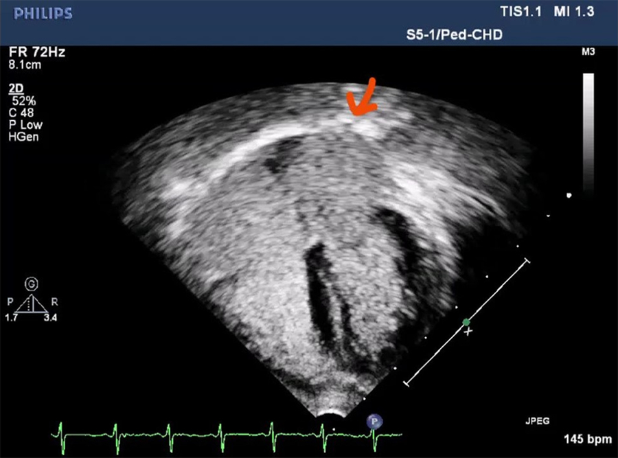
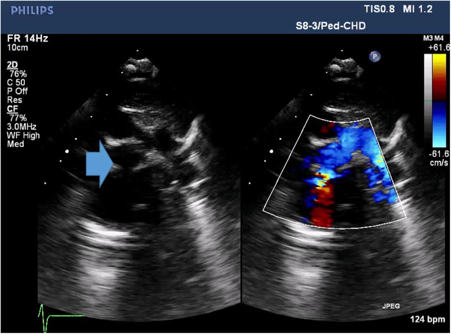
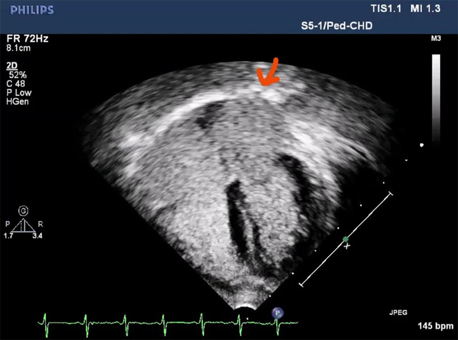
Eight-month-old female child presented to us with persistent desaturation right from her neonatal period. She was a pre-term neonate (1.5 kg birth weight), with no significant postnatal complications. She had persistent desaturation in range of 80–85%. Cardiovascular examination was within normal limit except her saturation was in between 80 and 85% even at rest. X-ray chest was also within normal limit as there was no cardiomegaly, lung vascularity appeared normal. Transthoracic echocardiography in high parasternal view showed abnormal communication from right pulmonary artery to left atrium (Fig 1), subsequently bubble contrast echo was done which showed significant filling of left atrium after 2–3 beats of injection (Fig 2). Child was then planned for cardiac catheterization and subsequent closure of right pulmonary artery to left atrium fistula by transcatheter closure by device.
Arrow showing right pulmonary artery to left atrium fistula.
Contrast echo image showing appearance of dense bubble contrast from roof of LA.
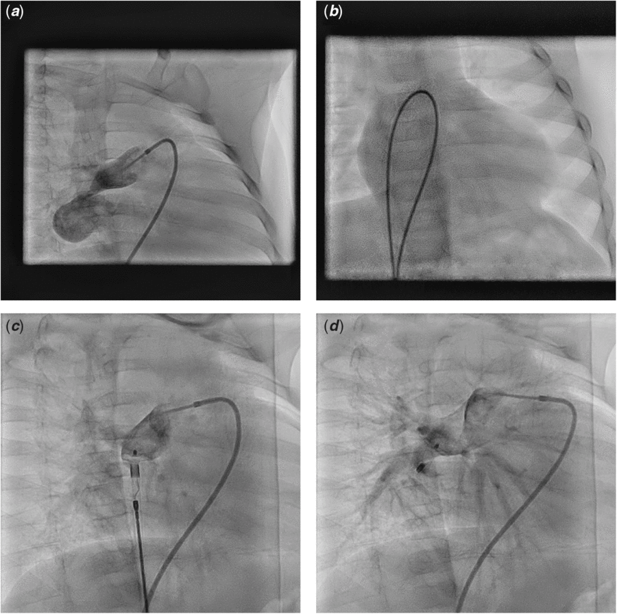
After informed consent, under general anesthesia, access from both femoral veins and right femoral artery were obtained. An angiogram was performed to profile the right pulmonary artery to left atrium communication (Fig 3a) in anteroposterior as well as right anterior oblique view. Right pulmonary artery to left atrium fistula sizing was done angiographically and the narrowest diameter measured was 2.5 mm. After careful planning, we opted to close this fistula using single disc device (Liftech duct occluder). From one venous access, angiogram was done and then wire was negotiated into left atrium through right pulmonary artery to left atrium fistula. Other venous access was used to snare wire from left atrium. In our case, the baby had an interatrial communication in the form of small atrial septal defect which facilitated the veno-venous loop formation without need of transseptal puncture, which could be hazardous in a small infant. Through fistula, a 0.035″ J tip angulated exchange length glide wire (Terumo Medical Corp., Somerset, United States of America) was advanced into the left atrium from the right pulmonary artery and snared in left atrium using a gooseneck snare (ev3 Endovascular Inc., Plymouth, MN, United States of America) leading to the formation of veno-venous loop from the left femoral vein–right pulmonary artery–left atrium–right atrium to the right femoral vein (Fig 3b). A 6F delivery sheath was advanced over the glide wire from the right femoral vein across the interatrial septum and then through the fistulous tract from left atrium to right pulmonary artery. A 6 mm/4 mm duct occluder (Heart R, Lifetech Scientific, Shenzhen, China) was selected to close the right pulmonary artery to left atrium fistula (Fig 3c). The retention disc of the device was positioned on the floor of the right pulmonary artery to left atrium and the remaining disc released in the fistulous tract. A selective right pulmonary artery angiogram showed a well-positioned device and no residual leak (Fig 3d). The systemic saturation improved from 85 to 96% immediately after closure of the fistula. There were no procedure-related complications. The child was observed for 48 hours prior to discharge and was asymptomatic at a follow-up after 1 month.
( a ) Right pulmonary artery to left atrium fistula – in right anterior oblique view. ( b ) Veno-venous loop formation through interatrial communication. ( c ) 6 mm/4 mm duct occlude was selected to close the right pulmonary artery to left atrium fistula, passed antegradely and retention disc positioned at floor of RPA. ( d ) final device deployment and subsequent angiogram.

Discussion
Clinical presentation of right pulmonary artery to left atrium fistula is varied. It may present in neonatal period as intense cyanosis requiring urgent intervention. If desaturation is less severe, presentation may be delayed to adulthood. The usual clinical presentation is cyanosis, clubbing, and exertional dyspnea.Reference Krishnamoorthy and Rao 2 Since the Pulmonary artery communicates with left atrium, bypassing the pulmonary filter function, the patient may present with cerebral or systemic embolization or abscess. Other common symptoms and signs are dyspnea, neurological symptoms, telangiectasia, and cyanosis.
The diagnosis of this rare anomaly should be suspected in patient with cyanosis and structurally normal heart. Clinical examination and routine investigations may not be contributory. Electrocardiogram may reveal left atrial or left ventricular enlargement, as well as cardiomegaly on chest radiograph, depending on the haemodynamic burden. Contrast echo will definitely help to confirm a pulmonary artery to pulmonary venous or left atrial fistula, the pathognomonic being early appearance of dense contrast in the left atrium (within 2–3 cardiac cycles) when contrast is injected in arm veins.Reference Narula, Choudhury, Chowdhary and Kiran 3 The large diameter and short length of these fistula results in low resistance in the fistulous channel and facilitates an obligatory large right to left shunt.
In our case, baby had only mild desaturation in range of 80–85%. There was no evidence of any other complications. Baby had persistent desaturation which was detected 1 month back and thoroughly investigated and referred to us. All causes of persistent cyanosis were considered like pulmonary artery hypertension, persistent left superior venacava to unroofed coronary sinus, pulmonary arteriovenous malformation, or pulmonary artery to left atrium fistula.
Atrial septal defect is the most common associated anomaly. Other associated cardiac malformation are rare, but anomalies of right lung, as the absence of lower or middle lobe, right lung sequestration, and diverticulum of the right main stem bronchus, have been reported. In our case also there was small fossa ovalis atrial septal defect.
Embryological basis of pulmonary arteriovenous fistula is incomplete degeneration of the septum between the arterial and venous plexus of the pulmonary vascular bed and/or due to formation of thin-walled saccular lesions because of a defect in the capillary loops. During developmental process, pulmonary vein if connected to such a fistula, is then absorbed into LA, forms a right pulmonary artery to left atrium fistula.Reference Ohara, Ito and Kohguchi 4 , Reference Lucas, Lind and Edwards 5
Only about 100 cases of pulmonary artery to left atrium fistula have been reported, all of them from right pulmonary artery to left atrium.
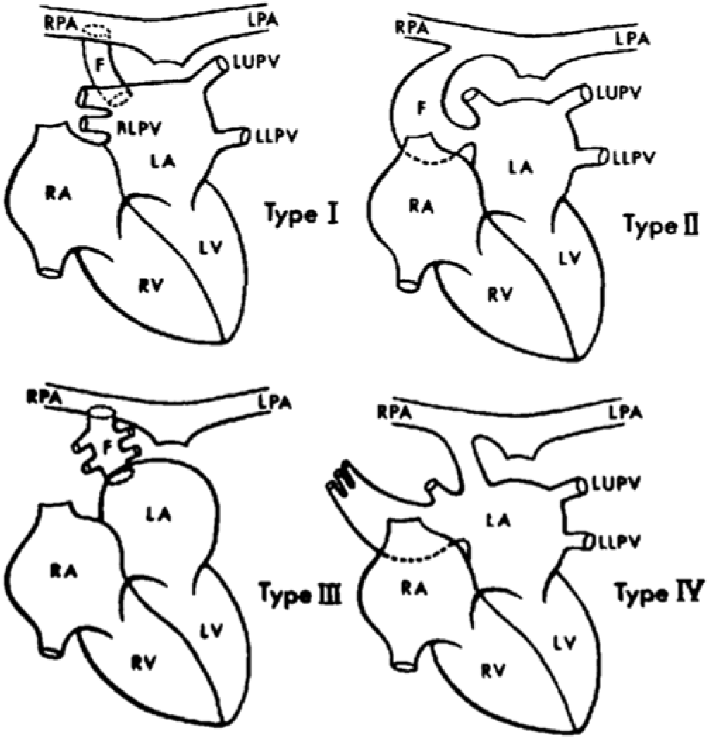
Based on the presence or absence of an aneurysm in the fistula and on the anatomy of pulmonary venous drainage pattern, de Souza e Silva et alReference DeSouza e Silva, Giuciani, Ritter, Davis and Pluth 6 categorized three types of connections. Type I is a normal pulmonary venous pattern; type II is an absent right inferior pulmonary vein with fistulous connection at the normal site of its ostium; and type III is when all the pulmonary veins are connected to the aneurysmal pouch. Subsequently, Ohara and coauthorsReference Ohara, Ito and Kohguchi 4 added a fourth type. Type IV is a right inferior pulmonary vein replaced by three small veins connected to the aneurysmal pouch. The left-sided pulmonary veins join the left atrium directly (Fig 4).
Classification of right pulmonary artery to left atrium fistula – de Souza e Silva et al, type IV by Ohara et al.
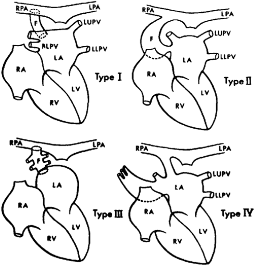
Our case was type II right pulmonary artery to left atrium fistula as clearly delineated in the cardiac catheterization images.
Early repair should always be performed to avoid complications, particularly systemic embolism, infective endarteritis, cerebral abscess, and rupture of aneurysmal communications. Surgical ligation of the fistula (with or without cardiopulmonary bypass) had been the preferred treatment of this condition until recently. Transcatheter coil occlusion of a right pulmonary artery to left atrium fistula was successfully performed in a neonate.Reference Slack, Jedeikin and Jones 7 The Amplatzer device offers an alternative occlusion for larger communications. Francis et alReference Francis, Sivakumar and Kumar 8 occluded a right pulmonary artery to left atrium fistula, the narrowest part of which was 8 mm by balloon sizing, using a 14–12 mm Amplatzer duct occluder in a 12-year-old girl. Uthaman et alReference Uthaman, Al-Qbandi, Abushaban and Rathinasamy 9 occluded a right pulmonary artery to left atrium fistula using the Amplatzer septal occluder in a 25-year-old male with cerebral abscess. Vadlamudi et alReference Vadlamudi, Verma, Christopher, Penumatsa and Koneti 10 reported successful closure of right pulmonary artery to left atrium fistula using two venous sheath, forming veno-venous loop through transseptal puncture in a 6-year-old child.
In our case, the right pulmonary artery to left atrium fistula was small 2.5 mm and type II. The aneurysmal sac is directly communicating with the superior aspect of left atrium. Initial plan was depending upon approach, if single venous access is to be taken then plan was to use either double disc device either atrial septal defect device/vascular plug or muscular ventricular septal defect device. But using these double devices, there was a definite risk of blocking right lower lobe pulmonary artery or pulmonary vein. Alternatively, if we can form veno-venous loop then we can use duct occluder but for that we need to have either interatrial communication or transseptal puncture to enter in the LA. Haemodynamically as right pulmonary artery pressure is always greater than left atrium so for this obligatory shunt, retention disc of device has to be kept on right pulmonary artery side. In our case, the baby had an interatrial communication in the form of small atrial septal defect which facilitated the venovenous loop formation without resort to trans-septal puncture, which could have hazardous in a small infant. We have successfully percutaneously closed this rare entity in such a young age group. Immediately after closure, saturation improved to 95–98%.
Conclusion
Although this entity is encountered as a rare anomaly, we should keep it in differential diagnosis while evaluating the patient with cyanosis. Early closure of fistula in such a young age will prevent complications of cyanosis. Management should be individualized and depends on type of fistula. Percutaneous transcatheter embolization of fistula is attractive alternative mode of therapy with lesser morbidity. Many types of devices could be used to occlude the fistula, choice of device depends upon access and size of baby, duct occluder I is good option for transcatheter closure of these fistula.
Acknowledgements
None.
Financial Support
This case report received no specific grant from any funding agency, commercial, or not for profit sectors.
Conflict of Interest
None.