I. INTRODUCTION
Titanium dioxide (TiO2), a wide band gap semiconductor, is a highly attractive material for a wide variety of industrial applications, because of its cheapness, easy availability, environmental friendliness, and high resistance to corrosion and photocorrosion in aqueous solution.Reference Bak, Nowotny, Rekas and Scorrell1 It has been reported in processes for photocatalytic degradation of various organic pollutantsReference Guillard, Horikoshi, Watanabe, Hidaka and Pichat2–Reference Doucet, Zahraa and Bouchy4 and as a part of gas sensors, solar cells,Reference Zakrzewska5 diluted magnetic semiconductors (DMSs),Reference Matsumoto, Murakami, Shono, Hasegawa, Fukumura, Kawasaki, Ahmet, Chikyow, Koshihara and Koinuma6, Reference Park, Ortega-Hertogs, Moodera, Punnoose and Seehra7 and numerous optical devices.Reference Kluson, Kment, Bartkova, Hubicka and Benjamin8
However, the large band gap (3.2 eV) of TiO2 lying in the ultraviolet (UV) region of solar radiation and the high recombination rate of the photoexcited electron-hole pairs hamper its utilization in photocatalysis.Reference Tang, Prasad, Sanjines, Schmid and Levy9, Reference Tong, Zhang, Tian, Chen and He10 Doping with foreign ions is one of the most efficient strategies for sensitizing TiO2 to visible-light region and also for forming charge traps to keep electron-hole pairs separate.Reference Fujishima, Zhang and Tryk11 Compared with the larger O2− anions, the smaller Ti4+ cations are much more easily substituted by other transition metal ions with similar ionic radius. Choi et al.Reference Choi, Termin and Hoffmann12 reported a systematic investigation of the photocatalytic activity of TiO2 doped with different transition metal ions. Among them, Fe and Mn ions doped into TiO2 were demonstrated to enhance the photocatalytic activities because they can not only extend the region of light response but also trap both photoexcited electrons and holes.
TiO2 is a nonmagnetic semiconductor. However, after being doped with some transition metal ions, it was discovered to exhibit ferromagnetism at and above room temperature, which makes them potential DMSs applied in spintronic devices.Reference Matsumoto, Murakami, Shono, Hasegawa, Fukumura, Kawasaki, Ahmet, Chikyow, Koshihara and Koinuma6, Reference Park, Ortega-Hertogs, Moodera, Punnoose and Seehra7 With the initial report of room temperature ferromagnetism in codoped TiO2, the TiO2-based DMSs have been extensively studied, such as Fe-TiO2,Reference Hong, Preltier, Sakai and Hassini13 Mn-TiO2,Reference Xu, Lin, Lu, Liu, Lu, Wamg, Zou, Lv, Zhang and Du14 Cr-TiO2,Reference Wang, Tang, Zhang, Golub, Spinu and Tung15 and so on.
Recently, considerable research interests were focused on low-dimensional nanoscalar materials because of their distinctive physical and chemical properties.Reference Henglein16 Metal oxide nanosheets, the new classes of nanostructured semiconductors, have been synthesized by the delamination of starting layered compounds into colloidal single sheets through soft-chemistry processes.Reference Treacy, Rice, Jacobson and Lewandowski17–Reference Fukuda, Akatsuka, Ebina, Ma, Takada, Nakai and Sasaki24 These two-dimensional (2D) crystallites are characterized by an extremely thin thickness (in the range of molecular dimensions), a large lateral size (from several hundred nanometers to several micrometers), and a well-defined chemical composition.
Among these oxide nanosheets, the titania nanosheet is one of the most important as well as extensively investigated materials because the titania nanosheet exhibits novel properties compared with other nanosized TiO2, such as sharp UV absorption band and enlarged band gap energy.Reference Sasaki and Watanabe19, Reference Sakai, Ebina, Takada and Sasaki25 The negatively charged nature retained from layered crystals makes titania nanosheets to be assembled into a well-organized multilayer organic/inorganic system by the layer-by-layer strategies with positively charged polymers, such as poly-ethylenimine and poly-dimethyldiallyl ammonium chloride (PDDA).Reference Sasaki, Ebina, Tanaka, Harada and Watanabe26
The intralayered titanium ions in precursor crystals can be substituted by other transition metal ions with similar ionic sizes, and exfoliation of these isotypic layered titanates into nanosheets is of great interest for achieving new nanostructured semiconductor for practical applications. Harada et al.Reference Harada, Sasaki, Ebina and Watanabe27 prepared Fe- or Ni-substituted titania nanosheets, which showed light response in visible region and exhibited efficient photocatalytic activities on photodegrading interlayer polymers. In addition, the ferromagnetism was also obtained in Co-, Fe-, or Mn-doped 2D titania nanosheets, and such (Fe/Co) codoped nanostructured ferromagnets strongly enhanced magneto-optical response, which stems from the d-d transition of Co2+-Fe3+.Reference Osada, Ebina, Takada and Sasaki28–Reference Dong, Osada, Ueda, Ebina, Kotani, Ono, Ueda, Kobayashi, Takada and Sasaki31 In this context, controlled doping or codoping of other transition metal ions is also of interest in photocatalysis and spintronics.
In this work, we reported the synthesis of new dual transition metal ion–doped titania nanosheets (Ti0.6Mnx /2Fe(0.8-x)/2O2) in which the (Mn/Fe) doping is controlled by adjusting the molar ratio of Mn and Fe in the initial mixture. In addition, the layer-by-layer self-assembly of the exfoliated (Mn/Fe) codoped titania nanosheets and cationic polymers into multilayer nanocomposite systems has been demonstrated.
II. EXPERIMENTAL
A. Synthesis of starting layered materials
The starting layered titanate, K0.8Ti1.2MnxFe0.8-xO4 (x = 0.1, 0.2, 0.3, 0.4), was synthesized by a traditional solid state reaction. A stoichiometric mixture of K2CO3, TiO2, Mn2O3, and Fe2O3 was heated at 1073 K for 1 h to be decarbonated and then calcined at 1273 K for another 20 h after grinding the mixture again.
B. Protonation of titanate
The obtained powder was converted to a protonated form by being treated with a 1 mol dm−3 HCl aqueous solution for 3 days. The acid solution was replaced with a fresh one every day. The resulting product, H0.8Ti1.2MnxFe0.8-xO4·H2O, was recovered by filtration, washed with copious amounts of water, and air-dried.
C. Exfoliation
To get a colloidal suspension of unilamellar nanosheets Ti0.6Mnx /2Fe(0.8-x)/2O2, 0.4 g of this protonated form was mixed with 100 ml of tetrabutylammonium hydroxide (TBAOH) solution in a flask, in which the molar ratio of TBA+/H+ was 10. After vigorously shaking the mixture in a mechanical shaker at 170 rpm for 10 days, a suspension containing exfoliated nanosheets was obtained.
D. Fabrication of multilayer thin films
Multilayer thin films of the prepared nanosheets were fabricated by a layer-by-layer assembly procedure similar to that described in published literatures.Reference Sasaki, Ebina, Tanaka, Harada and Watanabe26 Substrates, such as silicon wafers or quartz glass slides (1 cm × 5 cm), were treated in a bath of HCl/methanol (1/1) solution and concentrated H2SO4 for 30 min each. First, the substrates were immersed into a PDDA solution (pH = 9.0) for 20 min to introduce a positively charged surface, followed by washing with copious quantities of pure water, and then were dipped in a colloidal suspension of (Mn/Fe) codoped nanosheets (0.08 g dm−3; pH = 9.0) for 20 min and thoroughly washed with pure water again. These PDDA/nanosheet deposition steps were repeated for n times to get a multilayer film of [PDDA/Ti0.6Mnx /2Fe(0.8-x)/2O2]n. The obtained films were dried with N2 gas flow.
E. Characterizations
The morphology of the titanates was examined with a JEOL JSM-6700F scanning electron microscope (SEM, JEOL, Tokyo, Japan). X-ray diffraction patterns were collected on a Rigaku Rint-2000 diffractometer, (Rigaku, Tokyo, Japan) equipped with graphite monochromatized Cu Kα radiation (λ = 0.15405 nm). Ultraviolet–visible (UV–Vis) spectra were used to monitor the fabrication process of multilayer thin film on a Hitachi U-4000 spectrophotometer (Hitachi, Tokyo, Japan). A Seiko SPA-400 atomic force microscope (AFM, Seiko, Tokyo, Japan) was used to observe the topographical features of nanosheets on Si wafers. AFM images were acquired in tapping mode using a Si tip cantilever (force constant: 11 N m−1).
III. RESULTS AND DISCUSSION
A. Synthesis and characterization of the layered titanate materials
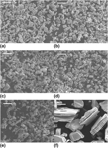
The different (Mn/Fe) doping contents can be controlled in the starting material K0.8Ti1.2MnxFe0.8-xO4 (x = 0.1, 0.2, 0.3, 0.4) via adjusting the molar ratio of Mn and Fe in the initial mixture. Figure 1 shows the SEM images of these (Mn/Fe) codoped titanates and the undoped titanate of K0.8Li0.27Ti1.73O4. From these images, we can clearly see a layered structure in the crystal particles. The sizes of K0.8Ti1.2MnxFe0.8-xO4 particles are of several micrometers; however, the size of undoped titanate is much larger, dozens of times than those of codoped samples, which indicates that the (Mn/Fe) doping hampers the growth of titanate crystals.
Scanning electron microscope images of (Mn/Fe) codoped titanates K0.8Ti1.2MnxFe0.8-xO4: (a) x = 0.0; (b) x = 0.1; (c) x = 0.2; (d) x = 0.3; (e) x = 0.4; and (f) the undoped titanate K0.8Li0.27Ti1.73O4. The scale bars in (a)–(e) are 10 μm, and the scale bar in (f) is 20 μm.

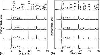
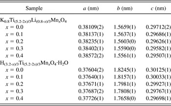
The powder x-ray diffraction (XRD) results [Fig. 2(a)] show that except a weak impurity peak of trace K2Ti6O13 phase in the pattern of x = 0.4, all diffraction peaks can be indexed as a C-base-centered orthorhombic single phase with similar refined lattice parameters (Table I) to those of previously published lepidocrocite-type titanate in which edge-sharing TiO6 octahedrons construct the layer of negatively charged titania and alkali metal ions locate in the interlayer space for the charge-neutrality requirement.Reference Sasaki, Kooli, Iida, Michiue, Takenouchi, Yajima, Izumi, Chakoumakos and Watanabe32 No impure peaks of Fe oxides and Mn oxides were discerned, demonstrating the high purity of these products in which Fe and Mn ions are fully incorporated into Ti sites in the host layers.
Powder x-ray diffraction (XRD) patterns of (a) layered titanates K0.8Ti1.2MnxFe0.8-xO4 and (b) their protonated forms H0.8Ti1.2MnxFe0.8-xO4·H2O. The weak peak denoted by an asterisk in (a) is attributed to the trace K2Ti6O13 phase.

Interlayer K ions were removed by an acid-exchange reaction in the 1 mol/dm3 HCl solution. Complete removal of alkali metal ions was achieved within 3 days of treatment; meanwhile, no Fe and Mn ions were exchanged by protons in such acidic solution.Reference Harada, Sasaki, Ebina and Watanabe27–Reference Dong, Osada, Ueda, Ebina, Kotani, Ono, Ueda, Kobayashi, Takada and Sasaki31 The XRD patterns [Fig. 2(b)] reveal that the acid-exchanged samples, H0.8Ti1.2MnxFe0.8-xO4·H2O, are highly crystalline and preserve a layered lepidocrocite-type structure from the starting materials. Because of the insertion of water molecule into the interlayer space, the basal spacing of the protonated titanates is expanded about 0.2–0.3 nm, compared with the corresponding potassium titanates (Table I). In addition, it should be noted that the weak peak of impurity in the x = 0.4 sample disappears after the acid-exchange treatment, demonstrating its trace amount in the sample.
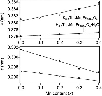
The refined in-planar lattice parameters (a and c) of protonated and unprotonated titanates as a function of Mn composition (x) are plotted in Fig. 3. It can be seen that these parameters nearly linear change upon the adjustment of Mn substitution, which further testifies the homogeneous distribution of Fe and Mn ions in the lattice of titanate layer. We note that the parameters a and c changed in opposite direction with increasing the Mn content, which may be ascribed to the John–Teller distortion of Mn3+ ions.
The linear change of the refined in-planar lattice parameters upon the Mn content (x).

B. Characterization of nanosheets
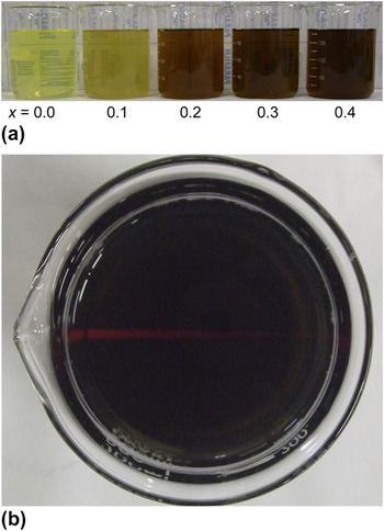
A colloidal suspension of Ti0.6Mnx /2Fe(0.8-x)/2O2 nanosheets was synthesized by exfoliating the layered titanate of H0.8Ti1.2MnxFe0.8-xO4·H2O with TBAOH. After shaking the protonated forms in TBAOH solution (TBA+/H+ = 10) for 10 days, transparent colloidal suspensions were yielded, and the color from yellow to dark-brown depended on the Mn content [Fig. 4(a)]. Clear Tyndall light scattering [Fig. 4(b)] is observed for the colloidal suspension, demonstrating the presence of exfoliated nanosheets dispersed in the aqueous suspension.
The photographs of (a) the colloidal suspension of Ti(5.2-2x)/6Mnx /2Fe(0.8-x)/2O2 and (b) the Tyndall effect on the colloidal suspension.

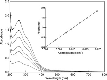
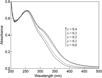
Figure 5 depicts the UV–Vis absorption spectra of the colloidal suspensions of Ti0.6Mnx /2Fe(0.8-x)/2O2 in various concentrations. A Lambert–Beer behavior, the linear relationship of absorbance upon the concentration, demonstrates that the Ti0.6Mnx /2Fe(0.8-x)/2O2 nanosheets are present in the suspension as individual colloidal sheets. A pronounced absorption band at around 260 nm in the spectra is similar to that of nonsubstituted titania nanosheets dispersed in a colloidal suspension, which is attributed to the transition from valence band (O 2p) to conduction band (Ti 3d).Reference Sasaki and Watanabe33 In contrast with non-substituted nanosheets, additional in-gap absorption located about 300–450 nm is checked in substituted nanosheets. Furthermore, some weak absorption bands contributed by the Fe and Mn d-d transitions appear in the visible region. A comparison of UV–Vis absorption spectra of Ti0.6Mnx /2Fe(0.8-x)/2O2 (x = 0.0, 0.1,0.2, 0.3, 0.4) nanosheets is shown in Fig. 6. With the increase in Mn content, the absorption edge gradually shifts to the visible-light region, which is in correspondence with the color change of the nanosheet suspension.
UV–Vis absorption spectra of the colloidal suspensions of Ti(5.2-2x)/6Mnx /2Fe(0.8-x)/2O2 in different concentrations. The inset shows the absorbance at peak top as a function of the concentration of suspension.

A comparison of UV–Vis absorption spectra of the colloidal suspensions of Ti(5.2-2x)/6Mnx /2Fe(0.8-x)/2O2.

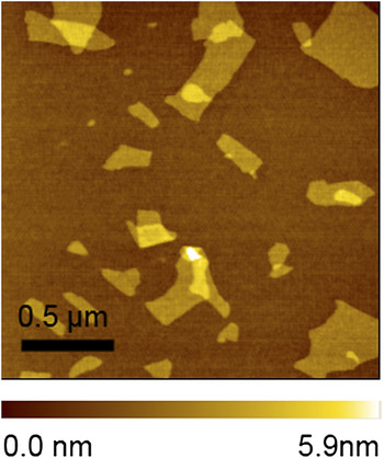
Figure 7 exhibits the tapping-mode AFM images of exfoliated nanosheets on silicon wafer substrates, which were immersed into the PDDA solution and nanosheets suspensions for 20 and 1 min, respectively. Exfoliated nanosheets of several hundred nanometers in lateral size have been deposited on the substrates by an electrostatic interaction although some small fragments below 100 nm and overlapped parts were also observed. The height profile indicated that the nanosheets have a quite flat terrace, exhibiting an average thickness of ∼1.5 nm. The thickness of nanosheets measured by AFM is much larger than that of titania nanosheet, 0.73 nm, determined by x-ray absorption fine structure (XAFS) analysis.Reference Fukuda, Nakai, Oishi, Nomura, Harada, Ebina and Sasaki34 This phenomenon, the thickness estimated from AFM deviating the real thickness, is present in negative oxide nanosheets,Reference Harada, Sasaki, Ebina and Watanabe27–Reference Dong, Osada, Ueda, Ebina, Kotani, Ono, Ueda, Kobayashi, Takada and Sasaki31 as well as in positive layered double hydroxides (LDH) nanosheetsReference Li, Ma, Ebina, Iyi and Sasaki35–Reference Ma, Takada, Fukuda, Iyi, Bando and Sasaki38 because of the adsorption of TBA, water, and other factors.
Tapping-mode atomic force microscope (AFM) image of exfoliated Ti(5.2-2x)/6Mnx /2Fe(0.8-x)/2O2 (x = 0.3) nanosheets deposited on a Si substrate.

C. Self-assembly of multilayer thin films
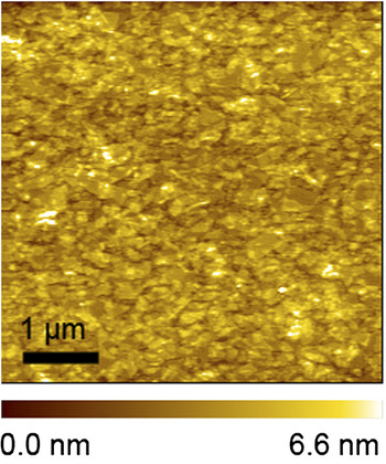
For possessing a negative surface after being treated in H2SO4, the substrates (Si wafer or quartz glass) were primed with a positively charged PDDA layer before the adsorption of negatively charged nanosheets. A prominent enhancement for surface coverage was achieved by prolonging the dipping time of nanosheets to 20 min, as shown in Fig. 8, the tapping-mode AFM image of the first deposited layer of Ti0.6Mnx /2Fe(0.8-x)/2O2 nanosheets on a Si substrate covered by PDDA. In this case, a fairly high coverage, above 90%, was observed, while overlapping area also increased to about 40%, which seems inevitable for these films fabricated by layer-by-layer strategies because of the large lateral dimensions of nanosheet crystallites.
AFM image of the first (Mn/Fe) codoped nanosheet layer on a Si wafer substrate precoated with poly-dimethyldiallyl ammonium chloride (PDDA).

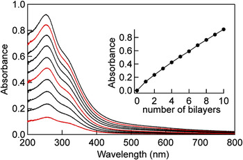
Multilayer films were fabricated by alternately immersing the substrates in a colloidal suspension of nanosheets and a PDDA solution. The growth of [PDDA/Ti0.6Mnx /2Fe(0.8-x)/2O2]n multilayer film was monitored by UV–Vis absorption spectra measured after each bilayer (Fig. 9). The spectral profile is similar to that of the colloidal suspension of nanosheets because the polycation of PDDA does not show any absorption in this spectral region. Thus, the nearly linear increase in absorbance at 260 nm as a function of the layer number reveals a stepwise and regular film.
UV–Vis absorption spectra for multilayer film of [PDDA/Ti(5.2-2x)/6Mnx /2Fe(0.8-x)/2O2]10 assembled on a quartz glass substrate. Nanosheet concentration was 0.08 g dm−3; PDDA concentration was 20 g dm−3; pH value of PDDA solution and nanosheet suspension was 9.0; and deposition time was 20 min. Inset: the observed absorbance at 260 nm as a function of the deposition cycle.

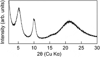
Figure 10 exhibits the XRD pattern of [PDDA/Ti0.6Mnx /2Fe(0.8-x)/2O2]10 thin film. The diffraction peak of 5.2° in 2θ indicates a 1.7 nm interplanar spacing, which is attributed to the reflection of periodically organic/inorganic nanostructure. The sharp (001) and (002) peaks and the barely resolved (003) peak demonstrate the high quality of these films. If we regard the thickness of Ti1-δO2 nanosheet, 0.73 nm, as that of (Mn/Fe)-substituted nanosheet, the layer height of PDDA in the multilayer thin film can be estimated to about 1.0 nm. The unilamellar nanosheets are characterized of a thickness in molecular scale and a lateral size in sub-micrometer scale, and layer-by-layer assembled into multi-layered thin films with the positively charged polycations.Reference Dong, Osada, Ueda, Ebina, Kotani, Ono, Ueda, Kobayashi, Takada and Sasaki31 The different water content in interlayer space maybe cause the interlayer expansion of 0.3 nm, which is the thickness of monolayer water molecule. Another possible reason is the different negative charge in the nanosheets. The charge in one molecule of Ti(5.2-2x)/6Mnx /2O2 (x = 0.0–0.4) is −(3.2−x)/6, larger than that in Ti0.6Mnx /2Fe(0.8-x)/2O2 (−0.4), which possibly induces the structure of repeating (PDDA/nanosheets) in [PDDA/Ti0.6Mnx /2Fe(0.8-x)/2O2]10 to become much looser.
The XRD pattern of [PDDA/Ti(5.2-2x)/6Mnx /2O2]10 (x = 0.3) film assembled on quartz glass substrates.

Refined lattice parameters of (Mn/Fe) codoped layered titanates and their protonated forms.

IV. CONCLUSIONS
In conclusion, a series of the controlled (Mn/Fe) codoped layered titanate crystals, K0.8Ti1.2MnxFe0.8-xO4 (x = 0.0, 0.1, 0.2, 0.3, 0.4), have been synthesized by a solid state reaction. Mn3+ and Fe3+ ions substitute partial Ti sites in the TiO6 octahedrons, which constitute the titania layers. The interlayer K+ ions can be acid-exchanged, yielding a protonated form, H0.8Ti1.2MnxFe0.8-xO4·H2O, which is delaminated into colloidal single sheets in the TBAOH solution. The tailored optical properties of titania nanosheet are achieved by the controlled (Mn/Fe) doping, which is demonstrated by the color of colloid, as well as the shift of absorption edge in absorption spectra. The unilamellar nanosheets are characterization of a thickness in molecular scale and a lateral size in submicrometer scale, and acting as building blocks, are layer-by-layer assembled. Such novel 2D nanosheets controllably doped with binary transition metal ions and their self-assembled systems are expected to be used in various applications, such as photocatalysis under visible light, titania-based DMS, and so on.
ACKNOWLEDGMENTS
The authors gratefully acknowledge the financial support from the National Natural Science Foundation of China (21001093, 20805043), the Zhejiang Provincial Natural Science Foundation of China (Y4090285), and the Science Foundation of Zhejiang Sci-Tech University (0913840-Y).