I. INTRODUCTION
The fabrication of regular arrays of single DNA molecules on a surface is important for the development of biosensors for diagnostics,Reference Kricka1 and for the further development of devices devoted to molecular electronics as well.Reference Seeman2 In fact, DNA is a very promising material to build chips with nanometric precision due to its unique recognition capacity, its physicochemical stability, and its mechanical rigidity.Reference Niemeyer3 More recently, integration of DNA-based origamis as programmable tracks for molecular machines appears as a possible route for guiding molecular robots.Reference Lund, Manzo, Dabby, Michelotti, Johnson-Buck, Nangreave, Taylor, Pei, Stojanovic, Walter, Winfree and Yan4
DNA chips are a very popular and powerful new tool that promises to be widely used in molecular biology and medicine in the years to come. At the single-molecule level, DNA has already been stretched for genome mappingReference Gerhold, Rushmore and Caskey5 and medical diagnostics.Reference Cuzin6 However, the current tendency is to be able to pattern individual stretched DNA molecules into a well-defined array so that a fast optical readout can be performed. The generation of such a controlled assembly could be a major building block for the construction of future devices. In fact, DNA chains can be metalizedReference Braun, Eichen, Sivan and Ben-Yoseph7–Reference Nguyen, Monteverde, Filoramo, Goux-Capes, Lyonnais, Jegou, Viel, Goffman and Bourgoin11 or functionalized with nanoparticlesReference Köhler, Csáki, Reichert, Möller, Straube and Fritzsche12 and this ability to generate well-aligned one-dimensional (1D) nanoscale templates onto desired locations on a solid support could provide further opportunities for the fabrication of assemblies with increasing complexity.
In the literature, a number of studies in molecular combingReference Bensimon, Bensimon and Heslot13–Reference Bensimon, Simon, Chiffaudel, Croquette, Heslot and Bensimon15 have been reported. The so-called “molecular combing” technique was introduced in 1993, and performed on short DNA molecules. This technique consists in obtaining from a coiled DNA molecule in its natural state, aligned and stretched DNA filaments that are easier to study than under their compacted state. For this purpose, a glass support chemically treated is immersed into a solution containing the DNA molecules. Each molecule gets fixed by one of its extremities. The glass surface is then gently removed and this movement provokes, by capillarity, the stretching of the molecules that stick to the glass support into stretched rods with same orientation. Other research groups have implemented the stretching of DNA molecules on surfaces of a different natureReference Lyubchenko, Gall, Shlyakhtenko, Harrington, Jacobs, Oden and Lindsay16–Reference Thundat, Allison, Warmack, Brown, Jacobson, Schrick and Ferrell19 with the aim of analyzing them and generating DNA chips. DNA molecules have also been separated thanks to nanopillars and under an electric field.Reference Kaji, Tezuka, Takamura, Ueda, Nishimoto, Nakanishi, Horiike and Baba20 Furthermore, DNA molecules have been immobilized in between metal electrodes. In Refs. Reference Maubach, Csaki, Seidel, Mertig, Pompe, Born and Fritzsche21–Reference Wolff, Leiterer, Csaki and Fritzsche23 the authors pin DNA in electrode gaps either by capillary-like approachesReference Maubach, Csaki, Seidel, Mertig, Pompe, Born and Fritzsche21 or dielectrophoresis,Reference Washizu and Kurosawa22, Reference Wolff, Leiterer, Csaki and Fritzsche23 for the integration of DNA molecules toward electronic applications.
DNA has also been combed by flowing the DNA solution through microfluidic channels.Reference Petit and Carbeck24 Opitz et al.Reference Opitz, Braun, Seidel, Pompe, Voit and Mertig25 have combed DNA molecules tethered to positively charged patterns. Other research groups have combed DNA molecules on surface-modified or unmodified polydimethylsiloxane (PDMS) stamps.Reference Gad, Sugiyama and Ohtani26–Reference Guan and Lee30
However, most of the time the combed molecules are randomly stretched onto the surface without any predetermined position, therefore they are not regularly distributed on the surface and this represents a real inconvenience for competitive readout.
Guan et al.Reference Guan and Lee30 are the first to have generated regular arrays of stretched DNA strands onto a surface. In this study in particular, they manually place in contact a structured PDMS stamp with a droplet of the DNA solution. They exert a pressure at the back of the stamp for a few seconds and then peel the stamp off. In this reference, the authors observed that the stretched DNA molecules are not single molecules and the technique presents the inconvenience of being manual and thus not perfectly reproducible. This is the reason why we have developed a new approach based on capillary assembly.Reference Malaquin, Kraus, Schmid, Delamarche and Wolf31 In fact, capillary assembly could represent a major alternative for these studies because it allows the experimenter’s control over the experimental parameters with high accuracy and precision. In the case of genomic applications for instance, where single molecules are required, the control of the experimental parameters allowing a selection of single molecular adsorption events is crucial. In this work, we present a methodology for accurate positioning and alignment of single DNA molecules onto a surface by a combination of capillary assembly with soft lithography.Reference Xia and Whitesides32 The control of the contact line velocity and the experimental conditions enable us to tightly direct the process taking place during the assembly of the DNA molecules. This technique is capable of micro- or nanopatterning stretched and perfectly aligned DNA molecules into highly ordered arrays. These arrays can then be transferred onto other surfaces such as cleaved mica or aminopropyltriethoxysilane (APTES)-coated glass slides or silicon wafers. As a further step in this development, we will present different extensions of the technique.
II. EXPERIMENTAL PROCEDURE
A. Materials
To prepare phage lambda DNA solution, 100-μL phage lambda DNA solution (Sigma, Sigma Aldrich, France, 48,502 bp, 329 μg/mL diluted to 50 μg/mL in 10 mM Tris-HCl/1 mM EDTA, pH 8) was heated at 65 °C for 5 min and dipped into ice water to avoid the molecules’ concatenation. The solution was then fluorescently labeled by adding 1.5 μL of YOYO-1 (100 μM); incubation was conducted in the dark at room temperature for a minimum of 2 h. Phage lambda DNA solution was further diluted to 10 μg/mL in a 1% v/v Triton X-100 solution in the same buffer. SmaI enzymes were provided by the LISBP laboratory (Toulouse, France). Phage lambda DNA has two specific sites for SmaI recognition. We ascertained the specificity of the phage lambda DNA cleavage by SmaI enzymes with an electrophoresis gel. The phage lambda DNA-SmaI enzymes reaction was conducted in solution for 3 h under favorable conditions and the solution was further diluted to a final concentration of 10 μg/mL (1% v/v Triton X-100). The 5100 bp linear plasmids (18 nM in purification kit buffer, 1% v/v Triton X-100) were provided by the University of Minnesota.
B. Fabrication of master and stamps
To direct the capillary assembly of DNA molecules, we used PDMS stamps with topographical cavities obtained from the replication of positive silicon masters (with protruding features). The silicon micropatterned master was achieved by ultraviolet photolithography and the pattern transfer by reactive ion etching. The PDMS prepolymer solution containing a mixture of 10:1 mass ratio of PDMS oligomers and a reticular agent from Sylgard 184 Kit (Dow Corning, Wilmington, DE) was then poured onto the silicon master and cured at a temperature of 80 °C during 12 h. The cured PDMS was peeled off and cut into 1 cm × 1 cm stamps.
In a general manner, the design of the topographic patterns requires a prior reflexion in terms of distribution, dimension, depth, and orientation. Indeed, the size of the patterns determines the number and the positioning of the objects to be assembled for assemblies of controlled geometry. Given the dispersion in size of the objects in solution, the patterns are usually intentionally enlarged to facilitate the assembly by compensating the fluctuations in size among the objects. The depth of the patterns is also a key geometrical parameter to take into consideration because it determines the number of layers to be deposited inside the patterns. To obtain a monolayer of objects and allow their subsequent transfer, the patterns’ depth has to be comparable to the diameter of the objects. When the stamp is used as support for capillary assembly, the design rule to keep in mind is that the deformation of the liquid contact line has to be minimized to avoid its premature disruption, and allow the forces involved to direct and gather the objects at the liquid front line to fill the cavities appropriately. Additionally, the periodicity of the patterns has to be large enough so the contact line can get pinned onto each row of patterns without missing one.
In the case of DNA molecules’ assembly, the silicon master was designed with protruding microfeatures 3 μm in diameter, 5 μm high, and with different periodicities (20, 25, 30, and 40 μm). Therefore, the corresponding PDMS stamps are the negatives of the master and consist of microcavities with the same sizes.
C. Experimental setup
The so-called directed assembly is carried out using a dedicated setup. The microstructured PDMS stamp where we want the DNA molecules to be assembled is placed on a motorized translation stage regulated in temperature, below a fixed glass slide at a distance of about 1 mm. A 30-μL droplet of DNA molecules in solution at a concentration of 10 μg/mL is injected between the glass and the substrate. The liquid contact line is therefore moved over the substrate at a constant velocity of 1 mm/s for the trapped DNA molecules to be stretched. The temperature was set to 25 ± 0.1 °C by a Peltier cell, which is monitored through an PID allowing high accuracy and fast temperature control. The experimental parameters (speed, temperature, concentration) were adjusted to enable the directed assembly and combing of single DNA molecules with high placement accuracy. It has to be noted that in the case of DNA, the temperature plays a minor role compared to the concentration of the solution or the displacement speed as this last parameter is set at a very high value. The assembly is performed throughout the entire surface of the PDMS stamp, approximately over an area of more than 1 cm2.
D. DNA molecules transfer
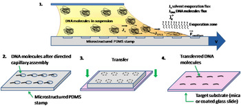
After the directed assembly process, to transfer the DNA arrays onto a solid surface, the stamp is brought into contact with the surface for 1 min without external pressure and then peeled away. The surface chosen for transfer is a glass slide coated with APTES molecules to enable electrostatic binding of DNA molecules (see Scheme 1). The adhesion force between DNA and the hydrophobic PDMS surface is low enough for transfer to occur to the coated glass slide. It has to be noted that another compatible surface for transfer could be mica. In fact, the only criteria to take into consideration are the use of a perfectly flat surface, hydrophilic (or even better positively charged), because the molecules have more affinity for the target surface than for the PDMS stamp’s surface.
Schematic representation of the procedure for generating and transferring the DNA arrays. (1) Capillary assembly of the DNA molecules in solution on a microstructured PDMS stamp. (2) The molecules assembled after the directed capillary assembly process are then (3) transferred onto the target surface by simple contact between the stamp and the surface. (4) We obtained well-ordered DNA molecule arrays on the surface after transfer.

E. Fluorescence imaging
The DNA arrays’ printing was controlled under an inverted epifluorescence microscope equipped with a ×100 oil immersion objective lens and imaged with an Andor Ixon (Belfast, N. Ireland) 512 × 512 camera.
F. AFM imaging
AFM images of DNA arrays on APTES-coated glass slides were obtained on a Nanowizard II from JPK Instruments (Berlin, Germany) in contact mode operated in air and liquid.
III. RESULTS AND DISCUSSION
A. Capillary assembly process
Figure 1 shows the results obtained after directed assembly of phage lambda DNA molecules on the microstructured PDMS stamp and transfer-printing on an APTES-coated glass slide. As it can be seen from the fluorescence and AFM images, we obtained arrays of DNA strands in which the strands of the same length and orientation are precisely positioned between adjacent patterns. Such arrays were obtained with high reproducibility over an area of several millimeters and with a strand coverage rate of 99%. Note that AFM imaging could also be performed in liquid conditions. DNA strands appear to be strongly grafted to the APTES surface, because they remain exactly in the same position even after rinsing several times. This suggests that there seems to be several anchoring points along the stretched DNA strands with the surface.
(a) Fluorescence image and (b) AFM image in air of highly ordered arrays of 22.9 μm (±1.9 μm) long and 1.71 nm (±0.35 nm) high phage lambda single DNA molecules printed on a (APTES)-coated glass slide after directed assembly onto a micropatterned PDMS stamp under optimized experimental conditions. (c) Zoom AFM image of a single DNA molecule with the corresponding cross section. Top right of the figure, histogram of the molecules’ length measured over 22 molecules on the same array.

In Figs. 1(b) and 1(c), the cross section measured over the stretched DNA strands on APTES reveals that the height of the strands is fairly uniform along the length of individual strands as well as among different strands. Measurements on several strands gave an average height of 1.71 nm (±0.35 nm) ranging from 1.42 to 2.08 nm. Thus, AFM images reveal that their height corresponds to the height of a single double-stranded DNA molecule (2 nm). We can thus affirm that the technique of directed capillary assembly allows us to control the experimental parameters and by extension the number of trapped molecules per cavity.
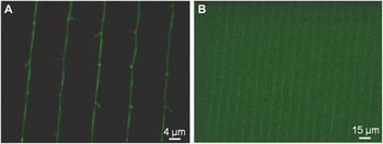
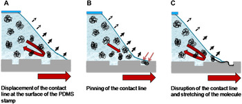
In our system, we use a glass slide mounted on a long flexible spreader to squeeze a thin film of liquid on the substrate where we want DNA molecules to be assembled. The spreader is fixed and the substrate can be moved using a translation stage.Reference Malaquin, Kraus, Schmid, Delamarche and Wolf31, Reference Kraus, Malaquin, Schmid, Riess, Spencer and Wolf33 The combination of capillary forces and convective flux taking place in the solution close to the liquid front line are used to confine the DNA molecules diluted in the solution on the substrate. The main parameters of our system, which control the assembly process, are the nominal macroscopic concentration of DNA molecules inside the solution, the velocity of the translation stage, the contact angle at the liquid/substrate/vapor interface, and the temperature. When the assembly takes place on a hydrophobic substrate, as it does in the present experiments, the assembly process can be directed if topographical patterns are predefined on the surface of the substrate. Indeed, in the presence of topographical patterns on the surface, due to the local modifications of the surface energy, the contact line is pinned onto the patterns. Under motion of the translation stage and with a surfactant system tuning appropriately the wetting behavior, the contact angle thus locally decreases and the front line of liquid exhibits a deformation, which allure depends on the shape of the surface patterns and their distribution on the surface. During this pinning time, due to the evaporation occurring at the contact line, a convective flux of DNA molecules nourishes the interface with the molecules present in the solution and the assembly takes place selectively on each pattern. The number of molecules assembled in the specific region principally depends on the pinning time, the concentration of DNA molecules in solution, and the evaporation rate. During this trapping step, the DNA molecules are still in solution and keep their globular shape. Under stretching of the contact line due to the motion of the stage, some elastic energy is stored. When this energy exceeds the pinning energy, the front line is abruptly disrupted and the contact line recovers its straightness. The stretching of the trapped DNA molecules takes place during this phase (Scheme 2). In our case, a high velocity and an appropriate concentration were required to induce the deposition of a single molecule inside the topographical cavities followed by an almost simultaneous stretching of the molecule. Different velocities and concentrations were tested above and below the optimal values (1 mm/s and 10 μg/mL, respectively). We observed that decreasing the substrate velocity or increasing the concentration leads to the formation of DNA ropes composed of a bundle of stretched DNA molecules with different numbers of strands. Conversely, increasing the substrate velocity or decreasing the concentration produces sparsely scattered DNA molecules arrays. Indeed, at 1μg/mL and below this concentration, the coverage rate of the strands array begins to drop linearly. Furthermore, the orientation of the DNA strands will be the same as the moving direction of the receding contact line. Thus, by tailoring the DNA concentration, the speed of the translation stage, the moving direction of the contact line, and the stamp microfeature (size, period, and arrangement of the patterns) we can create single DNA molecules arrays or more complex networks over a large area. As an example, if we reduce the periodicity between the patterns, we can create continuous lines of DNA over a large area (1 cm2) as can be seen in Fig. 2. This arrangement could be further used to create 1D conducting nanowires.
Gratings of continuous lines of DNA generated by using microstructured PDMS stamps with topographical structures with a shorter period (here 8-μm cavities at a pitch of 12 μm).

Schematic representation of the hypothetical assembly mechanism of DNA molecules. (a) DNA solution contact line is dragged over the PDMS stamp’s surface. (b) When the contact line encounters the topographical structures or cavities of the PDMS stamp, it gets pinned during a characteristic pinning time during which the coiled DNA molecules are directed toward the liquid front line by the hydrodynamic forces and trapped inside the cavities by the capillary forces exerted. (c) Finally, the contact line breaks and releases the molecules while stretching them.

Moreover, by two successive assembly and print steps on the same surface it is possible to generate more complex features of DNA single molecules. We can create for instance crossing lines by performing two prints rotated at 90° (data not shown).
B. Influence of surfactant
In this process, the surfactant is essential to obtain the right receding angle required to trigger the assembly of DNA molecules. The surfactant we chose is a nonionic surfactant, namely Triton X-100, which has a hydrophilic polyethylene oxide group and a hydrocarbon lipophilic or hydrophobic group. Experiments were carried out by adjusting the surfactant concentration in the buffer solution while maintaining constant the other parameters of the assembly process. The optimum concentration of Triton X-100 was found to be 1% where we measure a receding angle of the solution on the PDMS surface of 23° ± 3°. The exact interaction between the surfactant and the DNA molecules has not been fully elucidated in the literature, but we are hoping to be able to bring out new answers to this matter.
C. Influence of temperature
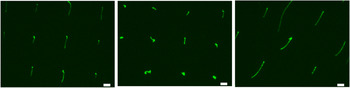
To study the influence of this parameter, several experiments were carried out by adjusting the temperature while maintaining a constant DNA concentration and translation stage speed. Note that we did not observe any significant influence of the temperature in the 25 to 28 °C range. However, for higher temperature values with substrate temperatures ranging from 32 to 75 °C, we obtained regular arrays of short and sometimes thornlike or double DNA molecules (Fig. 3). We have two hypotheses concerning the influence of the substrate temperature over the DNA molecules’ assembly. On the one hand, we suggest that by increasing the substrate’s temperature, which governs the evaporation rate of the suspension deposited on the hydrophobic substrate, it creates an increase of the convective flux that drags the molecules and nourishes the three-phase contact line. This leads to an increase in the local density of molecules close to the contact line during the same pinning time as the speed of the translation stage is kept constant. As a result, instead of having one single molecule trapped inside the pattern, there is a given amount of molecules located around the pinning point, undergoing the same capillary forces these molecules will be stretched at the same time when the contact line disrupts. On the other hand, we could assume that increasing the temperature of the substrate is equivalent to establishing a denaturation process. In fact, considering the small suspension volume squeezed between the fixed slide and the heated PDMS stamp, DNA molecules may start a denaturation process in solution. So, molecules at different states of dissociation undergo the capillary assembly process and then stretch, thus explaining the obtained results. In a more general view, it is clear that at higher temperatures, the assembly becomes more unpredictable. Nevertheless, it has to be noticed that the yield is not affected as we could have thought.
Temperature influence. Fluorescence image of the strand arrays obtained with a substrate temperature of 75 °C during the assembly process [Scale bar corresponds to 4 μm].

D. Influence of pattern geometry
If we further analyze the micrographs obtained (Fig. 1), we observe an extension of phage lambda DNA molecules to an average length of 22.9 μm (±1.9 μm) measured on 22 strands, ranging from 19.6 to 26.1 μm, which is much longer than the contour length of the unextended phage lambda (16.3 μm).Reference Smith, Perkins and Chu34 This high stretching factor (42%) induced by our technique is definitely worthwhile studying in more detail. It is generally admitted that TOTO and YOYO nucleic acid stains increase the nominal length by 30–35% in a 4:1 base pairs per intercalator ratio.Reference Perkins, Smith, Larson and Chu35, Reference Tegenfeldt, Prinz, Cao, Chou, Reisner, Riehn, Wang, Cox, Sturm, Silberzan and Austin36 However, in this study we observe an overstretching of the molecules of 40–50%, which is more important than the intercalators’ effect on DNA molecules reported. Thus, the overstretching observed is caused by other factors as well. We suggest that this overstretching is possibly due to the capillary forces created by the contact line joined to the high speed of movement. What we can certainly affirm is that in a process of flow stretching, the force exerted is the maximum at the anchoring point and minimum once the flow has passed. Thus, in such a case of figure, the force applied has a progressive nature where the hydrodynamic forces need to be added to the capillary forces and the high speed of the translation stage. Moreover, Malaquin et al.Reference Malaquin, Kraus, Schmid, Delamarche and Wolf31 demonstrated that the magnitude and the direction of the capillary forces induced by the contact line pinning depend strongly on the geometry of patterns. In fact, the combination of geometrical trapping of the contact line at the surface structures and capillary forces exerted during film breakage plays an important role in the selective trapping of DNA molecules inside the patterns, but may play an important role in the final stretching of the molecules as well. However, the forces really exerted are very difficult to quantify and would require a complete numerical simulation of the molecular dynamics.
This technique could be an important step forward for DNA chips readout and scanning diagnostics, but only if we are able to demonstrate that this overstretching does not significantly modify the intrinsic structure of the DNA molecule so that a subsequent hybridization of unstretched markers is still feasible. For these reasons, we are currently working to elucidate this phenomenon and determine the exact influence of each of the experimental parameters on the stretching of the molecules.
E. Extensions of the technique
All the different experiments performed, conducted to the observation that the stretched DNA molecules present unimodal size dispersion. This particular observation was worth studying more in detail to evidence whether the directed capillary assembly technique induces a selection in size of the objects in solution. To answer this question, we used the same experimental protocol, but this time assembling coiled DNA molecules in solution having undergone ultrasounds during 10 s. In this way, we expect to obtain a DNA solution containing DNA molecules with heterogeneous lengths resulting from a mechanical and random break of the molecules.
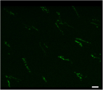
After capillary assembly of this heterogeneous DNA solution and transfer-printing on an APTES-coated glass slide, we obtained a DNA array with a lower coverage rate. In a same field of view, we obtained the assembly of DNA molecules with different lengths (Fig. 4). Therefore, at the micrometer scale, this experiment revealed there is no apparent or no detectable capillary-induced selection in size of the coiled DNA molecules assembled.
Fluorescence images of the results obtained after directed capillary assembly of a heterogeneous solution of DNA molecules (ultrasound agitation of phage lambda DNA) onto a microstructured PDMS stamp and transfer of the assembly onto an APTES-coated glass slide [Scale bars correspond to 4 μm].

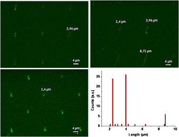
Schwartz et al. conducted a very powerful study by imaging long single DNA molecules (more than 100 kbp) combed on a glass surface and digested by restriction enzymes.Reference Armbrust, Berges, Bowler, Green, Martinez, Putnam, Zhou, Allen, Apt, Bechner, Brzezinski, Chaal, Chiovitti, Davis, Demarest, Detter, Glavina, Goodstein, Hadi, Hellsten, Hildebrand, Jenkins, Jurka, Kapitonov, Kroger, Lau, Lane, Larimer, Lippmeier, Lucas, Medina, Montsant, Obornik, Parker, Palenik, Pazour, Richardson, Rynearson, Saito, Schwartz, Thamatrakoln, Valentin, Vardi, Wilkerson and Rokhsar37, Reference Dimalanta, Lim, Runnheim, Lamers, Churas, Forrest, de Pablo, Graham, Coppersmith, Goldstein and Schwartz38 Stretching and tethering the DNA molecules on a glass slide is advantageous to perform a linear cartography of the spatial position versus the genomic position by preserving the haplotype information. The technique of Schwartz et al. suffers from an inhomogeneous stretching due to the DNA-surface interactions and from a disturbed enzymatic activity. It is clear that a solution to solve this issue would be to perform these reactions in solution rather than on a surface. In this perspective, the capillary assembly technique presented in this work offers other nonnegligible advantages. In fact, we demonstrate that if we are in the presence of a complex solution, we can find, after capillary assembly and transfer, a result that will account from the initial composition of the solution. To prove this, we assembled a solution of phage lambda DNA molecules digested by enzymatic reaction. We chose SmaI enzyme, which specifically cleaves phage lambda DNA in two well-known and identified restriction sites. The electrophoresis gel of digested phage lambda DNA by SmaI enzyme compared to the electrophoresis gel of undigested phage lambda DNA accounts for the correct activity of the enzyme and shows the three lengths we expect to obtain after digestion in solution: one segment of 8 kbp (equivalent to 2.47 μm), one segment of 12 kbp (equivalent to ~3.70 μm), and one segment of 28 kbp (equivalent to 8.66 μm). The solution is consequently three times more concentrated after digestion, so we diluted the solution to follow the experimental protocol previously described in the case of the assembly of single phage lambda DNA molecules. We then fluorescently labeled the resulting DNA with a YOYO-1 nucleic acid stain. After assembly of the solution and transfer-printing, Fig. 5 shows that we obtained the lengths we expected from the electrophoresis gel (2.40 μm ± 0.24 μm, 3.96 μm ± 0.24 μm, and 8.68 μm ± 0.24 μm in average). Thus, the result obtained after assembly, under the experimental conditions described (at high displacement speeds), and transfer is representative of the composition of the solution.
Fluorescence images of the results obtained after directed capillary assembly onto a microstructured PDMS stamp of a phage lambda DNA solution digested by a restriction enzyme (SmaI) and transfer. Bottom right corner, histogram showing the distribution of lengths measured over 64 molecules.

It has to be noted however that the histogram (Fig. 5) shows that over 64 molecules measured in different regions of the same substrate, we statistically found more molecules measuring 2.40 or 3.96 μm than molecules measuring 8.68 μm. Therefore, in this experiment, even though statistics have to be made at a larger scale, longer fragments might have been slightly slower to be directed toward the liquid front line by the hydrodynamic flows induced by the solvent evaporation. Another question that needs to be addressed is whether the overstretching observed in the case of phage lambda molecules is maintained in the case of shorter molecules. Here, the populations measuring 2.4 μm long in theory, measure 2.4 μm as well after the assembly process. The populations of 3.7 or 8.6 μm long in theory, measure around 4.0 and 8.8 μm in length after assembly, which corresponds to an overstretching of 8% and 2.3%, respectively. In any case, we do not obtain the high stretching factor observed in the case of 48 kbp molecules. We could ask ourselves whether the enzymatic activity plays a role in the final stretching of the molecules. Another hypothesis to explain these results could be that short molecules are less flexible or loose than long ones, so short molecules are likely to be more difficult to stretch.
As a further step in this development, we prove that the experimental protocol described in this paper works with phage lambda DNA molecules, so long DNA molecules but can be easily transposed to short DNA molecules as well. To demonstrate this statement, we used a solution of linear plasmids 18 nM in concentration (5100 bp so 1.7 μm nominal length in theory). Of course, in a rigorous manner, to assemble shorter individual DNA molecules, the size of the patterns should be tuned and properly adjusted. Here, we chose to use the same master, therefore the same microfeatures as the ones used in the case of phage lambda DNA molecules to compare the results.
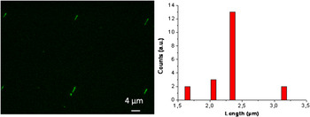
Figure 6 shows the results obtained after directed capillary assembly of plasmids in solution on a PDMS stamp and transfer. We observe that the results in terms of coverage rate and quality of the ordering are alike between plasmids and phage lambda DNA assemblies. The only noticeable difference lies in the length of the molecules, which measure this time 2.41 ± 0.11 μm in average measured over 20 molecules. This value is close to the nominal length of linear plasmids. However, it implies an elongation of 35–40% of the molecules with respect to the contour length of linear plasmids. This stretching factor is closer to the stretching factor obtained in the case of phage lambda DNA molecules, but does not correspond to the stretching factor observed in the case of digested DNA fragments previously discussed. This inconsistency in the stretching factor measured requires a complete and thorough study. Further experiments will need to be performed in this direction comparing fluorescently labeled or unlabeled molecules, the influence of the patterns’ size, and the influence of the translation speed along with numerical simulations to support the results and allow us to give a conclusive answer to this question. What needs to be highlighted in this study is that it is possible to assemble linear plasmids and by extension, DNA molecules ten times shorter in length than phage lambda DNA molecules with this technique. We could imagine, of course, adapting the technique to an even lower scale and assembling oligonucleotides just a few hundreds of base-pairs by simply replacing the microstructured PDMS stamp by a PDMS with appropriate nanometric cavities. This objective could be of great interest for the development of DNA and virus technology.
Fluorescence image of linear plasmids (5100 bp) assembled after directed capillary assembly onto a microstructured PDMS stamp and transfer-printing on an APTES-coated glass slide. On the right, histogram showing the distribution of lengths measured over 20 molecules.

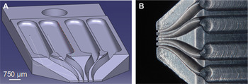
Finally, another major advantage of this directed capillary assembly technique that should be emphasized is that it can easily be adapted depending on the applications. As an example, we made the technique evolve toward multiplexing. One of the needs when assembling objects onto a surface can be the multiplexing of different solutions, therefore the assembly of different solutions in one step, and on the same substrate. The strategy we propose involves replacing the fixed glass spreader used for capillary assembly to drag the liquid contact line by a spreader with independent channels (Fig. 7). Here, the channels of the prototype, Fig. 7, are separated by 300 μm and the reservoirs capacity is only 5 μL, hence the working volumes are considerably reduced. The materials used need to be nonporous and hydrophilic, for example, polyurethane or aluminum. We have validated the design, material, and use of this prototype to stretch and align four different solutions of DNA molecules just the way the original glass spreader does but this time simultaneously. This multichannel spreader can be refilled with droplets of different molecules in solution, and used to dispense them simultaneously onto a substrate by capillary assembly over an area of less than 1 mm2. In this way, different substrates of different nature can be assayed simultaneously in a faster manner. This multiplexing can be transposed to any type of object and for a wide range of applications.
(a) 3D design of the multichannel spreader used to dispense four different solutions onto a substrate in one step. (b) Aluminum prototype of the multichannel spreader.

IV. CONCLUSION
This study proves that capillary forces and convective flows can be exploited on structured substrates to produce highly ordered arrays of single DNA molecules with a coverage rate of 99% in an automatized and highly reproducible way. The stretched DNA molecules are homogeneous in length and orientation and are precisely positioned over a millimeter-scale area in just a few seconds. The capillary assembly method is observed to extend the length of phage lambda DNA molecules to 40–50% of their contour length. The control of four experimental parameters, namely, the temperature, the contact angle, the substrate velocity, and the DNA molecules’ concentration, along with the design of the PDMS stamp’s features provides full control of the assembly process and permits an increase of the structural complexity for novel micro- and nanosystems. This technique does not induce a selection in size of the objects, thus it can handle heterogeneous solution, long (tens of kbp) but also short (a few thousands of bp) molecules, and bulk-functionalized molecules or complex objects directly in solution to allow the construction of future 1D nanoscale building templates. Finally, this technique is versatile compared to other existing techniques because it can easily be adapted to offer important and nonnegligible added functionalities such as multiplexing.
ACKNOWLEDGMENTS
This work was supported by the EC-funded project NaPa (Contract No. NMP4-CT-2003-500120). We thank the “Région Midi-Pyrénées” council, and the “Institut des Technologies Avancées en sciences du Vivant (ITAV)” for their financial support. We also thank Jean-Marie François and his research group, especially Marie-Ange Teste from the “Engineering of Biological Systems and Process Laboratory”.