Introduction
Cystic echinococcosis (CE), caused by Echinococcus granulosus, is a zoonotic disease that occurs mainly in sheep-grazing areas and has a global distribution (Budke et al., Reference Budke, Deplazes and Torgerson2006). Dogs and other carnivores are the definitive hosts, in which the adult parasites inhabit the intestine. Humans and other mammalian intermediate hosts, including sheep, pigs and cattle, are infected following ingestion of contaminated food and/or water (Beggs, Reference Beggs1985). Primary cystic echinococcosis occurs when an organ traps the larvae following intestinal absorption. Arrest occurs in the capillary beds of the liver or lungs in almost 90% of cases. Approximately 70% of hydatid cysts are located in the liver and one-quarter to one-third of cases have multiple cysts. The right lobe is affected in 85% of patients (Brunicardi, Reference Brunicardi, Brunicardi, Andersen and Billiar2005). Clinical features depend upon the site, size, stage of cyst, and development and viability of the cyst contents (Khuroo, Reference Khuroo2002). The disease may be asymptomatic for many years and may become symptomatic due to the enlargement or rupture of cysts. Various medical, surgical and percutaneous therapeutic techniques have been suggested.
Diagnosis is a basic component of population studies on echinococcosis and, other than careful necropsy, there is no gold standard. In the definitive host, techniques for direct parasite identification include copro-antigen and copro-DNA detection. In intermediate hosts, necropsy is typically used. In humans, diagnostic imaging and serology are widely employed (Pawlowski et al., Reference Pawlowski, Eckert, Vuitton, Ammann, Kern, Craig, Eckert, Gemmell, Meslin and Pawlowski2001). The use of multiple, parallel testing or additional confirmatory tests in a diagnostic strategy can overcome the lack of a gold standard. This will yield valuable information at both population and individual levels (Torgerson & Deplazes, Reference Torgerson and Deplazes2009).
The detection of anti-Echinococcus antibodies has been widely used in immunological assays such as indirect immunofluorescence (IFA), indirect haemagglutination (IHA), enzyme-linked immunosorbent assay (ELISA) (Kittelberger et al., Reference Kittelberger, Reichel, Jenner, Heath, Lightowlers, Moroe, Ibrahem, Craig and O'Keefe2002; Siavashi et al., Reference Siavashi, Taherkhani, Rezaei, Razavi Deligani and Assmar2005; Sadjjadi et al., Reference Sadjjadi, Abidi, Sarkari, Izadpanah and Kazemian2007), enzyme linked immunoelectrotransfer blots (EITB) and immunoblot assay (Sarkari et al., Reference Sarkari, Sadjjadi, Abidi, Izadpanah, Kazemian and Rafati2007). Antibody detection assays have many drawbacks as they neither distinguish between past and present infections nor assess the efficacy of treatments. Antigen detection assays may circumvent this problem (Doiz et al., Reference Doiz, Benito, Sbihi, Osuna, Clavel and Gomez-Lus2001) as hydatid cyst antigen can be detected in the serum or urine of patients with hydatidosis. Circulating hydatid antigens are present in the serum only during active infections, as antigen levels decrease following surgical cyst removal or successful chemotherapy (Devi & Parija, Reference Devi and Parija2003). The detection of circulating antigens rather than antibodies may be very useful in the immunodiagnosis of hydatid disease (Craig & Nelson, Reference Craig and Nelson1984; Carmena et al., Reference Carmena, Benito and Eraso2006). Accurate immunodiagnosis of the infection requires the use of highly specific and sensitive antigens in immunodiagnostic assays. The choice of an appropriate source of antigenic material is a crucial point in the improvement of the diagnostic features of tests and must be based on the developmental stage of the parasite and the host. The most common antigenic sources used for the immunodiagnosis of echinococcal disease are hydatid cyst fluid, somatic extracts and excretory–secretory products from protoscoleces or adults of E. granulosus (Gasser et al., Reference Gasser, Lightowlers and Rickard1989, Reference Gasser, Parada, Acuna, Burges, Laurenson, Gulland, Reichel and Paolillo1994; González et al., Reference González, Spinelli, Lorenzo, Hellman, Nieto, Willis and Salinas2000; Kittelberger et al., Reference Kittelberger, Reichel, Jenner, Heath, Lightowlers, Moroe, Ibrahem, Craig and O'Keefe2002; Ben Nouir et al., Reference Ben Nouir, Gianinazzi, Gorcii, Müller, Nouri, Babbaa and Gottstein2009).
The aims of the present study are to prepare and purify a 27.5 kDa protoscolex antigen (27.5 PA) of E. granulosus present in hydatid cysts and evaluate the diagnostic efficacy of raised IgG antibody against purified 27.5 PA in detection of Echinococcus antigen by sandwich ELISA for comparison with the antibody detection method in serum samples of naturally infected sheep and humans.
Materials and methods
Collection of blood and faecal samples
Blood and faecal samples were obtained from 200 sheep brought to be slaughtered at a local abattoir in Giza, Egypt from January to June 2011. Based on gross inspection and post-mortem examination of the livers of the sheep after slaughter and subsequent parasitological examination of stool samples using the direct smear method, merthiolate–iodine–formaldehyde concentration method (Sapero & Lawless, Reference Sapero and Lawless1953) and Kato–Katz concentration technique (Siongok et al., Reference Siongok, Mahmoud, Guma, Warren, Muller, Handa and Houser1976), sheep were classified into three groups, namely 100 sheep infected with Echinococcus, 50 infected with Fasciola gigantica, Ascaris vitulorum or Strongyloides papillosus and 50 uninfected controls. Blood from each sheep was collected, centrifuged at 760 × g at 4°C for 10 min and serum samples were stored at − 20°C until analysed.
Forty E. granulosus infected human patients from areas in Dakahlia Governorate, where the disease is highly endemic, were admitted to tropical and surgical departments of Zagazig and El-Azhar University hospitals and were diagnosed, by means of sonography, computed tomography (CT) and magnetic resonance (MR) imaging as having CE in their livers. In addition, 30 patients infected with other parasites (Schistosoma mansoni or F. gigantica) were included. In addition, 30 members of the medical staff at Theodor Bilharz Research Institute served as parasite-free, healthy, negative controls. Blood samples were collected from all cases and sera were separated, aliquoted and kept at − 70°C until used.
Preparation of protoscolex antigen and polyclonal antibodies
Protoscoleces were prepared using the method of Rafiei & Craig (Reference Rafiei and Craig2002). In brief, protoscoleces were collected from fresh, fertile, sheep liver cysts. Viability was determined by the vital colouration approach, with 0.2% eosin staining. Protoscoleces were subjected to three cycles of freezing and thawing, washed three times, each time by centrifugation (600 × g for 5 min), and suspended in ten times their volume of 0.15 m phosphate-buffered saline (PBS) (pH 7.2). Subsequently, protoscoleces were suspended in four times their volume of PBS containing 0.1 mg aprotinin/ml, then sonicated on ice in a 150 W ultrasonic disintegrator, in 10-s bursts with 5-s intervals, until no intact protoscoleces were visible microscopically. The sonicate was left standing on ice for 1 h, then sedimented at 10,000 × g for 30 min. The supernatant was concentrated using an Amicon 8400 ultrafiltration unit with membrane (3 kDa cut-off) (EMD Millipore Corporation, Bedford, Massachusetts, USA). The sample was applied to DEAE-sephadex A50 column (ion exchange column chromatography) followed by a Sephacryl S-200 HR column (gel filtration chromatography) equilibrated in 0.1 m Tris–HCl, pH 7. The purity of the produced antigen was assayed by sodium dodecyl sulphate-polyacrylamide gel electrophoresis (SDS-PAGE) under reducing conditions (Laemmli, Reference Laemmli1970).
Two New Zealand white 4-month-old rabbits, weighing approximately 2 kg each, were injected intramuscularly with an equal volume of 1 mg protoscolex antigen (PA) and complete Freund's adjuvant (priming dose). Two weeks later they were injected with an equal volume of 0.5 mg PA and incomplete Freund's adjuvant (booster dose) and the latter booster dose was repeated twice, with a 1-week interval. Sera were collected from an ear vein 4 days after the last injection, to detect the titre of antibodies produced. When the titre was high, the animal was killed and exsanguinated and the sera were separated and stored at − 20°C until used (Fagbemi et al., Reference Fagbemi, Obarisiagbon and Mbuh1995; Guohadia & Fagbemi, 1997).
Purification, labelling and identification of the polyclonal antibody
The produced IgG polyclonal antibody (pAb) was purified using 50% ammonium sulphate precipitation (Nowotny, 1979) and caprylic acid treatment (McKinney & Parkinson, Reference McKinney and Parkinson1987). The protein content was estimated by a Bio-Rad protein assay (Bio-Rad Laboratories, Hercules, California, USA) (Bradford, Reference Bradford1976) and the purity of the produced IgG was identified by SDS-PAGE according to Laemmli (Reference Laemmli1970). The IgG pAb was conjugated with horseradish peroxidase according to the periodate method of Tijssen & Kurstak (Reference Tijssen and Kurstak1984). The reactivity of anti-protoscolex antigen IgG against Echinococcus antigens was assessed using the indirect ELISA (Engval & Perlman, Reference Engvall and Perlman1971).
Identification and characterization of the chemical nature of anti-PA IgG was performed using indirect ELISA by testing its reactivity against the antigen before and after treatment with 20 mm sodium periodate (Sigma Aldrich, Gillingham, Dorset, UK) and 4% trichloroacetic acid (Sigma Aldrich) (Woodward et al., Reference Woodward, Young and Bloodgood1985).
Detection of anti-protoscolex antigen IgG in sera
This method was performed, with some modifications from the original method of Engvall & Perlman (Reference Engvall and Perlman1971). Briefly, wells of the microtitration plates (Thomas Scientific, Swedesboro, New Jersey, USA) were coated with 100 μl of purified 27.5 kDa PA (10 μg/ml in 0.1 m carbonate buffer pH 9.6) and incubated overnight at room temperature. Plates were washed thoroughly three times with washing buffer (0.1 m PBS pH 7.4), then blocked with 200 μl of 0.1% bovine serum albumin (BSA) (Sigma) in 0.1 m PBS pH 7.4 for 1 h at 37°C. Plates were washed five times with washing buffer, then 100 μl of serum samples (1:250 diluted in washing buffer) were added to each well and incubated for 1 h at 37°C. Plates were washed three times with washing buffer and 100 μl of either polyvalent-anti-sheep (1:1000) or anti-human (1:2000) peroxidase conjugate was added to plates and incubated at room temperature for 30 min. The enzyme substrate reaction was stopped by adding 50 μl/well of 8 n H2SO4. The absorbance was measured at 492 nm using an ELISA reader (Bio-Rad Microplate Reader) (Allam et al., Reference Allam, Bauomy, Hemyeda, Diab and Sakran2012).
Detection of circulating protoscolex antigen in sera
After several optimization trials, the following sandwich ELISA was performed for circulating protoscolex antigen (CPA) detection, using anti-27.5 PA IgG pAb as coating and peroxidase-conjugated antibody for capture of the antigen. For each step, 100 μl/well was added unless mentioned otherwise. Microtitration plates were coated with the anti-27.5 PA IgG pAb (20 μg/ml) in 0.1 m carbonate buffer, pH 9.6 and left overnight at room temperature. Plates were washed thoroughly three times with phosphate-buffered saline/Tween (PBS/T) 2 min each wash, and unbound sites were blocked with 200 μl/well of 2.5% fetal calf serum (FCS)/PBS/T and incubated for 2 h at 37°C. Following emptying the plates by suction, undiluted serum was added and incubated for 2 h at 37°C. After thorough washing as described above, peroxidase-conjugated anti-27.5 PA IgG pAb (1:1000 diluted in PBS/T) was added. Plates were incubated for 1 h at 37°C and washed with PBS/T. The substrate O-phenylenediamine dihydrochloride (Sigma) was added and the plates were incubated for 30 min in the dark at room temperature. The enzyme reaction was stopped with 50 μl/well of 8 n H2SO4. The absorbance was measured at 492 nm using an ELISA reader (Bio-Rad). The cut-off value was calculated as the mean optical density (OD) reading of negative controls + two standard deviations of the mean. The OD readings equal to or less than the cut-off value were considered negative, while those readings greater than the cut-off value were considered positive.
The specificity of the assays was determined as the sum of the data from the negative controls and the group of sheep or humans infected with other parasites.
Data analysis
Data were expressed as mean ± standard deviation and analysed using the SPSS computer program (Version 16 Windows; SPSS Inc., Chicago, Illinois, USA). Standard diagnostic indices were calculated as described by Galen (Reference Galen1980).
Results
SDS-PAGE analysis of PA showed a single band at 27.5 kDa. The specificity of the produced pAb against different parasite antigens, including S. mansoni, F. gigantica, hookworms and trichostrongyloids, was determined by indirect ELISA and was strongly reactive with PA of Echinococcus and non-reactive with the other parasite antigens. SDS-PAGE analysis of the produced pAb, showed two different bands, H- and L-chain, at 53 and 31 kDa, respectively. The chemical nature of anti-PA IgG recognized epitopes was defined following 20 mm sodium periodate treatment of the target antigen. A marked reduction in the reactivity of this antibody was detected, denoting that the reactive epitopes were glycoprotein in nature.
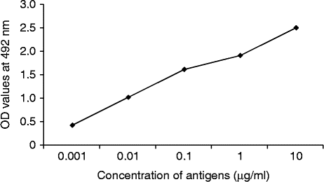
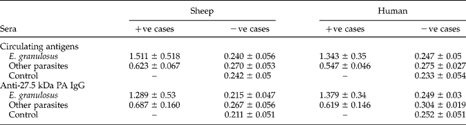
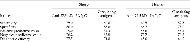
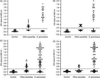
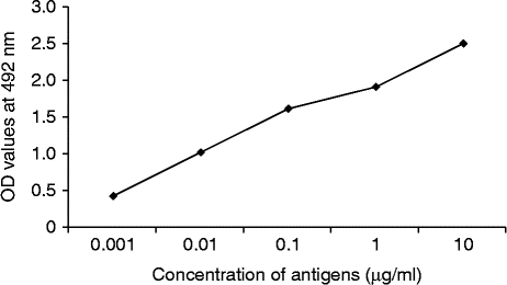
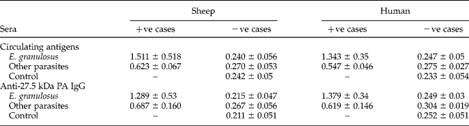
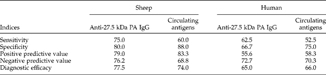
Using the standard curve (fig. 1), the lower detection limit of an ELISA assay was found to be 2.5 μg/ml and the cut-off values were 0.413 and 0.40 in sheep and human patients, respectively. In E. granulosus-infected sheep a high positive anti-27.5 PA IgG titre showed 75.0% sensitivity, compared with 60.0% for CPA. In humans, the sensitivity for anti-27.5 PA IgG was 62.5% compared with 52.5% for CPA (tables 1 and 2, fig. 2).
A standard curve for detecting the antigen limit (μg/ml) of protoscoleces using polyclonal antibody IgG. OD, mean optical density.
Mean optical density values (±SD) of anti-27.5 kDa protoscolex antigen IgG and circulating protoscolex antigens in sera of human subjects and sheep infected with E. granulosus, other parasites and controls; other parasites include S. mansoni and F. gigantica (human subjects) and F. gigantica, A. vitulorum and S. papillosus (sheep).
Diagnostic indices (%) of a sandwich ELISA used for the detection of anti-27.5 protoscolex antigen IgG and circulating protoscolex antigens in sheep and human sera of different studied groups.
Levels of anti-27.5 kDa protoscolex antigen IgG and circulating protoscolex antigens in sera of human (A and B) and sheep (C and D) infected with E. granulosus and other parasites and controls; mean optical density (OD) with cut-off values (horizontal dotted lines), other parasites include S. mansoni and F. gigantica (human subjects) and F. gigantica, A. vitulorum and S. papillosus (sheep).

All cases of negative controls had negative results in both sheep and humans. Twenty and 12 sheep of the other parasites group showed positive ELISA results for anti-27.5 PA IgG and CPA and were considered as false positives, therefore the specificity of the assay was 80.0 and 88.0%, respectively. In humans, 20 and 15 patients of other parasites group showed positive ELISA results for anti-27.5 PA IgG and CPA, so the specificity of the assay was 66.7 and 75.0%, respectively (tables 1 and 2, fig. 2).
In sheep, the positive and negative predictive values and diagnostic efficacy of ELISA assay in detecting anti-27.5 PA IgG were 79.0, 76.2 and 77.5%, respectively; and 83.3, 68.8 and 74.0% in detecting CPA, respectively. In humans the values were 55.6, 72.7 and 65.0% for anti-27.5 PA IgG and 58.3, 70.3 and 66.0% for CPA, respectively (table 2).
Discussion
Hydatid cyst fluid (HCF) is a complex mixture of glyco- and lipoproteins, carbohydrates and salts. Some components derive from the host, mainly albumin and immunoglobulins, while the remaining components are the products of the metacestode. HCF is considered to be the main antigenic source for the immunodiagnosis of human CE. In clinical practice, crude HCF has a high sensitivity, ranging typically from 75 to 95% (Zhang et al., Reference Zhang, Li and McManus2003). However, its specificity is often unsatisfactory and cross-reactivity with sera from patients infected with other cestode (89%), nematode (39%) and trematode (30%) species was commonly reported (Eckert & Deplazes, Reference Eckert and Deplazes2004).
In the present study, the protoscolex antigen (PA) was isolated from living hydatid cysts and purified, and the resulting 27.5 kDa PA was used in production of pAb. This anti-27.5 kDa PA IgG was used in the development of an antigen-based ELISA method for detection of CPA of E. granulosus in serum samples of naturally infected sheep and humans, for comparison with an antibody detection method. The identification of target antigens recognized by anti-27.5 kDa PA IgG showed that it was glycoprotein in nature. Similar to our antigen, a 29 kDa protein (P29) which was first described by González et al. (Reference González, Spinelli, Lorenzo, Hellman, Nieto, Willis and Salinas2000) appeared to be exclusively expressed on E. granulosus protoscoleces (Chemale et al., Reference Chemale, Van Rossum, Jefferies, Barrett, Brophy and Ferreira2003) and exhibited a good immunodiagnostic potential for detecting CE in patients before surgery (González et al., Reference González, Spinelli, Lorenzo, Hellman, Nieto, Willis and Salinas2000). Ben Nouir et al. (Reference Ben Nouir, Gianinazzi, Gorcii, Müller, Nouri, Babbaa and Gottstein2009) reported that recombinant P29 also exhibited good prognostic value in the follow-up monitoring of cured as opposed to non-cured CE patients who were able to develop a seropositive status against recP29 either before or shortly after diagnosis and/or initiation of a treatment protocol. In sheep, Ris et al. (Reference Ris, Hamel and Mackle1987) used two polysaccharide protoscolex and mouse HCF membrane antigens in detecting E. granulosus antibodies, and reported a sensitivity of 80%. Kittelberger et al. (Reference Kittelberger, Reichel, Jenner, Heath, Lightowlers, Moroe, Ibrahem, Craig and O'Keefe2002) reported that in naturally infected sheep a protoscolex-derived crude antigen preparation had higher sensitivity than purified 8 kDa protein (64.6 versus 11.2%) and had comparable specificities (98.2 versus 99.5%). They concluded that a protoscolex–based ELISA assay could be a useful tool for the identification of flocks of sheep infected with E. granulosus.
In the present investigation, the sensitivity of the indirect ELISA in detecting anti-27.5 kDa PA IgG was higher (75.0% in sheep and 62.5% in humans) than that recorded in detecting CPA (60.0% in sheep and 52.5% in humans). Antigen detection assays diagnosing hydatidosis resulted in varying rates of sensitivity and specificity (Craig & Nelson, Reference Craig and Nelson1984; Gottstein, Reference Gottstein1984; Shariff & Parija, Reference Shariff and Parija1993; Ravinder et al., Reference Ravinder, Parija and Rao1997). It has been shown that hydatid cyst antigen can be detected in sera of 33–85% of hydatidosis patients (Gottstein, Reference Gottstein1984; Ravinder et al., Reference Ravinder, Parija and Rao1997). Sadjjadi et al. (Reference Sadjjadi, Sedaghat, Hosseini and Sarkari2009) used IgG pAb against hydatid cyst fluid in development of a simple antigen-based ELISA method for diagnosis of human hydatidosis for comparison with antibody detection methods. The reported sensitivity and specificity were 25.7 and 98.0% in the antigen detection assay and 94.2 and 81.6%, respectively, in the antibody detection method. The difference in the sensitivity recorded in this study and that of Sadjjadi et al. (Reference Sadjjadi, Sedaghat, Hosseini and Sarkari2009), which was in favour of our work, is that they used crude hydatid cyst fluid while we used a more purified 27.5 kDa PA.
The low sensitivity of the antigen detection assay in this study and other studies could be related to the fact that most of the antigens in sera are immune complexes of antigens and antibodies which cannot be easily detected by serological assays. Moreover, the intact cysts, small cysts and cysts in privileged sites do not release enough antigens in the serum to elicit more than a minimally detectable response, whereas previously ruptured or leaking cysts are associated with strong responses (Moro & Schantz, Reference Moro and Schantz2009). Other factors generally affect the diagnostic efficacy of assays, e.g. the type of antigen used, whether they are crude or purified native antigens. The difficulties are associated with the standardization of the antigen preparations and techniques (Gottstein, Reference Gottstein1984; Sunita et al., Reference Sunita, Dubey, Khurana and Malla2007) and the type of intermediate host species, which affects the nature and quality of these antigens (Poretti et al., Reference Poretti, Felleisen, Grimm, Pfister, Teuscher, Zuercher, Reichen and Gottstein1999). Also, in antibody detection assays, many authors attributed the false negative results to the antibody titre which is raised slowly during the course of infection, especially in old persons and infants. Also in cerebral, ocular and calcified cysts, the antibody titre is low and cannot be easily detected (Ravinder et al., Reference Ravinder, Parija and Rao1997).
In this study, the specificity of ELISA in detecting CPA was higher (88.0% in sheep and 75.0% in humans) than that recorded in detecting anti-27.5 kDa PA (80.0% in sheep and 66.7% in humans). Although the sensitivity of the antibody detection assay was high, its specificity was rather low. This may be due to the reduced host-specificity of this parasite, which increases the possibility of cross-reaction with other parasites (Lymbery & Thompson, Reference Lymbery and Thompson1996; Eckert & Thompson, Reference Eckert and Thompson1997; Rafiei & Craig, Reference Rafiei and Craig2002). Also, Sadjjadi et al. (Reference Sadjjadi, Sedaghat, Hosseini and Sarkari2009) in their work on an antigen detection assay, attributed the cross-reaction with leishmaniasis to the high level of antibodies in the visceral leishmaniasis patient, which might interfere with the antigen detection assay. Sadjjadi et al. (Reference Sadjjadi, Abidi, Sarkari, Izadpanah and Kazemian2007) used antigen B of E. granulosus in ELISA for detecting anti-hydatid cyst antibodies in human patients and the reported sensitivity and specificity were 92.5 and 97.3%, respectively.
In the present investigation, the greater sensitivity and specificity of both assays in sheep than in humans could be attributed to the fact that sheep are considered to be the main intermediate host while humans are paratenic hosts, i.e. accidentally infected intermediate hosts. In conclusion, an antibody detection assay is still superior to and more sensitive than an antigen detection assay, especially in diagnosing active infection in which hydatid cysts are predominant, while the antigen detection assay might be a useful approach for assessment of the efficacy of treatment, especially after removal of the cyst. Further studies are recommended to improve the diagnostic efficacy of the antigen-based ELISA method by using a highly purified recombinant antigen.
Conflict of interest
None.
Ethical standards
The authors assert that all procedures contributing to this work comply with Theodor Bilharz Research Institute-Institutional Review Board (TBRI-IRB; FWA00010609), following Declaration of Helsinki (2013), which reviewed and approved the study (approval number 6/2011).