Implications
The relationship between nutritional status and reproductive success in animals has been studied for many years. Considerable evidence has been accumulated to implicate the existence of a common endocrine system that controls metabolism and the reproductive system. It is suggested that orexins belong to the group of hormones, which regulate metabolic status and reproduction. In this study, we demonstrated differences in orexin gene and orexin proteins in the porcine pituitary during the estrous cycle. The results imply the impact of hormonal milieu on orexins’ expression in the porcine pituitary. A sound knowledge of mechanisms that control energy homeostasis and reproduction is needed in animal breeding practice.
Introduction
Orexin A and B (OXA, OXB), also known as hypocretin 1 and 2, are hypothalamic peptides with a 46% sequence homology. OXA is a 33-amino acid peptide with a molecular mass of ~3.5 kDa, which has an N-terminal pyroglutamyl residue and C-terminal amidation. OXB is a 28-amino acid peptide with a molecular mass of ~2.9 kDa and C-terminal amidation. Orexins are cleaved from a 130-amino acid common precursor molecule, prepro-orexin (PPO), which is synthesized mainly in neurons inside and in the area of the lateral and posterior hypothalamus. Orexin action is mediated by two different receptors, termed orexin receptor 1 (OX1R) and orexin receptor 2 (OX2R), both coupled with protein-G. OXA is a more selective ligand for OX1R, whereas OX2R binds both orexins with similar affinity (De Lecea et al., Reference De Lecea, Kilduff, Peyron, Gao, Foye, Danielson, Fukuhara, Battenberg, Gautvik, Bartlett, Franlel, Van den Pol, Bloom, Gautvik and Sutcliffe1998; Sakurai et al., Reference Sakurai, Amemiya, Ishii, Matsuzaki, Chemelli, Tanaka, Williams, Richardson, Kozlowski, Wilson, Arch, Buckingham, Haynes, Carr, Annan, McNulty, Liu, Terret, Elshourbagy, Bergsma and Yanagisawa1998). The orexins were originally identified as regulators of food intake and energy homeostasis (Sakurai et al., Reference Sakurai, Amemiya, Ishii, Matsuzaki, Chemelli, Tanaka, Williams, Richardson, Kozlowski, Wilson, Arch, Buckingham, Haynes, Carr, Annan, McNulty, Liu, Terret, Elshourbagy, Bergsma and Yanagisawa1998) and later as key modulators of the sleep-wake cycle and arousal (Chemelli et al., Reference Chemelli, Willie, Sinton, Elmquist, Scammell, Lee, Richardson, Williams, Xiong, Kisanuki, Fitch, Nakazato, Hammer, Saper and Yanagisawa1999), but multiple lines of evidence also demonstrate that orexins participate in the control of different endocrine axes, including the hypothalamic–pituitary–adrenal axis (Voisin et al., Reference Voisin, Rouet-Benzineb, Reuter and Laburthe2003; Spinazzi et al., Reference Spinazzi, Andreis, Rossi and Nussdorfer2006) and the hypothalamic–pituitary–ovarian axis (Silveyra et al., Reference Silveyra, Lux-Lantos and Libertun2007a and Reference Silveyra, Catalano, Lux-Lantos and Libertunb; Nitkiewicz et al., Reference Nitkiewicz, Smolinska, Przala and Kaminski2010; Kaminski et al., Reference Kaminski, Smolinska, Nitkiewicz and Przala2010a and Reference Kaminski, Smolinska, Nitkiewicz and Przala2010b).
Orexins regulate the secretion of pituitary hormones by acting on hypothalamic neurons and/or directly on the pituitary. It has been found that ∼30% of GnRH neurons in pigs (Su et al., Reference Su, Lei, Zhang, Ning and Ping2008) and 75% to 85% in rats were contacted by orexin fibers and that 85% of GnRH neurons coexpressed OX1R (Campbell et al., Reference Campbell, Grove and Smith2003). OXA was shown to stimulate the secretion of corticotropin-releasing hormone (CRH) and GnRH from rat hypothalamic explants in vitro (Russell et al., Reference Russell, Kim, Small, Abbott, Morgan, Taheri, Murphy, Todd, Ghatei and Bloom2000; Small et al., Reference Small, Goubillon, Murray, Siddiqui, Grimshaw, Young, Sivanesan, Kalamatianos, Kennedy, Coen, Bloom and Wilson2003). Moreover, OXB enhanced the release of basal and GnRH-stimulated LH from porcine pituitary cells (Barb and Matteri, Reference Barb and Matteri2005), whereas OXA suppressed the secretion of CRH-stimulated ACTH and basal LH release from rat pituitaries (Russell et al., Reference Russell, Small, Kennedy, Stanley, Seth, Murphy, Taheri, Ghatei and Bloom2001; Samson and Taylor, Reference Samson and Taylor2001). Intracerebroventricular (i.c.v.) injection of both orexins stimulated LH release in ovariectomized (ovx) rats pretreated with estradiol and progesterone. However, orexins decreased LH secretion levels in unprimed ovx rats (Pu et al., Reference Pu, Jain, Kalra and Kalra1998). The above results suggest that the animals’ hormonal milieu affects the orexin system. The expression of orexins and their receptors in the pituitary of humans (Blanco et al., Reference Blanco, Lopez, Garcia-Caballero, Gallego, Vazquez-Boquete, Morel, Senaris, Casanueva, Dieguez and Beiras2001 and Reference Blanco, Gallego, Garcia-Caballero, Dieguez and Beiras2003), rats (Date et al., Reference Date, Mondal, Matsukura, Ueta, Yamashita, Kaiya, Kangawa and Nakazato2000; Jöhren et al., Reference Jöhren, Bruggemann, Dendorfer and Dominiak2003; Silveyra et al., Reference Silveyra, Catalano, Lux-Lantos and Libertun2007b) and pigs (receptors only) (Kaminski et al., Reference Kaminski, Smolinska, Nitkiewicz and Przala2010a) also supports the possibility that orexin participates in autocrine and/or paracrine control of the pituitary gland. OXA was found in lactotrophs, thyreotrophs, somatotrophs and gonadotrophs but not in corticotrophs in the human anterior pituitary. By contrast, OXB was observed in all corticotrophs of the adenohypophysis (Blanco et al., Reference Blanco, Gallego, Garcia-Caballero, Dieguez and Beiras2003). The expression of orexin receptors in the pituitary is also determined by gender, which once again emphasizes the importance of the animals' hormonal status. The expression of the OX1R gene was higher in the pituitary of male rats than in female rats (Jöhren et al., Reference Jöhren, Neidert, Kummer, Dendorfer and Dominiak2001).
Research advances over the past decade have confirmed that the orexin system participates in the control of rat and human pituitary. In the existing body of research, there are no studies investigating the presence of orexins in the porcine pituitary and possible impact of hormonal status of the animals connected with the phase of the estrous cycle on orexin concentrations. For this reason, the objective of this study was to compare the expression levels of: (1) PPO gene by quantitative real-time PCR, (2) PPO protein by Western blotting and (3) OXA and OXB protein by fluorescent immunohistochemistry in anterior (AP) and posterior pituitary (NP) on days 2 to 3, 10 to 12, 14 to 16, and 17 to 19 of the estrous cycle.
Material and methods
Experimental animals
The studies were carried out in accordance with the principles and procedures of the Animal Ethics Committee at the University of Warmia and Mazury in Olsztyn. Mature gilts (Large White×Polish Landrace), 7 to 8 months old, weighing 120 to 130 kg, were obtained from private breeders. The pigs were last fed the afternoon of the day before their slaughter. Twenty gilts were assigned to one of four experimental groups (n=5 per group) as follows: (1) days 2 to 3 of the estrous cycle: the early luteal phase, development of new corpora lutea; (2) days 10 to 12: the mid-luteal phase, fully active corpora lutea; (3) days 14 to 16: the late-luteal phase, luteolysis; and (4) days 17 to 19: the follicular phase of the estrous cycle. Females were monitored daily for estrus behaviour in the presence of an intact boar. The day of onset of the second estrus was designated as day 0 of the estrous cycle. The phase of the estrous cycle was also confirmed on the basis of the morphology of the ovaries (Akins and Morrissette, Reference Akins and Morrissette1968) and plasma progesterone concentration during the cycle. Within a few minutes after slaughter the pituitary gland was removed and separated into anterior and posterior lobes. All the samples were frozen in liquid nitrogen and maintained at −80°C until RNA and protein analysis were performed. Moreover, blood samples were collected into heparinized tubes, centrifuged (2.500×g, 15 min, 4°C) and the obtained plasma was stored at −80oC until progesterone measurements.
Measurement of plasma progesterone level
To confirm correctness of the evaluation of the estrous cycle phase, the level of progesterone (P4) was determined according to the method described by Ciereszko et al. (Reference Ciereszko, Petroff, Ottobre, Guan, Stokes and Ottobre1998). Validity of the assay was confirmed by parallelism between the standard curves and a series of dilutions of the samples. The sensitivities of the assays for P4 was 2 pg per tube. The plasma level of P4 on days 2 to 3, 10 to 12, 14 to 16 and 17 to 19 was as follows: 4±2 ng/ml, 19±3.4 ng/ml, 8±2.2 ng/ml and 0.2±0.03 ng/ml, respectively, and corresponds with earlier published data pertaining to the steroid concentration in pig plasma during the estrous cycle (Henricks et al., Reference Henricks, Guthrie and Handlin1972). Intra-assay coefficients of the variations of P4 were 1.46% and samples were done in one assay.
Total RNA isolation, cDNA synthesis and quantitative real-time PCR
Total RNA was extracted from all collected tissues using the Absolutely RNA Miniprep Kit (Stratagene, La Jolla, CA, USA). RNA concentration and quality were determined spectrophotometrically (NanoDrop ND-1000, NanoDrop Technologies Inc., Wilmington, DE, USA). Approximately 1 μg of RNA was reverse-transcribed into cDNA in a total volume of 20 μl with 0.5 μg oligo(dT)15 primer (Roche, Meylan, France) using the Omniscript RT Kit (Qiagen, Valencia, CA, USA) at 37°C for 1 h and was terminated by incubation at 93°C for 5 min. Quantitative real-time PCR analysis was performed using a PCR System 7300 (Applied Biosystems, Grand Island, NY, USA) with SYBR Green. Selected forward and reverse primers were: PPO, forward: 5′-AAG ACG ACA CCC TTC CTG GAG AC-3′, reverse: 5′-TGA TTG CCA GCG CCG TGT AGC A-3′; GAPDH, forward: 5′-CCT TCA TTG ACC TCC ACT ACA TGG T-3′, reverse: 5′-CCA CAA CAT ACG TAG CAC CAG CAT C-3′. PPO primers (access No: EF434655) were complementary to positions 41-63 (F) and 240-261 (R) of the pig PPOX gene sequence, and GAPDH primers (access No: U48832) encompassed positions 61-85 (F) and 219-243 (R) of porcine GAPDH gene sequence. A constitutively expressed gene, GAPDH, was used as the internal control to verify the quantitative real-time PCR. The PCR reaction included 100 ng cDNA, 0.5 μM primers, 25 μl SYBR Green PCR Master Mix (Applied Biosystems), and RNase free water in a final volume of 50 μl. Quantitative real-time PCR cycling conditions were as follows: initial denaturation and enzyme activation at 95°C for 10 min, followed by 40 cycles of denaturation at 95°C for 15 s, annealing at 59°C for 1 min. The hypothalamus was used as a positive control for the gene expression. Negative controls were performed in which water was substituted for cDNA, or reverse transcription was not performed before PCR. All samples were amplified in duplicate. The specificity of amplification was tested at the end of the PCR by melting-curve analysis. Product purity was confirmed by electrophoresis. Calculation of relative expression level of PPO was conducted based on the comparative cycle threshold method (ΔΔC
T; Livak and Schmittgen, Reference Livak and Schmittgen2001). Expression of PPO mRNA was calculated by the equation
 $2^{{{\minus}\Delta \Delta C_{{\rm T}} }} $
, where ΔC
T was obtained by subtracting the corresponding GAPDH C
T value from the specific C
T of the target (PPO), and ΔΔC
T was determined by subtracting the ΔC
T of each experimental sample from ΔC
T of the reference sample, called the calibrator (the tissue with the lowest expression). ΔC
T was calculated to normalize the differences in the amount of total nucleic acid added to cDNA reaction mixture and the efficiency of reverse transcription reaction. To note, no significant differences (P<0.05) in C
T values were observed for GAPDH in AP and NP derived from different stages of the estrous cycle. Therefore, GAPDH was an acceptable housekeeping gene for this study. PCR-amplified DNA was electrophoresed on 1.5% agarose in Tris-acetate buffer. After isolation from gel, DNA was sequenced (ABI PrismTM BigDyeTM Terminator Cycle Sequencing kit, ABI Prism 3777 DNA sequencer, USA) in both directions to confirm the accuracy of amplification. Comparison of the PCR-amplified DNA sequences to those in the database indicated 100% homology at the nucleotide level.
Western blotting
Western blotting analysis was performed as described by Smolinska et al. (Reference Smolinska, Kaminski, Siawrys and Przala2007). Briefly, equal amounts of porcine pituitary (anterior and posterior parts, 10 μg) were resolved by SDS-PAGE (12.5%) for separating PPO and actin and transferred to nitrocellulose membranes (Whatman, Piscataway, NJ, USA). Membranes were blocked for 5 h at 4°C in Tris-buffered saline Tween-20 containing 5% skimmed milk powder, then overnight at 4°C with rabbit polyclonal antibodies to mouse PPO (Merck Millipore, Billerica, MA, USA) at a dilution of 1:250 or rabbit polyclonal antibodies to human actin (Sigma-Aldrich, Saint Louis, MO, USA) at a dilution of 1:200, which were used as an internal control for equal loading as well as to quantify porcine PPO proteins. To identify immunoreactive bands, membranes were incubated for 1.5 h at room temperature (RT) with mouse anti-rabbit IgG conjugated with alkaline phosphatase (Sigma-Aldrich; diluted 1:500 for PPO protein or 1:5000 for actin protein). Non-specific fetal calf serum (MP Biomedicals, Santa Ana, CA, USA) was used instead of primary antibodies to produce negative control blots. The immunocomplexes were visualized using 4-nitroblue tetrazolium chloride (NBT) and 5-bromo-4-chloro-3-indolyl phosphate (BCIP). The results of Western blotting were quantified by densitometric scanning of immunoblots with GelScan for Windows ver. 1.45 software (Kucharczyk, Warsaw, Poland). Data were expressed as a ratio of PPO protein relative to actin protein in arbitrary optical density units.
Fluorescent immunohistochemistry
Fluorescent immunohistochemistry was performed as described by Nitkiewicz et al. (Reference Nitkiewicz, Smolinska, Maleszka, Kiezun and Kaminski2012). Porcine anterior pituitary and posterior pituitary were sectioned on a cryostat CM3050 (Leica, Wetzlar, Germany) to 10 µm thickness and thaw-mounted onto poly-l-lysine-coated glass microscope slides (Menzel-Glaser, Braunschweig, Germany). Slides were brought to RT and a hydrophobic marking pen (Dako Cytomation, Glostrup, Denmark) was used to encircle the tissue to prevent reagent run-off. Slides were immersed in a freshly prepared solution of 4% paraformaldehyde (Sigma-Aldrich) in 0.01 M phosphate-buffered saline (PBS, pH 7.4) for 10 min at 4ºC then washed three times in 0.01 M PBS. After being fixed and washed endogenous proteins were blocked in 10% normal goat serum (Sigma-Aldrich) diluted in 0.01 M PBS with 0.1% bovine serum albumin (BSA, Sigma-Aldrich) and 1% triton X-100 (Sigma-Aldrich) for 1 h at RT, and rinsed three times in wash buffer (0.01 M PBS containing 0.1% BSA, 0.2% bovine gelatin (Sigma-Aldrich) and 0.05% saponin (Sigma-Aldrich)). The sections were then incubated with rabbit anti-OXA or rabbit anti-OXB primary antibodies (1:50; Phoenix Pharmaceuticals Inc., Burlingame, CA, USA) diluted in antibody diluent (0.01 M PBS containing 0.1% BSA and 0.3% triton X-100) at 4°C overnight. After primary incubation, the sections were washed three times in wash buffer and incubated with biotinylated anti-rabbit IgG (1:100; Vector Laboratories, Burlingame, CA, USA) diluted in antibody diluent for 1 h at RT. The sections were then washed three times in 0.01 M PBS and incubated in darkness with fluorescein-conjugated streptavidin (1:50; Vector Laboratories) diluted in 0.01 M PBS for 1 h at RT. After a final wash in 0.01 M PBS, the sections were stained with propidium iodide to identify the nucleus. As a negative control, 0.01 M PBS or rabbit universal negative control (Dako Cytomation) instead of primary antibodies were used. The slides were coverslipped with fluorescent mounting medium (Sigma-Aldrich) and stored in the dark at 4°C.
The labeled tissues were photographed using a DS.-5Mc-U2 Camera mounted on an Eclipse 80i fluorescence microscope using a dual filter cube for FITC and TRITC (Nikon Corporation, Tokyo, Japan). Fluorescence intensity was analyzed in each section using NIS-Elements BR image analysis software (Nikon Corporation). The spectral data were automatically converted to optical density (OD) units by taking the negative log of the transmitted image divided by the illumination. Ten pictures of the stained tissue were taken randomly from each coverslip. Data from each coverslip were archived and expressed as the intensity staining (arbitrary units; range: 0 to 255).
Statistical analysis
All data were analyzed by two-way ANOVA and least significant difference (LSD) post hoc test and are reported as the mean±s.e.m. from five independent observations. Statistical analyses were performed using the Statistica program (StatSoft Inc., Tulsa, OK, USA). Values for P<0.05 were considered statistically significant.
Results
Quantitative real-time PCR
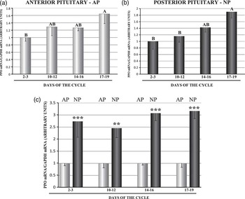
In AP, the expression levels of PPO mRNA were 1.6 times higher (P<0.05) on days 17 to 19 of the cycle than on days 2 to 3, and in NP, PPO gene expression was 1.9 and 1.7 times more pronounced (P<0.05) on days 17 to 19 than on days 2 to 3 and 10 to 12, respectively. The differences between the remaining stages of the estrous cycle were not significant in AP and NP (Figures 1a and b). The PPO gene was more highly expressed (P<0.01) in NP than in AP during the entire cycle (Figure 1c).
A comparison of prepro-orexin (PPO) mRNA expression determined by quantitative real-time PCR in porcine anterior (a) and posterior (b) pituitary glands between days 2 to 3, 10 to 12, 14 to 16 and 17 to 19 of the estrous cycle and (c) between anterior and posterior pituitary glands on days 2 to 3, 10 to 12, 14 to 16 and 17 to 19 of the cycle. Results are means±s.e.m. (n=5). Bars with different superscripts are significantly different. Uppercase letters indicate P<0.05; **P<0.01, ***P<0.001.

Western blotting
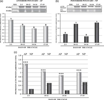
In AP, PPO protein content was from about 1.4 to 1.5 times significantly (P<0.05) higher on days 2 to 3 than in the remaining stages of the estrous cycle (Figure 2a). In NP, PPO protein was observed from about 2.1 to 3.2 times in greater abundance (P<0.05) on days 10 to 12 and 17 to 19 than on days 2 to 3 and 14 to 16 of the cycle (Figure 2b). Contrary to the PPO gene, the expression of protein was higher (P<0.001) in AP than in NP during all phases of the estrous cycle (Figure 2c).
A comparison of prepro-orexin (PPO) protein concentration determined by Western blotting analysis in porcine anterior (a) and posterior (b) pituitary glands between days 2 to 3, 10 to 12, 14 to 16 and 17 to 19 of the estrous cycle and (c) between anterior and posterior pituitary glands on days 2 to 3, 10 to 12, 14 to 16 and 17 to 19 of the cycle. Upper panels: representative immunoblots (molecular marker (MM)); lower panels: densitometric analysis of PPO protein relative to actin protein. Values are expressed as means±s.e.m. in arbitrary optical density units (n=5). Bars with different superscripts are significantly different. Uppercase letters indicate P<0.05; ***P<0.001.

Fluorescent immunohistochemistry
OXA and OXB proteins were localized in the porcine AP and NP on days 2 to 3, 10 to 12, 14 to 16 and 17 to 19 of the cycle. No staining was detected in the porcine pituitary when 0.01 M PBS or rabbit universal negative control was used instead of primary antibodies (Supplementary Figure S1).
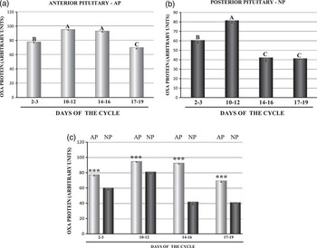
In AP, OXA immunoreactivity was from about 1.2 to 1.3 times higher (P<0.05) on days 10 to 12 and 14 to 16 of the estrous cycle than on days 2 to 3 and 17 to 19. The lowest (P<0.05) OXA protein concentrations were noted during the follicular phase of the estrous cycle (Figure 3a). In NP, from about 1.3 to 2.0 times markedly higher (P<0.05) expression of OXA protein was observed on days 10 to 12, and the lowest (P<0.05) expression levels were reported on days 14 to 16 and 17 to 19 (Figure 3b). Similarly to the PPO protein, the expression of OXA protein was higher (P<0.001) in AP than in NP throughout the entire cycle (Figure 3c).
A comparison of orexin A (OXA) protein immunoreactivity in porcine anterior (a) and posterior (b) pituitary glands between days 2 to 3, 10 to 12, 14 to 16 and 17 to 19 of the estrous cycle and (c) between anterior and posterior pituitary glands on days 2 to 3, 10 to 12, 14 to 16 and 17 to 19 of the cycle. Values are expressed as means±s.e.m. in arbitrary optical density units (n=5). Bars with different superscripts are significantly different. Uppercase letters indicate P<0.05; ***P<0.001.

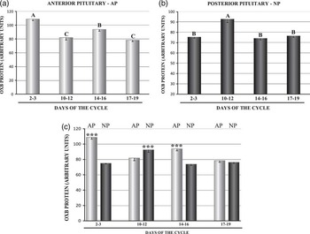
In AP, OXB immunoreactivity levels were highest (P<0.05; from about 1.2 to 1.4 times higher) on days 2 to 3 and lowest (P<0.05) on days 10 to 12 and 17 to 19 of the cycle (Figure 4a). Similarly to the OXA protein, the highest (P<0.05; from about 1.2 to 1.3 times higher) content of OXB protein was noted in NP on days 10 to 12 (Figure 4b). OXB protein concentrations were higher (P<0.05) in AP than in NP on days 2 to 3 and 14 to 16 of the cycle, whereas OXB protein expression levels were higher in NP than in AP on days 10 to 12 (Figure 4c).
A comparison of orexin B (OXB) protein immunoreactivity in porcine anterior (a) and posterior (b) pituitary glands between days 2 to 3, 10 to 12, 14 to 16 and 17 to 19 of the estrous cycle and (c) between anterior and posterior pituitary glands on days 2 to 3, 10 to 12, 14 to 16 and 17 to 19 of the cycle. Values are expressed as means±s.e.m. in arbitrary optical density units (n=5). Bars with different superscripts are significantly different. Uppercase letters indicate P<0.05; ***P<0.001.

Discussion
Our study was the first experiment to demonstrate the expression of PPO mRNA and PPO, OXA and OXB proteins in both AP and NP of cyclic gilts. To our knowledge, this is the first study that quantifies PPO gene expression and the concentrations of orexin peptides in the porcine pituitary during the estrous cycle. The expression of PPO mRNA was higher during the follicular phase in both AP and NP. The highest PPO protein content was noted on days 2 to 3 of the estrous cycle in AP and on days 10 to 12 and 17 to 19 in NP. In AP, OXA immunoreactivity was highest on days 10 to 12 and 14 to 16 of the cycle, and the lowest immunoreactivity levels were reported during the follicular phase of the cycle. In NP, OXA protein content was highest on days 10 to 12 and lowest on days 14 to 16 and 17 to 19. OXB immunoreactivity was highest on days 2 to 3 in AP and on days 10 to 12 in NP. The lowest content of OXB protein in AP was observed during mid-luteal and the follicular phases of the cycle. The above results indicate that gene and protein expression levels were determined by the stage of the estrous cycle. The differences between the expression patterns of PPO and OXA or OXB proteins may be due to the fact that the PPO is a common precursor of both orexins with different half-lives. It is worth noting that the expression of the PPO gene was lower in AP than in NP, whereas the highest PPO protein concentrations were noted in AP. The cause of the above variations remains unknown.
To date, OXA and OXB have been identified in the pituitaries of rats (Date et al., Reference Date, Mondal, Matsukura, Ueta, Yamashita, Kaiya, Kangawa and Nakazato2000) and humans (Blanco et al., Reference Blanco, Gallego, Garcia-Caballero, Dieguez and Beiras2003). The concentrations of both peptides, measured by reverse-phase HPLC, were 13-fold higher in NP than in AP of rats. OXB protein concentrations were higher than OXA levels in AP and NP. The results of immunohistochemical analyze revealed the presence of OXA and OXB fibers in NP, but orexin fibers were not determined in AP or the intermediate pituitary (Date et al., Reference Date, Mondal, Matsukura, Ueta, Yamashita, Kaiya, Kangawa and Nakazato2000). Immunoreactivity to OXA was detected in a high number of human lactotrophs, and it was determined in a smaller percentage of thyrotrophs, somatotrophs and gonadotrophs. Corticotrophs were negative for OXA. By contrast, OXB immune-positive cells were present in all corticotrophs, whereas the remaining cell types were negative. Immunostaining for both peptides was weak in human NP (Blanco et al., Reference Blanco, Gallego, Garcia-Caballero, Dieguez and Beiras2003). The above results indicate that NP is the main source of orexins in rats, whereas human lactotrophs are the main source of OXA and corticotrophs are the main source of OXB. The presence of OX1R and OX2R was demonstrated in rat (Date et al., Reference Date, Mondal, Matsukura, Ueta, Yamashita, Kaiya, Kangawa and Nakazato2000; Jöhren et al., Reference Jöhren, Neidert, Kummer, Dendorfer and Dominiak2001, Reference Jöhren, Bruggemann, Dendorfer and Dominiak2003), human (Blanco et al., Reference Blanco, Lopez, Garcia-Caballero, Gallego, Vazquez-Boquete, Morel, Senaris, Casanueva, Dieguez and Beiras2001), sheep (Zhang et al., Reference Zhang, Blache, Vercoe, Adam, Blackberry, Findlay, Eidne and Martin2005) and porcine pituitaries (Kaminski et al., Reference Kaminski, Smolinska, Nitkiewicz and Przala2010a). In rats, OX1R and OX2R genes were found in AP and NP. Intense hybridization signals were detected in the intermediate pituitary and AP, whereas weak signals were noted in NP. In AP, the expression of OX1R mRNA was higher than that of OX2R (Date et al., Reference Date, Mondal, Matsukura, Ueta, Yamashita, Kaiya, Kangawa and Nakazato2000). In humans, OX1R was also more abundant in AP than in NP, and in the human AP, the cells immunostained for OX1R were much more numerous than the cells expressing OX2R protein. In the human AP, OX1R protein was expressed by somatotrophs and OX2R by corticotrophs (Blanco et al., Reference Blanco, Lopez, Garcia-Caballero, Gallego, Vazquez-Boquete, Morel, Senaris, Casanueva, Dieguez and Beiras2001). In pigs, OX1R protein levels were higher in AP than in NP on days 10 to 12 of the cycle and OX2R protein expression was higher in AP thorough the entire cycle (Kaminski et al., Reference Kaminski, Smolinska, Nitkiewicz and Przala2010a). The presence of orexins in AP and NP provides evidence that the pituitary gland is capable of synthesizing the discussed hormones locally. The local production of orexins and the presence of specific receptors suggest that OXA and OXB exert a direct effect on the pituitary with possible autocrine and/or paracrine action of neuropeptides in the gland.
Some lines of evidence suggest that orexins regulate pituitary hormone secretion and control the pituitary branch of the hypothalamic–pituitary–gonadal (HPG) axis. In a study of prepubertal gilts, OXB stimulated the secretion of basal and GnRH-induced LH from AP cells in a dose-dependent manner (Barb and Matteri, Reference Barb and Matteri2005). In immature female rats, OXA stimulated the release of LH from primary culture of pituitary cells (Martynska et al., Reference Martynska, Wolinska-Witort, Chmielowska, Bik and Baranowska2011). OXA dose dependently inhibited the secretion of GnRH-induced LH from pituitary cells in proestrous females rats (Russell et al., Reference Russell, Small, Kennedy, Stanley, Seth, Murphy, Taheri, Ghatei and Bloom2001). Orexins control the secretion of LH not only by acting directly on pituitary cells, but also by influencing GnRH neurons at the hypothalamic level. Campbell et al. (Reference Campbell, Grove and Smith2003) reported that 75% to 85% of hypothalamic GnRH neurons in rats have direct synaptic contacts with orexin fibers and ∼85% of GnRH neurons express OX1R. In pigs, around 30% of GnRH neurons were in close contact with OXB immunoreactive fibers (Su et al., Reference Su, Lei, Zhang, Ning and Ping2008). An in vivo experiment demonstrated that OXA has a dual effect on LH release. The stimulatory effect was observed after injection into the rostral preoptic area, and the inhibitory effect was noted when OXA was injected into the medial preoptic area or the median eminence of the hypothalamus (Small et al., Reference Small, Goubillon, Murray, Siddiqui, Grimshaw, Young, Sivanesan, Kalamatianos, Kennedy, Coen, Bloom and Wilson2003). In vitro studies have shown that OXA can enhance GnRH release from hypothalamic explants harvested from intact male rats and proestrous female rats (Russell et al., Reference Russell, Small, Kennedy, Stanley, Seth, Murphy, Taheri, Ghatei and Bloom2001). A stimulatory effect of OXA on GnRH transcription and release from immortalized GT1-7 hypothalamic neurons was also observed by Sasson et al. (Reference Sasson, Dearh, White, Chappell and Mellon2006). In addition, i.c.v. injected orexins modulated LH secretion in an ovarian-steroid-dependent manner (Pu et al., Reference Pu, Jain, Kalra and Kalra1998; Small et al., Reference Small, Goubillon, Murray, Siddiqui, Grimshaw, Young, Sivanesan, Kalamatianos, Kennedy, Coen, Bloom and Wilson2003). Orexins enhanced LH release in estradiol- and progesterone-pretreated ovx rats, but they suppressed LH secretion in unprimed rats (Pu et al., Reference Pu, Jain, Kalra and Kalra1998). Both the stimulatory and inhibitory effects of OXA were antagonized by SB334867A, a selective OX1R antagonist (Small et al., Reference Small, Goubillon, Murray, Siddiqui, Grimshaw, Young, Sivanesan, Kalamatianos, Kennedy, Coen, Bloom and Wilson2003).
In addition to exerting an influence on the HPG axis, the orexin system is characterized by a gender-specific pattern of expression. Significantly higher levels of hypothalamic PPO mRNA (Jöhren et al., Reference Jöhren, Neidert, Kummer and Dominiak2002) and OXA protein (Taheri et al., Reference Taheri, Mahmoodi, Opacka-Juffry, Ghatei and Bloom1999) were reported in female than in male rats. OX1R mRNA expression in the pituitary was much higher in male than in female rats. The expression of OX1R mRNA was enhanced in the hypothalamus of female rats (Jöhren et al., Reference Jöhren, Neidert, Kummer, Dendorfer and Dominiak2001). The expression of PPO and orexin receptors genes as well as OXA concentrations in the hypothalamus were regulated by the estrous cycle, pregnancy, parturition and lactation (Russell et al., Reference Russell, Small, Kennedy, Stanley, Seth, Murphy, Taheri, Ghatei and Bloom2001; Wang et al., Reference Wang, Murata, Narita, Honda and Higuchi2003). Thus, it seems that gonadal steroid hormones may regulate the expression of orexin system. In ovx rats, the expression of the pituitary OX1R gene was suppressed after estradiol treatment (Jöhren et al., Reference Jöhren, Bruggemann, Dendorfer and Dominiak2003). On the other hand, in a study by Silveyra et al. (Reference Silveyra, Catalano, Lux-Lantos and Libertun2007b) the expression of PPO, OX1R and OX2R genes in rat hypothalami and pituitaries was enhanced during proestrous. In the case of both receptors, mRNA concentrations increased in rat ovaries during proestrous, and the above coincided with the hormone peaks (Silveyra et al., Reference Silveyra, Lux-Lantos and Libertun2007a). These data are in line with our present and recent findings that indicate that the expression of orexins, orexin receptors genes and proteins fluctuate throughout the estrous cycle. In our previous study, higher expression levels of OX1R and OX2R genes and proteins in porcine hypothalamic structures during the follicular phase could suggest that estrogens have a stimulatory effect on both receptors (Kaminski et al., Reference Kaminski, Smolinska, Nitkiewicz and Przala2010b). In the porcine pituitary, enhanced levels of OX1R mRNA during the luteal phase and higher concentrations of PPO and OX2R mRNAs during the follicular phase of the cycle imply that OX1R gene expression is stimulated by progesterone, whereas the expression of PPO and OX2R genes is influenced by estradiol (Kaminski et al., Reference Kaminski, Smolinska, Nitkiewicz and Przala2010a; present study). Similarly, variation in the expression of OX1R and OX2R genes in the porcine ovary during luteal and follicular phases of the cycle may imply that progesterone and estradiol affect orexin receptor expression (Nitkiewicz et al., Reference Nitkiewicz, Smolinska, Przala and Kaminski2010). Summarizing, the above studies clearly suggest the impact of the physiological hormonal milieu on the orexinergic system.
Conclusions
In this study, the expression of the PPO gene and the presence of PPO, OXA and OXB proteins were determined in the porcine pituitary. mRNA and protein concentrations are influenced by the phase of the estrous cycle. Our present and previous findings expand our knowledge of the role played by orexin system in the control of the pituitary gland.
Acknowledgment
This research was supported by National Science Centre (project no: NN308122134) and by Ministry of Science and Higher Education (project no: 528-0206.0805).