Introduction
Living cetaceans primarily use auditory cues to hunt and communicate, because the physical properties of water restrict the use of their other senses (Pihlström Reference Pihlström, Thewissen and Nummela2008). Their ear is highly adapted for underwater hearing at either infrasonic frequencies in mysticetes or ultrasonic frequencies in odontocetes (e.g., Ketten Reference Ketten1997, Reference Ketten, Au, Popper and Fay2000; Nummela et al. Reference Nummela, Thewissen, Bajpai, Hussain and Kumar2007; Churchill et al. Reference Churchill, Martinez-Caceres, de Muizon, Mnieckowski and Geisler2016; Ekdale Reference Ekdale2016a,Reference Ekdaleb; Park et al. Reference Park, Fitzgerald and Evans2016). Previous studies of extinct cetaceans show that the first underwater hearing morphologies were acquired in archaeocetes, for example, Remingtonocetidae and Basilosauridae (Nummela et al. Reference Nummela, Thewissen, Bajpai, Hussain and Kumar2004, Reference Nummela, Thewissen, Bajpai, Hussain and Kumar2007), and it was proposed that the ancestor of the Neoceti (Mysticeti + Odontoceti) had low-frequency (but likely not infrasonic) hearing (Ekdale and Racicot Reference Ekdale and Racicot2015; Ekdale Reference Ekdale2016a,Reference Ekdaleb; Mourlam and Orliac Reference Mourlam and Orliac2017; Park et al. Reference Park, Evans, Gallagher and Fitzgerald2017a). Odontocetes have a high-frequency hearing range, and their inner ear is characterized by a short cochlear canal, a stiff basilar membrane with extensive bony support, and a reduced vestibular system, among other characteristics (Oelschläger Reference Oelschläger1986; Ketten and Wartzok Reference Ketten, Wartzok, Thomas and Kastelein1990; Ketten Reference Ketten, Au, Popper and Fay2000; Gutstein et al. Reference Gutstein, Figueroa-Bravo, Pyenson, Yury-Yañez, Cozzuol and Canals2014; Churchill et al. Reference Churchill, Martinez-Caceres, de Muizon, Mnieckowski and Geisler2016; Park et al. Reference Park, Fitzgerald and Evans2016; Costeur et al. Reference Costeur, Grohe, Aguirre-Fernandez, Ekdale, Schulz, Muller and Mennecart2018).
Riverine dolphins are a paraphyletic group that includes four extant species: Platanista gangetica (Platanistidae), Lipotes vexillifer (Lipotidae), Inia geoffrensis (Iniidae), and Pontoporia blainvillei (Pontoporiidae; e.g., de Muizon Reference de Muizon1988; Cassens et al. Reference Cassens, Vicario, Waddell, Balchowsky, Van Belle, Ding, Chen, R, Mohan, Simoes-Lopes, Bastida, Meyer, Stanhope and Milinkovitch2000; Fordyce and de Muizon Reference Fordyce, de Muizon, Mazin and De Buffrénil2001; Nikaido et al. Reference Nikaido, Matsuno, Hamilton, Brownell, Cao, Ding, Zuoyan, Shedlock, Fordyce, Hasegawa and Okada2001). They are a textbook example of convergent evolution (e.g., Geisler et al. Reference Geisler, McGowen, Yang and Gatesy2011), with their long rostrum, small eyes, and unfused cervical vertebrae, among other anatomic characteristics (Page and Cooper Reference Page and Cooper2017; Fordyce Reference Fordyce, Würsig, Thewissen and Kovacs2018; Rommel and Reynolds Reference Rommel, Reynolds, Würsig, Thewissen and Kovacs2018). Riverine dolphins also share acoustic characteristics, with a high-frequency narrow-banded click structure; high echolocation peak energy (>100 kHz) sounds; and periotic characteristics like a rounded, slender, and high pars cochlearis (Ketten and Wartzok Reference Ketten, Wartzok, Thomas and Kastelein1990; Gutstein et al. Reference Gutstein, Figueroa-Bravo, Pyenson, Yury-Yañez, Cozzuol and Canals2014). Generally, their cochleae have a nearly planar spiral in less than 2 full turns, longer outer bony lamina than most other odontocetes, and compressed cochlear ducts. However, some authors have suggested that cochlear morphology does not appear to be convergent (Park et al. Reference Park, Mennecart, Costeur, Grohé and Cooper2019; but see Costeur et al. Reference Costeur, Grohe, Aguirre-Fernandez, Ekdale, Schulz, Muller and Mennecart2018). Riverine species rely on echolocation sounds to sort their complex environment and detect small objects at shorter average distances than pelagic species (Ketten and Wartzok Reference Ketten, Wartzok, Thomas and Kastelein1990; Jensen et al. Reference Jensen, Rocco, Mansur, Smith, Janik and Madsen2013).
One living riverine species, P. gangetica, warrants special attention. This species is the sole extant representative of an otherwise diverse fossil group, the Platanistoidea (sensu de Muizon Reference de Muizon1987), whose contents and phylogenetic relationships are still debated (e.g., Fordyce and de Muizon Reference Fordyce, de Muizon, Mazin and De Buffrénil2001; Boersma et al. Reference Boersma, McCurry and Pyenson2017; Tanaka and Fordyce Reference Tanaka and Fordyce2017 and references therein; Viglino et al. Reference Viglino, Buono, Fordyce, Cuitiño and Fitzgerald2018a,Reference Viglino, Buono, Gutstein, Cozzuol and Cuitiñob, Reference Viglino, Gaetán, Cuitiño and Buono2020; Gaetán et al. Reference Gaetán, Buono and Gaetano2019; Bianucci et al. Reference Bianucci, de Muizon, Urbina and Lambert2020). The earliest fossil record of this group comes from the late Oligocene, reaching a peak in diversity by the early Miocene and a marked decrease by the middle–late Miocene (e.g., de Muizon Reference de Muizon1987; Fordyce and de Muizon Reference Fordyce, de Muizon, Mazin and De Buffrénil2001). Several clades are usually recovered within Platanistoidea: upper Oligocene outcrops from New Zealand record some of the oldest platanistoids, such as Waipatia spp., Otekaikea spp., and Awamokoa tokarahi (Waipatiidae; Fordyce Reference Fordyce1994; Tanaka and Fordyce Reference Tanaka and Fordyce2017 and references therein). Allodelphinids are recorded in lower Miocene outcrops of the eastern and western margins of the North Pacific (Boersma and Pyenson Reference Boersma and Pyenson2016; Kimura and Barnes Reference Kimura and Barnes2016). In Patagonia, lower Miocene outcrops (Cuitiño et al. Reference Cuitiño, Buono, Viglino, Farroni and Bessone2019) have produced two platanistoid species, the squalodelphinid Notocetus vanbenedeni and the platanistoid Aondelphis talen (Moreno Reference Moreno1892; Viglino et al. Reference Viglino, Buono, Gutstein, Cozzuol and Cuitiño2018b; Viglino Reference Viglino2019); and two putative members of this group, Phoberodon arctirostris and Prosqualodon australis (Viglino et al. Reference Viglino, Buono, Fordyce, Cuitiño and Fitzgerald2018a; Gaetán et al. Reference Gaetán, Buono and Gaetano2019). Lower Miocene outcrops in Italy, France, Peru, and the United States hold other squalodelphinid taxa; and Miocene outcrops of mainly Europe and the United States record other extinct Platanistidae species (Paleobiology Database 2020; e.g., Barnes et al. Reference Barnes, Kimura, Godfrey, Ruiz-Garcia and Shostell2010; Lambert et al. Reference Lambert, Bianucci and Urbina2014; Godfrey et al. Reference Godfrey, Barnes and Lambert2017). Platanista gangetica is a critically endangered (Braulik et al. Reference Braulik, Barnett, Odon, Islas-Villanueva, Hoelzel and Graves2015; Braulik and Smith Reference Braulik and Smith2019) species that possesses several unique morphological characteristics, such as pneumatized maxillary crests containing a pterygoid sinus extension, a long mandibular symphysis, and a closed optic foramen, among others (Anderson Reference Anderson1878; Jensen et al. Reference Jensen, Rocco, Mansur, Smith, Janik and Madsen2013). Curiously, the evolutionary patterns that led to Platanista's bizarre morphology are still unknown, but see Boersma et al. (Reference Boersma, McCurry and Pyenson2017).
There are several hypotheses and much uncertainty surrounding the most important question for Platanistoidea: what are the causes that led diversity to decrease significantly, resulting in a single extant riverine representative today? Some authors have proposed that platanistoids were ecologically replaced by delphinids (e.g., Fordyce and Barnes Reference Fordyce and Barnes1994; Hamilton et al. Reference Hamilton, Caballero, Collins and Brownell2001; Steeman et al. Reference Steeman, Hebsgaard, Fordyce, Ho, Rabosky and Nielsen2009); that the ancestor of platanistids inhabited epicontinental seas and remained in fluvial basins through the sea-level regression during the middle–late Miocene (e.g., Cassens et al. Reference Cassens, Vicario, Waddell, Balchowsky, Van Belle, Ding, Chen, R, Mohan, Simoes-Lopes, Bastida, Meyer, Stanhope and Milinkovitch2000; Hamilton et al. Reference Hamilton, Caballero, Collins and Brownell2001); or that the temperature decline after the middle Miocene climatic optimum negatively affected shallow-water species (e.g., Marx et al. Reference Marx, Lambert and Uhen2016). Similarly, other vertebrate groups also comprise a majority of extinct species and a few extant representatives, for example, the pygmy right whale (e.g., Marx et al. Reference Marx, Park, Fitzgerald and Evans2018), tuatara (e.g., Meloro and Jones Reference Meloro and Jones2012), coelacanth (e.g., Cavin and Guinot Reference Cavin and Guinot2014), and red panda (e.g., Salesa et al. Reference Salesa, Peigné, Antón, Morales and Glatston2011). These groups illustrate complex but interesting macroevolutionary patterns which need to be analyzed from different perspectives.
Despite the increasing amount of contemporary information on the phylogenetic relationships of platanistoids, the inner ear morphology and evolutionary pattern associated with this group have never been reported, and it is here described and analyzed for the first time. Thus, the goal of the present contribution is to characterize the inner ear morphology of some late Oligocene–early Miocene extinct marine platanistoids, compare them with their living riverine representative P. gangetica, and propose a scenario for the evolution of their morphology. The sample includes, for the first time, platanistoid species from the late Oligocene of New Zealand and early Miocene of Patagonia, a key time in the diversification and ecological expansion of the group. Having representatives of the most frequently recovered families within Platanistoidea allow us to study, on a broader scale, how the inner ear morphology of the group evolved. Our initial hypothesis is that extinct marine platanistoids did not have a derived inner ear like P. gangetica, and thus their morphology and inferred hearing abilities were more closely related to pelagic odontocete species. Therefore, P. gangetica would represent a unique morphological ecomorphotype specialized for the riverine environment it inhabits.
Materials and Methods
Institutional Abbreviations
AMNH, American Museum of Natural History, New York, USA; MNHN, Museum National d'Histoire Naturelle, Paris, France; MPEF-PV, Colección de Paleovertebrados, Museo Paleontológico “Egidio Feruglio,” Trelew, Argentina; NHMUK, Natural History Museum, London, United Kingdom; NMB, Natuurmuseum Brabant, Tilburg, Holland; NMVC, Museum Victoria Mammalogy Collection, Melbourne, Australia; OU, Geology Museum, University of Otago, Dunedin, New Zealand; USNM, Department of Paleobiology and Department of Vertebrate Zoology (Division of Mammals), National Museum of Natural History, Smithsonian Institution, Washington, D.C., USA.
Sample
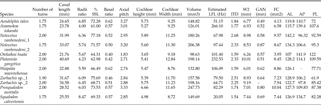
To perform this study, we obtained micro-computed tomography (microCT) scans of the periotics (the bone containing the inner ear) of 10 platanistoid genera by imaging osteological and palaeontological specimens from museum collections (total number of specimens = 11) and utilizing data from previous studies (Park et al. Reference Park, Mennecart, Costeur, Grohé and Cooper2018, Reference Park, Mennecart, Costeur, Grohé and Cooper2019; see Table 1). With these data, we reconstructed 3D models of the inner ears using the segmentation and thresholding editors in Avizo v. 9.0 (Visualization Sciences Group 2016) and then cleaned the resulting 3D models using Geomagic Wrap (3D Systems 2017).
List of specimens included in the present study, with their corresponding collection numbers and groups as used in statistical analyses.

For the purposes of this study, we have defined the clade Platanistoidea as including the genera Waipatia, Otekaikea, Awamokoa, Zarhachis, Notocetus, Aondelphis, and Platanista (Viglino et al. Reference Viglino, Buono, Fordyce, Cuitiño and Fitzgerald2018a,Reference Viglino, Buono, Gutstein, Cozzuol and Cuitiñob). These genera represent families that are usually recovered within this clade: Waipatiidae, Squalodelphinidae, and Platanistidae (for details on specimens used, see Table 1) (but note that some authors recovered waipatiids as stem Odontoceti; e.g., Lambert et al. Reference Lambert, de Muizon, Malinverno, Di Celma, Urbina and Bianucci2018; Bianucci et al. Reference Bianucci, de Muizon, Urbina and Lambert2020). For further details on the still-debated systematic and phylogenetic aspects of Platanistoidea, see, for example, Fordyce (Reference Fordyce1994), Lambert et al. (Reference Lambert, Bianucci and Urbina2014), Tanaka and Fordyce (Reference Tanaka and Fordyce2015, Reference Tanaka and Fordyce2017), Boersma and Pyenson (Reference Boersma and Pyenson2016), Boersma et al. (Reference Boersma, McCurry and Pyenson2017), Viglino et al. (Reference Viglino, Buono, Fordyce, Cuitiño and Fitzgerald2018a,Reference Viglino, Buono, Gutstein, Cozzuol and Cuitiñob, Reference Viglino, Gaetán, Cuitiño and Buono2020), Gaetán et al. (Reference Gaetán, Buono and Gaetano2019), and Bianucci et al. (Reference Bianucci, de Muizon, Urbina and Lambert2020), as well as references therein. The sample also included representatives of two putative platanistoids, Squalodon calvertensis and Prosqualodon australis (for details on the phylogenetic position of these taxa, please see de Muizon [Reference de Muizon1994] and Gaetán et al. [Reference Gaetán, Buono and Gaetano2019]).
For the statistical analyses (see “3D Geometric Morphometric and Statistical Analyses”), we also included one species as a representative of each of the remaining extant odontocete families (n = 9) for comparative purposes, as well as one archaeocete representative (n = 1) of the family Basilosauridae (see Table 1). This resulted in a total dataset of 21 specimens.
Measurements
We took multiple measurements of internal structures of the cochlea and semicircular canals postulated to be related to hearing physiology (Table 2; e.g., Ketten and Wartzok Reference Ketten, Wartzok, Thomas and Kastelein1990; Luo and Marsh Reference Luo and Marsh1996; Manoussaki et al. Reference Manoussaki, Chadwick, Ketten, Arruda, Dimitriadis and O'Malley2008; Ekdale and Racicot Reference Ekdale and Racicot2015; Mourlam and Orliac Reference Mourlam and Orliac2017). These were taken using the Measure, Slice, Spline Probe, and Surface Area Volume tools in Avizo, following the protocols of Park et al. (Reference Park, Fitzgerald and Evans2016). These measurements include: (1) cochlear height; (2) cochlear width; (3) number of turns; (4) cochlear canal length (measured along the midline); (5) extent of the secondary spiral lamina (SSL); (6) cochlear volume; (7) basal radius; (8) apical radius; (9) inter-turn distance (ITD); (10) shorter diameter of the basal turn (W2); (11) diameter of the spiral ganglion canal (GAN); (12) area of the fenestra cochleae (FC); and (13) anteroposterior (AP), (14) anterolateral (AL), and (15) posterolateral (PL) angles of the semicircular canals. The extension (%) of the SSL was measured by dividing the length of the cochlear canal at the apical-most point of the SSL by the total length of the cochlear canal, then multiplying by 100. The ITD is the thickness of the bony wall separating the first turn from the second turn at the proximal end of the basal turn (Ekdale and Racicot Reference Ekdale and Racicot2015). The GAN was measured in the first quarter turn, following Churchill et al. (Reference Churchill, Martinez-Caceres, de Muizon, Mnieckowski and Geisler2016) and Mourlam and Orliac (Reference Mourlam and Orliac2017).
Measurements of internal structures of the cochlea of the platanistoid and stem odontocete species included. SSL, secondary spiral lamina; LFL, low-frequency hearing limit; ITD, inter-turn distance; W2, shorter diameter of the basal turn; GAN, diameter of the spiral ganglion canal; FC, area of the fenestra cochleae; AL, anterolateral angle of the semicircular canals; AP, anteroposterior angle of the semicircular canals; PL, posterolateral angle of the semicircular canals.

From these measurements, we additionally calculated several previously established ratios, which together form a quantitative description of cochlear morphology (Ketten and Wartzok Reference Ketten, Wartzok, Thomas and Kastelein1990): (1) axial pitch (height of the cochlea divided by the number of turns); (2) the basal ratio (height of the cochlea divided by its basal diameter), which is negatively proportional to frequency (Ketten and Wartzok Reference Ketten, Wartzok, Thomas and Kastelein1990); and (3) radii ratio (radius of the cochlea at its base divided by the radius at its apex), which is strongly correlated with low-frequency hearing limits (Manoussaki et al. Reference Manoussaki, Chadwick, Ketten, Arruda, Dimitriadis and O'Malley2008). However, see Ritsche et al. (Reference Ritsche, Fahlke, Wieder, Hilger, Manke and Hampe2018) for issues with different measurement methods in this metric. Finally, we estimated the low-frequency hearing limit (LFL) for all specimens following Manoussaki et al. (Reference Manoussaki, Chadwick, Ketten, Arruda, Dimitriadis and O'Malley2008):
 $$f = {\rm \;}1507\,{\rm exp}\lpar {-0.578 [ {{\rm \rho }-1} \rsqb } \rpar $$
where f is the low-frequency hearing limit at 60 dB re 20 μPa in air and 120 dB re 1 μPa in water, and ρ is the radii ratio value. However, this equation was derived mainly from terrestrial mammals in air and should therefore be considered tentative (Manoussaki et al. Reference Manoussaki, Chadwick, Ketten, Arruda, Dimitriadis and O'Malley2008; Ekdale and Racicot Reference Ekdale and Racicot2015).
3D Geometric Morphometric and Statistical Analyses
We landmarked the digital models of the inner ear with one fixed landmark and 68 sliding semilandmark curves comprising a total of 625 landmarks (see Supplementary Material), using IDAV Landmark (Wiley 2005). The position of these curves followed the protocols of Costeur et al. (Reference Costeur, Grohe, Aguirre-Fernandez, Ekdale, Schulz, Muller and Mennecart2018).
Using these landmarks, we generated and analyzed three different datasets: cochlea and fenestra vestibuli landmark configuration for Platanistoidea only (P_Coch); the complete inner ear landmark configuration for Platanistoidea and comparative Odontoceti and Basilosauridae (P+O_All); cochlea and fenestra vestibuli landmark configuration for Platanistoidea and comparative Odontoceti and Basilosauridae (P+O_Coch). These datasets will facilitate comparisons of the inner ear morphology both among platanistoids and also with other cetacean clades, as well as revealing any potential evolutionary patterns. All of the statistical analyses were conducted for each of these datasets, as described in the following paragraphs.
After placing the landmarks as described, we conducted a generalized Procrustes analysis with sliding curves for the semilandmarks using the gpagen function. To test for the effect of size (allometry), we performed regressions of shape on size (log of centroid size) with 10,000 iterations using the procD.lm function. Both analyses used the geomorph package (Adams and Otárola-Castillo Reference Adams and Otárola-Castillo2013) in R software (R Core Team 2017). Given that there was no allometric effect (see “Results” section), Procrustes coordinates were used for subsequent statistical analyses.
Then, to explore variation of inner ear shape, we performed a principal component analysis (PCA) using the plotTangentSpace function of the geomorph package (Adams and Otárola-Castillo Reference Adams and Otárola-Castillo2013), and deformation 3D grids were generated to show shape variation among the PC axes. Next, we conducted canonical variate analysis (CVA), which is an ordination method that maximizes between-group variation relative to within-group variation. Deformation 3D grids were generated in these analyses as well. For these analyses, the CVA function of the Morpho package (Schlager Reference Schlager, Zheng, Li and Szekely2017) with 10,000 iterations was used. It should be noted that the basilosaurid specimen was excluded from this analysis, as it requires a minimum of two specimens. To evaluate the presence of a phylogenetic signal in shape variation of the inner ear in our sample, we performed the physignal function of the geomorph package (Adams and Otárola-Castillo Reference Adams and Otárola-Castillo2013) with 1000 permutations. This function uses the Kmult-statistic (Adams Reference Adams2014) that measures similarity of trait values relative to a Brownian motion model of evolution. Finally, we plotted the first two PC axes including a phylogenetic hypothesis using the plotGMPhyloMorphoSpace function of the geomorph package (Adams and Otárola-Castillo Reference Adams and Otárola-Castillo2013) to show a possible evolutionary trajectory for inner ear shape variation. The phylogenetic hypothesis used follows Viglino et al. (Reference Viglino, Buono, Fordyce, Cuitiño and Fitzgerald2018a,Reference Viglino, Buono, Gutstein, Cozzuol and Cuitiñob) and Gaetán et al. (Reference Gaetán, Buono and Gaetano2019), given that it includes all platanistoid species used in our study, and we have thoroughly revised the coding of the matrix used in those publications.
Results
Descriptions of Cochlear Anatomy
Aondelphis talen
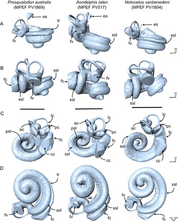
The cochlea of A. talen (Table 1) is well preserved, with the exception of minor damage to the basal half of the apical turn. The right cochlea completes approximately 1.75 turns (Fig. 1). The overall shape of the cochlea most closely resembles the spiral shape of ziphiids and differs from delphinids. In anterior view, the cochlear spiral is tall, a feature seen in mysticetes, archaeocetes, and Paleogene odontocetes. The fenestra cochleae is large relative to most extant odontocete taxa. There is no tympanal recess. The cochlear aqueduct is large, similar to Zygorhiza. In vestibular view, the first quarter of the basal turn is more loosely coiled than the remaining 1.5 turns. The apical-most half-turn slightly overlaps the basal turn, enclosing a small open space, rather than being fully closed like in stem cetacean taxa (e.g., Zygorhiza). The axial pitch is 3.27, and the basal ratio is 0.62 (Table 2). The secondary spiral lamina extends along the radial wall of the cochlear canal for 72% of its total length. The radii ratio is 6.85, a value that is higher than those calculated for the crown odontocetes (except ziphiids) (Park et al. Reference Park, Fitzgerald and Evans2016), resulting in an estimated low-frequency hearing limit of 51.15 Hz.
3D reconstruction of the cochlea of Patagonian stem Odontoceti and platanistoids analyzed in this study. A, anterior; B, lateral; C, dorsal; and D, vestibular views. Scale bars, 5 mm. Abbreviations: ac, anterior canal; ant, anterior; cc, cochlear canal; dor, dorsal; es, endolymphatic sac; fc, fenestra cochleae; fv, fenestra vestibuli; lat, lateral; lc, lateral canal; med, medial; pc, posterior canal; pos, posterior; psl, primary spiral lamina; ssl, secondary spiral lamina; tr, tympanal recess.

Notocetus vanbenedeni
The left cochlea of N. vanbenedeni (Table 1) is well preserved, with only minor damage to the cochlear canal and the semicircular canals. The cochlea completes approximately 2 turns (Fig. 1). The overall shape of the cochlea most closely resembles the lower, flatter spiral shape of delphinids (Ekdale and Racicot Reference Ekdale and Racicot2015). The fenestra cochleae is approximately the same size as in most extant odontocete taxa (Table 2). There is no tympanal recess. The cochlear aqueduct is large. In vestibular view, the cochlear canal is loosely coiled, displaying no overlap of the basal and apical turns. This produces a large open space at the apex of the cochlea. The axial pitch is 2.95, and the basal ratio is 0.52 (Table 2). The secondary spiral lamina extends along the radial wall of the cochlear canal for 77% of its total length. The radii ratio is 6.36, a value that is higher than those calculated for crown odontocetes (except ziphiids) (Park et al. Reference Park, Fitzgerald and Evans2016), resulting in an estimated low-frequency hearing limit of 67.98 Hz.
Prosqualodon australis
The left cochlea of P. australis (Table 1) is well preserved. The cochlea completes approximately 2 turns (Fig. 1). The overall shape of the cochlea most closely resembles that of stem odontocetes, being tightly coiled with a tall spire. As in A. talen, the cochlear spiral is tall in anterior view, a feature also seen in mysticetes, archaeocetes, and Paleogene odontocetes. The fenestra cochleae is the same size as in most extant odontocete taxa. There is a distinct tympanal recess (sensu Park et al. Reference Park, Marx, Fitzgerald and Evans2017c), with the scala tympani being inflated radially along the first half turn. The cochlear aqueduct is large. In vestibular view, the cochlear canal is tightly coiled, with the first half of the basal turn only slightly separated from the remaining 1.5 turns. The apical-most half-turn overlaps the cochlear canal dorsal to it. There is no open space at the apex of the cochlea. The axial pitch is 3.33, and the basal ratio is 0.57 (Table 2). The secondary spiral lamina extends along the radial wall of the cochlear canal for 74% of its total length. The radii ratio is 6.03, a value that is higher than those calculated for crown odontocetes (except ziphiids) (Park et al. Reference Park, Fitzgerald and Evans2016), resulting in an estimated low-frequency hearing limit of 82.29 Hz.
Awamokoa tokarahi
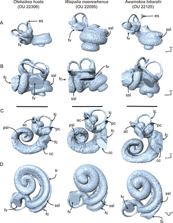
The cochlea of A. tokarahi (Table 1) is quite well preserved, although the cochlear canal has been slightly damaged by sediment, resulting in a more textured endocast, as opposed to a smooth one. The left cochlea completes approximately 1.75 turns (Fig. 2). The overall shape of the cochlea is similar to that of crown odontocetes, having a lower spiral than Zygorhiza and Waipatia, most closely resembling Otekaikea huata in its spiral shape. In anterior view, the cochlear spiral is tall, a feature seen in mysticetes, archaeocetes, and Paleogene odontocetes. The fenestra cochleae is approximately the same size as that of most extant odontocete taxa. There is no tympanal recess. The cochlear aqueduct is large, similar to Zygorhiza. In vestibular view, the first quarter of the basal turn is more loosely coiled than the remaining 1.5 turns. The apical-most half-turn slightly overlaps the basal turn, enclosing a small open space, rather than being fully closed like in stem cetacean taxa (e.g., Zygorhiza). The axial pitch is 3.01, and the basal ratio is 0.57 (Table 2). The secondary spiral lamina extends along the radial wall of the cochlear canal for 61% of its total length. The radii ratio is 4.00, a value that is similar to those calculated for the crown odontocetes (except ziphiids) (Park et al. Reference Park, Fitzgerald and Evans2016), resulting in an estimated low-frequency hearing limit of 266.10 Hz.
3D reconstruction of the cochlea of New Zealand platanistoids analyzed in this study. A, anterior; B, lateral; C, dorsal; and D, vestibular views. Scale bars, 5 mm. Abbreviations: ac, anterior canal; ant, anterior; cc, cochlear canal; dor, dorsal; es, endolymphatic sac; fc, fenestra cochleae; fv, fenestra vestibuli; lat, lateral; lc, lateral canal; med, medial; pc, posterior canal; pos, posterior; psl, primary spiral lamina; ssl, secondary spiral lamina; tr, tympanal recess.

Otekaikea huata
The cochlea of O. huata (Table 1) is well preserved, although the cochlear canal has been slightly damaged by sediment, resulting in a more textured endocast, as opposed to a smooth one. The left cochlea completes approximately 2 turns (Fig. 2). The overall shape of the cochlea is similar to that of crown odontocetes. It has a lower spiral than Zygorhiza and Waipatia, most closely resembling A. tokarahi in its spiral shape. The cochlear spiral is tall in anterior view, a feature seen in mysticetes, archaeocetes, and Paleogene odontocetes. The fenestra cochleae is approximately the same size as in most extant odontocete taxa. Unlike A. tokarahi, there is a tympanal recess present (sensu Park et al. Reference Park, Marx, Fitzgerald and Evans2017c), with the scala tympani being inflated radially along almost the entire first half turn. The cochlear aqueduct is small. In vestibular view, the first quarter of the basal turn is more loosely coiled than the remaining 1.5 turns. The apical-most half-turn slightly overlaps the basal turn, enclosing a small open space, rather than being fully closed like in stem cetacean taxa (e.g., Zygorhiza). The axial pitch is 1.83, and the basal ratio is 0.40 (Table 2). The secondary spiral lamina extends along the radial wall of the cochlear canal for 64% of its total length. The radii ratio is 5.67, a value that is similar to those calculated for the crown odontocetes (except ziphiids) (Park et al. Reference Park, Fitzgerald and Evans2016), resulting in an estimated low-frequency hearing limit of 101.4 Hz.
Waipatia maerewhenua
The cochlea of W. maerewhenua (Table 1) is quite well preserved, although the cochlear canal has been damaged by sediment, as were the other New Zealand species included in this study, resulting in a textured endocast, as opposed to a smooth one. The left cochlea completes approximately 2 turns (Fig. 2). The overall shape of the cochlea is similar to that of Zygorhiza. In anterior view, the cochlear spiral is tall, a feature seen in mysticetes, archaeocetes, and Paleogene odontocetes. The fenestra cochleae is approximately the same size as that of P. australis and Squalodon calvertensis. There is no tympanal recess. The cochlear aqueduct is small. In vestibular view, the first quarter of the basal turn is more loosely coiled than the remaining 1.5 turns, but not as loose as in Otekaikea or Awamokoa. The apical-most three-quarter turn slightly overlaps the basal turn, enclosing a very small open space, which is almost fully closed like in stem cetacean taxa (e.g., Zygorhiza). The axial pitch is 2.74, and the basal ratio is 0.62 (Table 2). The secondary spiral lamina extends along the radial wall of the cochlear canal for 66% of its total length. The radii ratio is 5.59, a value that is similar to those calculated for crown odontocetes (except ziphiids) (Park et al. Reference Park, Fitzgerald and Evans2016), resulting in an estimated low-frequency hearing limit of 106.1 Hz.
3D Geometric Morphometric Analyses
We initially analyzed the allometric effect on shape variation in each dataset, to test whether size had a significant effect. For all datasets, the effect of size was not significant (P+O_All p = 0.68; P+O_Coch p = 0.80; P_Coch p = 0.90), so all subsequent statistical analyses were performed on the Procrustes coordinates. We will focus our analyses and discussion on the results from the P+O_All dataset only; for the remaining dataset results, please see Supplementary Material.
Principal Component Analysis
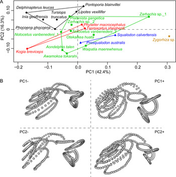
The PCA for the P+O_All dataset showed the highest variation in the first 12 PCs (95% total variance), but we will focus on the first two (58.7% total variance) (Fig. 3). When plotting PC 1 vs. PC 2, there is a slight overlap between Platanistoidea (Platanista gangetica) and Delphinida in both axes (for species included in each taxonomic group, see Table 1). The stem Odontoceti representatives overlap with the Platanistoidea morphospace in PC 1, while Zygorhiza sp. is on the extreme positive side of PC 1. Curiously, Physeter macrocephalus and Tasmacetus shepherdi overlap with platanistoids, while Kogia breviceps is on the negative extreme of PC 1. The morphospace occupied by the Platanistoidea is the largest of any of the groups included in this study. The main variables in PC 1 are the width of the cochlea, especially the basal turn, and the position and area of the fenestra vestibuli; on PC 2, the main shape variation includes the tightness of the cochlear spiral, the width of the basal turn, and the height of the cochlea.
A, Principal component analysis (PCA) plot of the complete inner ear (P+O_All dataset) of Basilosauridae and Odontoceti species included in our study. Green square, Platanistoidea; blue triangle, stem Odontoceti; red polygon, Physeteroidea + Ziphiidae; black circle, Delphinida; brown star, Basilosauridae. B, Extreme configurations for each PC axis. (Color online.)

The phylomorphospace (Fig. 4) shows that all extant lineages arose from a small portion of the morphospace of the first two axes and that the hypothetical shape of the most common recent ancestor of Odontoceti is more closely related to the Basilosauridae type. Also, W. maerewhenua, A. tokarahi, A. talen, and K. breviceps would have a more distant morphology with respect to their corresponding clades.
Phylomorphospace plot of the complete inner ear (P+O_All dataset) of Basilosauridae and Odontoceti species included in our study. Black squares denote the position of each taxa, while open squares represent the inferred positions of the internal nodes.

Canonical Variate Analysis
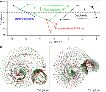
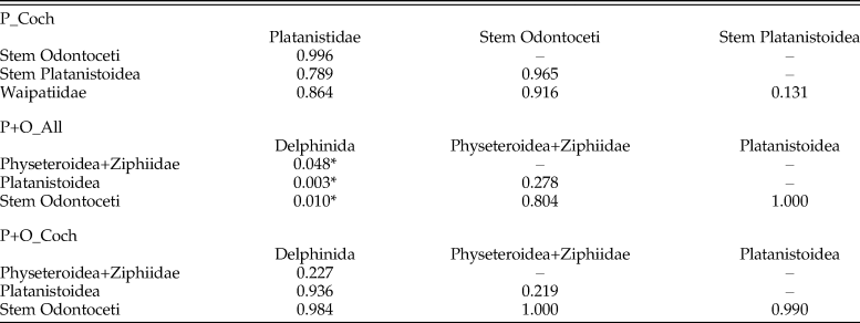
The CVA (excluding Zygorhiza sp.), on the other hand, further supports a clear separation between Platanistoidea and Delphinida, with an overlap between stem Odontoceti and Platanistoidea on CV 1 (Fig. 5). The Physeteridae and Ziphiidae species overlap on CV 1 with Platanistoidea as well. The first two CV axes account for 95% of the variation on the sample, so it has higher explanatory power than the PCA. The Mahalanobis distance between Delphinida and the remaining groups are statistically different (Table 3).
A, Canonical variate analysis (CVA) plot of the complete inner ear (P+O_All dataset) of all Odontoceti species included in our study (except Basilosauridae, see “Materials and Methods” for explanation). Green square, Platanistoidea; blue triangle, stem Odontoceti; red polygon, Physeteroidea + Ziphiidae; black circle, Delphinida. B, Extreme configurations for each CV axis, red dots represent the positive extreme and green dots the negative extreme configurations. (Color online.)

p-value of Mahalanobis distance of CVA analyses with the different datasets. Note that asterisks indicate a statistically significant distance (p < 0.05). Datasets: P_Coch, cochlea and fenestra vestibuli landmark configuration for Platanistoidea only; P+O_All, the complete inner ear landmark configuration for Platanistoidea and comparative Odontoceti and Basilosauridae; P+O_Coch, cochlea and fenestra vestibuli landmark configuration for Platanistoidea and comparative Odontoceti and Basilosauridae. The Basilosauridae specimen was excluded from these analyses, please see “Materials and Methods.”

Phylogenetic Signal
Both the complete inner ear as well as the cochlea datasets presented a statistically significant phylogenetic signal (P+O_All p = 0.01, K = 0.81; P+O_Coch p = 0.03, K = 0.65).
Comparisons
Among platanistoids, there is a wide morphological diversity recorded, and we could not identify a typical “platanistoid” inner ear (Figs. 3, 6). However, some common patterns were observed. All of the platanistoids studied have 1.75–2 number of turns, >60% extent of the secondary bony lamina, and 0.40–0.62 basal ratio. All of these measurements fall within the ranges reported for extant odontocete cochleae (Ketten and Wartzok Reference Ketten, Wartzok, Thomas and Kastelein1990; Ketten Reference Ketten, Webster, Fay and Popper1992; Park et al. Reference Park, Fitzgerald and Evans2016). The radii ratio for platanistoids (4.00–6.45) is within ranges reported for most odontocetes, except for A. talen and stem Odontoceti S. calvertensis, which are more similar to ziphiids, as well as archaeocete and mysticete values (Ekdale and Racicot Reference Ekdale and Racicot2015; Park et al. Reference Park, Fitzgerald and Evans2016). The fenestra cochleae is widest in stem odontocete P. australis and narrowest in A. talen, and all of the platanistoids have smaller fenestra cochleae than those reported for extant mysticetes (Park et al. Reference Park, Evans, Gallagher and Fitzgerald2017a). The cochlea is higher in stem odontocete Prosqualodon, and platanistoids Waipatia, Zarhachis, and Otekaikea, while it is wider in Notocetus, Platanista, Awamokoa, and Aondelphis. Cochlear volumes for platanistoids are within those reported for odontocetes (Ekdale and Racicot Reference Ekdale and Racicot2015; Park et al. Reference Park, Fitzgerald and Evans2016) and mammalodontid mysticetes (Park et al. Reference Park, Evans, Gallagher and Fitzgerald2017a), and again stem odontocete P. australis showed the highest value. The diameter of the spiral ganglion canal in platanistoids is higher than in toothed mysticetes but lower than in extant baleen whales (Park et al. Reference Park, Evans, Gallagher and Fitzgerald2017a), and their values are similar to the Type I morphology of Ketten and Wartzok (Reference Ketten, Wartzok, Thomas and Kastelein1990). The estimated low-frequency limit for all fossil platanistoids is distinctly lower than in P. gangetica but higher than those of mysticetes (Park et al. Reference Park, Fitzgerald and Evans2016, Reference Park, Evans, Gallagher and Fitzgerald2017a).
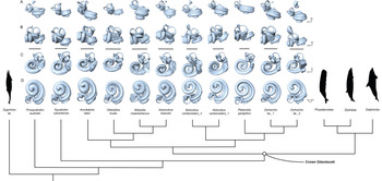
3D models representing stem Odontoceti and Platanistoidea species used in our study in a reduced phylogenetic hypothesis (see “Materials and Methods” for details) in: A, anterior; B, lateral; C, dorsal; and D, vestibular views. The phylogenetic hypothesis used follows Viglino et al. (Reference Viglino, Buono, Fordyce, Cuitiño and Fitzgerald2018a,Reference Viglino, Buono, Gutstein, Cozzuol and Cuitiñob) and Gaetán et al. (Reference Gaetán, Buono and Gaetano2019). Silhouettes are from PhyloPic, by Chris Huh, used under a Creative Commons Attribution-ShareAlike 3.0 Unported License (http://creativecommons.org/licenses/by-sa/3.0). Scale bars, 5 mm.

Regarding the remaining odontocetes included in our sample, some brief comparisons are made here. In agreement with Park et al. (Reference Park, Mennecart, Costeur, Grohé and Cooper2019), but in contrast to Gutstein et al. (Reference Gutstein, Figueroa-Bravo, Pyenson, Yury-Yañez, Cozzuol and Canals2014) and Costeur et al. (Reference Costeur, Grohe, Aguirre-Fernandez, Ekdale, Schulz, Muller and Mennecart2018), we found that Platanista, Inia, Lipotes, and Pontoporia do not fall into the same position in the morphospace (Figs. 3, 4). Moreover, like Costeur et al. (Reference Costeur, Grohe, Aguirre-Fernandez, Ekdale, Schulz, Muller and Mennecart2018), we found no morphological similarity between Lipotes and Inia, although our sample is limited in extant representatives and might therefore be masking such characteristics, though a single specimen should be indicative of morphology (Martins et al. Reference Martins, Park, Racicot and Cooper2020). Therefore, although numerous common adaptations are reported in the skull and postcranial skeleton of riverine dolphins (Page and Cooper Reference Page and Cooper2017; Fordyce Reference Fordyce, Würsig, Thewissen and Kovacs2018; Rommel and Reynolds Reference Rommel, Reynolds, Würsig, Thewissen and Kovacs2018), in agreement with Park et al. (Reference Park, Mennecart, Costeur, Grohé and Cooper2019), we found no “river dolphin” convergent morphology for the inner ear. The pygmy sperm whale (K. breviceps) was consistently recovered in the morphospace as distantly positioned with respect to Physeter and the ziphiids included in our analyses (Figs. 3, 4). These results would support the different cochlear morphological regimes recently proposed by Park et al. (Reference Park, Mennecart, Costeur, Grohé and Cooper2019), the relationship between cochlear morphology and deep-diving capabilities suggested for the group, and the unique echolocation system of Kogia (Thornton et al. Reference Thornton, McLellan, Rommel, Dillaman, Nowacek, Koopman and Pabst2015).
Finally, the position of Zygorhiza sp. (Basilosauridae) on the extreme positive end of PC 1 and PC 2 (Fig. 3) on our analyses suggests that the cochlea was higher, more tightly coiled, and wider compared with the odontocete species included in our sample, characteristics that have been related to a low-frequency hearing capability (Ketten Reference Ketten, Webster, Fay and Popper1992; Ekdale and Racicot Reference Ekdale and Racicot2015). Current knowledge suggests that the common ancestor of Neoceti had low-frequency hearing (Ekdale and Racicot Reference Ekdale and Racicot2015), which was retained by stem mysticetes and even potentially some stem odontocetes (Park et al. Reference Park, Evans, Gallagher and Fitzgerald2017a; Racicot et al. Reference Racicot, Boessenecker, Darroch and Geisler2019).
Discussion
The scope and position of Platanistoidea remain controversial, but recent anatomic and phylogenetic analyses have increased our understanding of the group (e.g., Lambert et al. Reference Lambert, Bianucci and Urbina2014; Boersma et al. Reference Boersma, McCurry and Pyenson2017; Tanaka and Fordyce Reference Tanaka and Fordyce2017; Viglino et al. Reference Viglino, Buono, Fordyce, Cuitiño and Fitzgerald2018a,Reference Viglino, Buono, Gutstein, Cozzuol and Cuitiñob; Gaetán et al. Reference Gaetán, Buono and Gaetano2019; Bianucci et al. Reference Bianucci, de Muizon, Urbina and Lambert2020). We present, for the first time, qualitative and quantitative descriptions, comparisons, and analyses of the inner ear of fossil and extant platanistoids, allowing us to better understand how the hearing abilities (and by extension, echolocation abilities) of this group have evolved. This, in turn, has broadened our understanding of cochlear evolution in odontocetes as a whole.
Cochlear Evolution in Platanistoidea
Both descriptions and measurements taken indicate that all extinct platanistoids had high-frequency hearing broadly similar to that of extant odontocetes (Ketten and Wartzok Reference Ketten, Wartzok, Thomas and Kastelein1990; Ketten Reference Ketten, Webster, Fay and Popper1992; Park et al. Reference Park, Fitzgerald and Evans2016). Like all cetaceans, except for pakicetids, the semicircular canals are smaller than the cochlea, which is thought to be related to a reduced sensitivity to rotational movements (Spoor et al. Reference Spoor, Bajpai, Hussain, Kumar and Thewissen2002; Ketten and Wartzok Reference Ketten, Wartzok, Thomas and Kastelein1990; Ketten Reference Ketten, Webster, Fay and Popper1992, Reference Ketten, Au, Popper and Fay2000; Costeur et al. Reference Costeur, Grohe, Aguirre-Fernandez, Ekdale, Schulz, Muller and Mennecart2018). Despite having characteristically odontocete cochleae, there are some intra- and interclade differences between platanistoids and other extant odontocetes.
PCA describing the cochlear shape variation revealed that Platanistoidea has the largest area of morphospace in the first two PC axes of all groups, indicating a higher diversity in inner ear morphology than the other groups in our sample (Fig. 3). In accordance with the Type I morphology of high-frequency hearing by Ketten and Wartzok (Reference Ketten, Wartzok, Thomas and Kastelein1990), all platanistoids included in our study have 2 or fewer turns, as well as >60% extension of the secondary bony lamina. This morphological type was associated with nearshore or freshwater odontocetes of narrower frequency range (Ketten and Wartzok Reference Ketten, Wartzok, Thomas and Kastelein1990; Ketten Reference Ketten, Webster, Fay and Popper1992). Conversely, the basal ratio of platanistoids (except Otekaikea huata, Zarhachis sp._1, and Platanista gangetica) is within the range defined for the Type II cochlear morphology (Ketten Reference Ketten1997); it is higher than for xenorophids (Churchill et al. Reference Churchill, Martinez-Caceres, de Muizon, Mnieckowski and Geisler2016; Park et al. Reference Park, Fitzgerald and Evans2016) but lower than the value reported for an undescribed squalodontid (Luo and Eastman Reference Luo and Eastman1995). Therefore, marine extinct platanistoids inner ear morphology was not clearly associated with any known cochlear type (sensu Ketten and Wartzok Reference Ketten, Wartzok, Thomas and Kastelein1990) and suggests that this clade might have its own pattern.
The presence of a long secondary bony lamina and reduced number of turns in platanistoids, key characteristics of odontocete cochlear morphology (Ketten and Wartzok Reference Ketten, Wartzok, Thomas and Kastelein1990; Ketten Reference Ketten, Webster, Fay and Popper1992; Ritsche et al. Reference Ritsche, Fahlke, Wieder, Hilger, Manke and Hampe2018), supports the hypothesis that high-frequency hearing capabilities were acquired early in the evolution of toothed whales (Churchill et al. Reference Churchill, Martinez-Caceres, de Muizon, Mnieckowski and Geisler2016; Park et al. Reference Park, Fitzgerald and Evans2016). However, these morphological characteristics are already far more specialized than those seen in older and earlier-diverging odontocetes (Churchill et al. Reference Churchill, Martinez-Caceres, de Muizon, Mnieckowski and Geisler2016; Park et al. Reference Park, Fitzgerald and Evans2016; Racicot et al. Reference Racicot, Boessenecker, Darroch and Geisler2019). These adaptations likely represent an improved ability to navigate complex acoustic environments, which could be related to occupying more nearshore habitats, as has been hypothesized for several platanistoids (Dowsett and Wiggs Reference Dowsett and Wiggs1992; Fordyce Reference Fordyce1994; Gottfried et al. Reference Gottfried, Bohaska and Whitmore1994; Tanaka and Fordyce Reference Tanaka and Fordyce2015, Reference Tanaka and Fordyce2017; Cuitiño et al. Reference Cuitiño, Buono, Viglino, Farroni and Bessone2019), producing shorter-range echolocation signals (Madsen et al. Reference Madsen, Kerr and Payne2004; Johnson et al. Reference Johnson, Hickmott, Aguilar Soto and Madsen2008; Jensen et al. Reference Jensen, Rocco, Mansur, Smith, Janik and Madsen2013), or potentially feeding on smaller prey items. The latter has already been found to be correlated with skull shape (McCurry and Pyenson Reference McCurry and Pyenson2018), and we suggest that paleoecology is the primary driving factor here. It is also noteworthy that during the late Oligocene–early Miocene, a variety of rostra and associated tooth morphologies among platanistoids coexisted (Tanaka and Fordyce Reference Tanaka and Fordyce2017; Viglino et al. Reference Viglino, Buono, Fordyce, Cuitiño and Fitzgerald2018a, Reference Viglino, Gaetán, Cuitiño and Buono2020; Bianucci et al. Reference Bianucci, de Muizon, Urbina and Lambert2020), implying a range of feeding strategies existed within Platanistoidea, similar to extant odontocetes (Werth Reference Werth2006; Hocking et al. Reference Hocking, Marx, Park, Fitzgerald and Evans2017a,Reference Hocking, Marx, Park, Fitzgerald and Evansb). Together with the more-derived inner ear morphology found here, it might indicate that feeding and ecological niches were powerful factors acting on the evolution of echolocating abilities and diverse skull morphologies in early odontocetes (Gutstein et al. Reference Gutstein, Figueroa-Bravo, Pyenson, Yury-Yañez, Cozzuol and Canals2014; Mourlam and Orliac Reference Mourlam and Orliac2017; Costeur et al. Reference Costeur, Grohe, Aguirre-Fernandez, Ekdale, Schulz, Muller and Mennecart2018).
Some peculiarities are also worth discussing from our results. Consistently among all analyses, Zarhachis sp._1 is distantly located in a unique region of morphospace with respect to the remaining platanistoids (Figs. 3–5). It is unclear what separates this specimen from the others, as its overall morphology appears to be similar and measurements fall within the range observed for the group, or even for odontocetes (Ketten and Wartzok Reference Ketten, Wartzok, Thomas and Kastelein1990; Ketten Reference Ketten, Webster, Fay and Popper1992; Churchill et al. Reference Churchill, Martinez-Caceres, de Muizon, Mnieckowski and Geisler2016; Park et al. Reference Park, Fitzgerald and Evans2016). Stem odontocete Squalodon calvertensis and platanistoid Aondelphis talen possess the lowest limit of hearing frequency among our sample (Table 2), higher than Zygorhiza and closely related to the values reported for some mysticetes (Park et al. Reference Park, Evans, Gallagher and Fitzgerald2017a). For both species, basal ratio and extent of secondary bony lamina fall within odontocete ranges reported (Ketten and Wartzok Reference Ketten, Wartzok, Thomas and Kastelein1990; Ketten Reference Ketten, Webster, Fay and Popper1992; Park et al. Reference Park, Fitzgerald and Evans2016). In the phylomorphospace, these species do not cluster together and are actually on opposite sides of the PC and CV axes (Figs. 3–5). Therefore, S. calvertensis and A. talen seem to have had a unique morphology either related to a wider range of frequencies heard or to a shorter and lower total frequency range than the remaining platanistoids compared. Future analyses including a wider sample of stem Neoceti or other measurements related to hearing frequency might help explain the pattern observed.
Finally, it is worth mentioning that Physeter and Tasmacetus overlapped within the Platanistoidea morphospace (Fig. 3). As outlined by Park et al. (Reference Park, Mennecart, Costeur, Grohé and Cooper2019), these extant species share an open bony contact (i.e., not closed or fused) between the tympanoperiotic and the skull, unique middle-ear morphology, and the presence of a tympanal recess on the cochlea. On the other hand, Platanista is also characterized by having an open bony suture between the periotic and the skull (Anderson Reference Anderson1878). It could be possible to hypothesize then, that the shared plesiomorphic characteristic of an open bony suture between the tympanoperiotic and the skull in all these taxa is reflected in the cochlear morphology and thus results in the observed overlap on the morphospace. A thorough understanding of the consequences of an open bony contact between the tympanoperiotic and the skull in the underwater hearing capabilities in extant odontocetes is needed to fully comprehend and estimate them in extinct species.
Odontocetes from the Lower Miocene of Patagonia
Among the Patagonian specimens, inner ear morphology varied, indicating differential hearing abilities. The platanistoid Notocetus vanbenedeni shows the most derived cochlear shape (Figs. 4, 6), in accordance with its phylogenetic position as more closely related to P. gangetica (e.g., Lambert et al. Reference Lambert, Bianucci and Urbina2014, Reference Lambert, de Muizon, Malinverno, Di Celma, Urbina and Bianucci2018; Viglino et al. Reference Viglino, Buono, Fordyce, Cuitiño and Fitzgerald2018a,Reference Viglino, Buono, Gutstein, Cozzuol and Cuitiñob; Bianucci et al. Reference Bianucci, de Muizon, Urbina and Lambert2020). Notocetus and Platanista share a loosely coiled and wide cochlea, a low number of widely separated turns, and >60% extension of the secondary bony lamina. They additionally have a relatively high cochlear volume compared with most other odontocetes in our sample (Table 2; Figs. 1, 6). It is also worth noting that both Notocetus specimens included in our study were not clustered together in the morphospace (Figs. 3–4; see also Supplementary Material), possibly indicating a wider disparity in cochlear morphology for the species than previously thought or potentially even separate taxa, given that intraspecific variation is thought to be low in odontocetes (Martins et al. Reference Martins, Park, Racicot and Cooper2020). Ontogenetic effects on the shape of the cochlea can be discarded, as the cochlea in odontocetes attains adult proportions in early gestation stages (Thean et al. Reference Thean, Kardjilov and Asher2017).
Prosqualodon australis is the first Patagonian stem odontocete known to possess a tympanal recess (sensu Park et al. Reference Park, Marx, Fitzgerald and Evans2017c; Figs. 1, 6). This structure has been speculatively suggested as a specialization for lower-frequency sound reception via a vibroacoustic duct mechanism (March et al. Reference March, Brown, Gray, Curthoys, Wong and Higgins2016; Park et al. Reference Park, Fitzgerald and Evans2017b). However, its function is currently unclear, and its widespread and apparently stochastic distribution across multiple mysticete and odontocete lineages (Ekdale Reference Ekdale2016b; Park et al. Reference Park, Fitzgerald and Evans2017b,Reference Park, Marx, Fitzgerald and Evansc, Reference Park, Mennecart, Costeur, Grohé and Cooper2019) makes it uncertain whether there is a single function for the structure. Therefore, we cannot hypothesize on the effects of the tympanal recess in underwater hearing capabilities, and a thorough understanding of this structure and function in living cetaceans is decidedly needed. Nonetheless, the cochlear morphology of P. australis indicates that this species had high-frequency hearing, similar to extant odontocetes.
As for the platanistoid A. talen, the spiral and tightly coiled cochlea most closely resembles that of ziphiids and stem cetaceans (see “Descriptions of Cochlear Anatomy”; Figs. 2, 3, 5). The tighter coiling is also a feature shared with the New Zealand taxa Awamokoa and Waipatia, both of which are very close to A. talen in the morphospace. Morphologically, the periotics of all three genera share some characteristics; for example: a vestigial dorsal crest (plesiomorphic characteristic), an elongated anterior bullar facet and a larger and squared posterior bullar facet, and an anterointernal sulcus on the dorsal surface of the anterior process (Fordyce Reference Fordyce1994; Tanaka and Fordyce Reference Tanaka and Fordyce2017; Viglino et al. Reference Viglino, Buono, Gutstein, Cozzuol and Cuitiño2018b). Curiously, the inferred low-frequency limit is similar to that of stem mysticetes, Eschrichtius robustus, and balaenopterids (Ketten Reference Ketten1997; Ekdale and Racicot Reference Ekdale and Racicot2015; Park et al. Reference Park, Evans, Gallagher and Fitzgerald2017a); significantly lower than the frequency range for extant odontocetes, except ziphiids (e.g., Ketten and Wartzok Reference Ketten, Wartzok, Thomas and Kastelein1990; Ketten Reference Ketten1997, Reference Ketten, Au, Popper and Fay2000; Park et al. Reference Park, Fitzgerald and Evans2016); and even lower than in P. gangetica (Kelkar et al. Reference Kelkar, Dey, Deshpande, Choudhary, Dey and Morisaka2018). Therefore, the inferred low-frequency limit for A. talen would be in agreement with its more basal inner ear morphology (Fig. 6), and we could then hypothesize that either this species had a wider frequency range, or it was uniquely less specialized in high-frequency hearing than its coeval Patagonian platanistoids and stem Odontoceti species.
Anatomic and phylogenetic studies have suggested that Patagonian platanistoids and stem odontocete species employed niche separation to maximize resource use (Viglino et al. Reference Viglino, Buono, Fordyce, Cuitiño and Fitzgerald2018a,Reference Viglino, Buono, Gutstein, Cozzuol and Cuitiñob, Reference Viglino, Gaetán, Cuitiño and Buono2020; Gaetán et al. Reference Gaetán, Buono and Gaetano2019). The morphological differences among Patagonian platanistoid cochleae would support this hypothesis. Diverse feeding apparatuses and hearing abilities could have resulted in avoidance of direct ecological competition between species (Madsen et al. Reference Madsen, Kerr and Payne2004; Johnson et al. Reference Johnson, Hickmott, Aguilar Soto and Madsen2008; Jensen et al. Reference Jensen, Rocco, Mansur, Smith, Janik and Madsen2013). Future studies integrating other feeding and auditory anatomic structures of Patagonian species, as well as paleoenvironmental information (e.g., Galatius et al. Reference Galatius, Olsen, Steeman, Racicot, Bradshaw, Kyhn and Miller2018), might help in analyzing and proposing paleoecological interactions between them more clearly.
Late Oligocene Platanistoids from New Zealand
Similar to the Patagonian platanistoids studied, species included here from New Zealand also show some variation in their inner ear morphology. Awamokoa tokarahi, Waipatia maerewhenua, and O. huata shared an endocast slightly damaged by sediment or diagenetic mineral precipitation, possibly related to their depositional environment in the upper Oligocene Otekaike Limestone Fm. (Fordyce Reference Fordyce1994; Tanaka and Fordyce Reference Tanaka and Fordyce2015, Reference Tanaka and Fordyce2017). Also, the three species share a tall cochlear spiral, similar to stem cetaceans, mysticetes, and stem odontocetes (e.g., Ekdale and Racicot Reference Ekdale and Racicot2015; Park et al. Reference Park, Fitzgerald and Evans2016, Reference Hocking, Marx, Park, Fitzgerald and Evans2017a; Ritsche et al. Reference Ritsche, Fahlke, Wieder, Hilger, Manke and Hampe2018), as well as a long secondary spiral lamina similar to that of extant odontocetes (e.g., Ketten and Wartzok Reference Ketten, Wartzok, Thomas and Kastelein1990; Ketten Reference Ketten, Webster, Fay and Popper1992; Costeur et al. Reference Costeur, Grohe, Aguirre-Fernandez, Ekdale, Schulz, Muller and Mennecart2018; Ritsche et al. Reference Ritsche, Fahlke, Wieder, Hilger, Manke and Hampe2018).
When comparing the inner ear morphology of all New Zealand platanistoids, W. maerewhenua represents a more basal morphology (e.g., size of fenestra cochleae, high overlap of apical turn, less loosely coiled basal turn, narrow cochlea; Fig. 6), and O. huata is the only species that presents a tympanal recess, like the Patagonian stem Odontoceti P. australis (Fig. 2). On the other hand, A. tokarahi shares an estimated low-frequency hearing similar to that of the extant representative P. gangetica, while the limit in W. maerewhenua and O. huata is lower (Table 2). Nonetheless, all species are within the estimated ranges for odontocetes (e.g., Ketten and Wartzok Reference Ketten, Wartzok, Thomas and Kastelein1990; Ketten Reference Ketten1997, Reference Ketten, Au, Popper and Fay2000; Park et al. Reference Park, Fitzgerald and Evans2016).
In summary, platanistoids from the late Oligocene of New Zealand present inner ear morphological characteristics that indicate they had high-frequency hearing, such as a short cochlear canal length, 2 or fewer turns, long secondary bony lamina, and low radii ratio (Fig. 6; Table 2). This inference is also supported by their skull morphology, where osteological correlates of the nasal sac and pterygoid sinus system were described (Fordyce Reference Fordyce1994; Tanaka and Fordyce Reference Tanaka and Fordyce2015, Reference Tanaka and Fordyce2017). Even though these species were probably less specialized than modern odontocetes (Figs. 2, 6), they could hear high-frequency sounds, supporting an early-acquired ability for odontocetes during their evolutionary history (e.g., Churchill et al. Reference Churchill, Martinez-Caceres, de Muizon, Mnieckowski and Geisler2016; Park et al. Reference Park, Fitzgerald and Evans2016; Mourlam and Orliac Reference Mourlam and Orliac2017). Continuing efforts to study hearing abilities in fossil odontocetes are key to understanding how and when early odontocetes diversified and expanded in marine and riverine habitats, as modern forms do today.
Survival of Platanista
The combination of Type I and Type II features observed in the fossil platanistoid cochleae in this study only partially supports our initial hypothesis that they would have a cochlear morphology dissimilar to extant Platanista. However, the cochlea of Platanista was consistently overlapping with delphinoid odontocetes in the morphospace (Figs. 3–6), indicating it has the most derived morphology among platanistoids. It was also always distant in the morphospace from its sister taxon Zarhachis sp., particularly from Zarhachis sp._1 (Figs. 3–5; also see Supplementary Material), consistent with a >9 Myr separation between these genera (Paleobiology Database 2020). Platanista gangetica is characterized by loosely coiled turns in a wide and short cochlea (Fig. 6). The echolocation signals of this species were reported to be adapted to search for prey at short distances and in lower-frequency levels than marine delphinids (Jensen et al. Reference Jensen, Rocco, Mansur, Smith, Janik and Madsen2013).
Cochlear morphology, in addition to their highly autapomorphic maxillary crests that might aid in sound directionality during echolocation (Jensen et al. Reference Jensen, Rocco, Mansur, Smith, Janik and Madsen2013), overall skull morphology (Anderson Reference Anderson1878; Purves and Pilleri Reference Purves and Pilleri1973; Pilleri et al. Reference Pilleri, Gihr, Purves, Zbinden and Kraus1976), and pterygoid sinus system (Fraser and Purves Reference Fraser and Purves1960), demonstrates that Platanista still represents a unique and bizarre morphological type among platanistoids, and indeed even Cetacea, reflecting a disparate and highly specialized echolocation system. Interestingly, Boersma et al. (Reference Boersma, McCurry and Pyenson2017) found that crest enlargement first evolved in platanistoids in the Oligocene, but only the latest-surviving platanistids (including Platanista) possessed highly pneumatized crests, thought to function in sound production (Purves and Pilleri Reference Purves and Pilleri1973; Jensen et al. Reference Jensen, Rocco, Mansur, Smith, Janik and Madsen2013; Boersma et al. Reference Boersma, McCurry and Pyenson2017). Here, we propose that the evolution of these anatomic adaptations would have allowed platanistids to survive during the course of the late Neogene when other platanistoid taxa went extinct, resulting in the unique morphotype of P. gangetica. Moreover, the significant reorganization of the major tributaries of the Ganges and Indus Rivers around 0.5 Myr ago affected these species populations’ distribution and represented a strong ecological factor of adaptation (Braulik et al. Reference Braulik, Barnett, Odon, Islas-Villanueva, Hoelzel and Graves2015). New findings of fossil platanistoids, particularly from the middle–late Miocene, will help test these hypotheses.
Conclusions
The inner ear anatomy of fossil Platanistoidea is described for the first time. Our comparisons reveal there is no “typical” platanistoid cochlear type, as the group displays a disparate range of cochlear anatomies that possess a combination of basal and derived features. Nonetheless, we found that early-diverging platanistoid cochleae have morphological characteristics that are clearly better adapted for high-frequency hearing than the coeval earliest stem Odontoceti, thus indicating a rapid evolution of this functional complex. Notocetus and Platanista share a loosely coiled and wide cochlea, a low number of widely separated turns, and >60% extension of the secondary bony lamina. Stem odontocete Prosqualodon australis and platanistoid Otekaikea huata are the only species in our sample that present a tympanal recess in their cochleae, of yet unknown specific function in the underwater hearing mechanism in cetaceans. Also, Aondelphis talen's basal inner ear morphology and inferred frequency range indicate it had lower high-frequency hearing than other odontocetes. Finally, Platanista has the most-derived cochlear morphology, a finding in accordance with its unique skull anatomy, adding to evidence that it is an outlier within the group. The cochlear morphology of Platanista is also in accordance with its distant position in the morphospace from its sister genus Zarhachis. The evolution of this singular sound production system within Platanistidae may have facilitated the survival of Platanista to the present day.
Acknowledgments
We would like to thank M. Cipollone from Y-TEC (Argentina) for all his help during microCT scanning of Patagonian specimens. We also would like to thank the following organizations for financial support: Cetacean Society International (CSI) to M.V. and M.R.B.; Sociedad Argentina para el Estudio de Mamíferos (SAREM) to M.V.; Ernst Mayr Grant from the Museum of Comparative Zoology of Harvard University (MCZ) to M.V.; grants in aid of research from the Society for Marine Mammalogy (SMM) to M.V. and M.G.; and Agencia Nacional de Promoción Científica y Tecnológica (grant number PICT 0792) to M.R.B. T.P. was funded by a Marie Skłodowska-Curie Individual Fellowship (748167/ECHO), ERC Starting Grant (677774/TEMPO) and a Leverhulme Research Project Grant (RPG-2019-323). We also thank the Paleobiology Database, particularly M. D. Uhen, for the extremely useful online cetacean records information. Finally, we would also like to thank editor N. Froebisch and two reviewers for their helpful comments that improved this article.
Data Availability Statement
Data available from the Dryad Digital Repository: https://doi.org/10.5061/dryad.g4f4qrfpm.