Introduction
Mucoceles are epithelium-lined cavities containing mucus which completely occupy a paranasal sinus,Reference Gavioli, Grasso, Carinci, Amoroso and Pastore1 most commonly the frontal and ethmoidal. They occur due to obstruction of the sinus ostium, usually as a result of chronic sinusitis, trauma or endoscopic sinus surgery.Reference Picavet and Jorissen2 An online literature search using PubMed confirmed that the current report is the first documented case of frontal mucocele arising due to an ectopic maxillary sinus tooth.
Case report
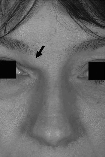
A 39-year-old woman presented with an intermittent, painful swelling above her right eyelid (Figure 1). She had a history of recurrent, right-sided maxillary sinusitis, treated unsuccessfully with steroid nasal sprays and oral antibiotics.
Clinical photograph demonstrating a right frontal mucocele (arrow).

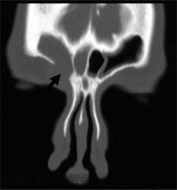
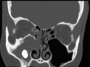
A computed tomography (CT) scan of the patient's sinuses showed a soft tissue attenuation filling the right frontal sinus and recess, with a well defined bony defect in the sinus floor and soft tissue extending into the superomedial aspect of the right orbit anteriorly (Figure 2). These findings were consistent with a ‘Pott's puffy tumour’ or frontal mucocele. Of note was an ectopic, upper right third molar tooth in the right maxillary antrum as a focus for the patient's maxillary sinusitis and consequent frontal mucocele (Figure 3).
Computed tomogram showing a bony defect in the floor of the right frontal sinus (arrow), through which the mucocele extended into the superomedial aspect of the right orbit.

Computed tomogram demonstrating an ectopic tooth in the right maxillary antrum.

Under general anaesthesia, a Caldwell–Luc procedure was performed, as the tooth was embedded too laterally to be amenable to endoscopic removal. It was apparent that the upper right third molar tooth had developed in an abnormal position in association with its alveolar bone. The tooth was surgically removed. The patient made an uneventful recovery, and her frontal mucocele and symptoms of maxillary sinusitis resolved without further intervention.
Discussion
This is the first documented report of a frontal mucocele arising due to an ectopic maxillary sinus tooth. Even more surprising is the fact that the condition disappeared following removal of the offending tooth. Left untreated, erosion of the bony sinus wall by such a mucocele can lead to anterior cranial fossa and/or orbital invasion. Endoscopic sinus surgery is an effective conventional treatment,Reference Khong, Malhotra, Wormald and Selva3 and the external approach, with or without obliteration of the sinus, also has a useful role.Reference Gavioli, Grasso, Carinci, Amoroso and Pastore1 It is clear in this case that maxillary sinusitis predisposed to the development of the frontal mucocele, as a result of the ectopic tooth.
Ectopic teeth, in general, are common. Numerous sites for ectopic teeth have been reported, including the nasal cavity, chin, mandibular condyle, coronoid process, palate and maxillary sinus.Reference Hasbini, Hadi and Ghafari4–Reference Kim, Kim, Chae, Hwang, Lee and Lee10 Ectopic eruption of teeth into areas other than the oral cavity is thought to be due to an abnormal interaction between the oral epithelium and the underlying mesenchyme.Reference Goh9 Rhinosinusitis resulting from ectopic teeth in the maxillary sinusReference Di Felice and Lombardi7, Reference Goh9, Reference Buyukkurt, Tozoglu, Aras and Yolcu11 or in the floor of both nasal cavitiesReference Sokolov, Jecker and Roth12 has been reported, due to osteomeatal complex blockage by the teeth, and has been demonstrated clearly by CT scanning.Reference Jude, Horowitz and Loree13
This is the first case report to describe a frontal mucocele arising secondary to maxillary sinusitis caused by an ectopic maxillary tooth. It demonstrates the ease with which this patient's symptoms resolved following surgical removal of the aberrant ectopic tooth, thus avoiding an endoscopic sinus procedure. The case also highlights a more unusual cause of frontal mucocele.
• This is the first documented case of a frontal mucocele arising in association with maxillary sinusitis due to an ectopic maxillary sinus tooth
• The ectopic tooth was removed via a sub-labial approach, following which the maxillary sinusitis and the frontal mucocele resolved