Bovine mastitis, an inflammatory reaction of the mammary gland that is usually caused by a microbial infection, is recognised as the most costly disease for dairy farmers and industry (Halasa et al. Reference Halasa, Nielen, Huirne and Hogeveen2009). Intramammary infection (IMI) reduces the number and activity of epithelial cells and consequently contributes to decrease milk production, mostly associated with tissue damage. The mechanisms responsible for mammary epithelium and tissue damage during mastitis include bacterial and host factors that are still not well defined. However, it is recognised that these mechanisms can lead to cell death both by necrosis and apoptosis, which can be distinguished by morphological, biochemical, and molecular changes in dying cells (Zhao & Lacasse, Reference Zhao and Lacasse2008).
Staphylococcus aureus, one of the most prevalent contagious pathogenic bacteria causing IMI, often presents as chronic subclinical mastitis characterised by poor response to conventional antibiotic therapy (Zecconi & Scali, Reference Zecconi and Scali2013). Classical histopathological studies have shown that S. aureus IMI causes necrosis of secretory tissues that are replaced by nonsecretory tissue (Zhao & Lacasse, Reference Zhao and Lacasse2008). In vitro studies have demonstrated that S. aureus can induce apoptosis of neutrophils (Sladek et al. Reference Sladek, Rysanek, Ryznarova and Faldyna2005), lymphocytes (Slama et al. Reference Slama, Sladek, Rysanek and Langrova2009), macrophages (Wang et al. Reference Wang, Zhou and He2010) and bovine mammary epithelial cells (Bayles et al. Reference Bayles, Wesson, Liou, Fox, Bohach and Trumble1998; Hu et al. Reference Hu, Cui, Tao, Xiu, Wang and Wang2014). These effects can compromise the antimicrobial immune response, lead to indirect tissue damage and facilitate bacterial spread in vivo (Zhao & Lacasse, Reference Zhao and Lacasse2008). However, there is only limited information about cell's death mechanisms in mammary tissue of cows with chronic naturally acquired S. aureus IMI (Dallard et al. Reference Dallard, Baravalle, Ortega, Ruffino, Heffel and Calvinho2008).
During involution the mammary gland undergoes extensive tissue remodelling involving coordinated cell death, extracellular matrix degradation and adipose tissue regeneration (Norgaard et al. Reference Norgaard, Theil, Sorensen and Sejrsen2008). S. aureus IMI acquired during lactation can persist during the involution period (Sordillo & Nickerson, Reference Sordillo and Nickerson1988) and therefore alters physiological processes, including mammary cell apoptosis (Dallard et al. Reference Dallard, Baravalle, Ortega, Ruffino, Heffel and Calvinho2008). There are two major pathways leading to apoptosis: an intrinsic and an extrinsic pathway. The intrinsic pathway involves the mitochondria and can be triggered by intracellular stressors such as oxidants. Mitochondrial release of cytochrome c, promotes apoptosome formation which in turn actives pro-caspase-9, triggering downstream effectors, such as caspase-3, -6, and -7 (Yin, Reference Yin2000). The extrinsic pathway is induced by activation of death receptors located on the cell surface. The Fas antigen is a member of the tumour necrosis factor receptor superfamily that can mediate apoptotic cell death in various cells types (Kikuchi et al. Reference Kikuchi, Kuribayashi and Imajoh-Ohmi2014), including bovine mammary epithelial cells (Hu et al. Reference Hu, Cui, Tao, Xiu, Wang and Wang2014).
Members of the Bcl-2 family are known to be involved in the regulation of apoptosis. This family includes anti-apoptotic and pro-apoptotic proteins, like Bcl-2 and Bax respectively (Colitti, Reference Colitti2012). Bax accumulation on mitochondrial membrane precedes activation of caspase-3, an apoptosis executor (Zhang et al. Reference Zhang, Heim and Meyhack1998). Variations in the expression of pro- and anti-apoptotic proteins network and signalling events involved in cell apoptosis in the bovine mammary gland have not yet been fully explored (Zarzynska & Motyl, Reference Zarzynska and Motyl2008).
Mammary gland remodelling depends on a dynamic equilibrium between mitosis and apoptosis. Previous studies have quantified cell turnover in uninfected (Norgaard et al. Reference Norgaard, Theil, Sorensen and Sejrsen2008) and S. aureus-infected bovine mammary gland (Dallard et al. Reference Dallard, Baravalle, Ortega, Ruffino, Heffel and Calvinho2008), using different cellular proliferation markers in situ. Dallard et al. (Reference Dallard, Baravalle, Ortega, Ruffino, Heffel and Calvinho2008) using Ki-67 and PCNA (proliferation cell nuclear antigen) immunostaining, demonstrated that chronic S. aureus IMI in cows induce a significant increment of epithelial and stromal cells proliferation during early involution, which could be associated with repair mechanisms directed to limit host injury. The objective of this study was to determine whether S. aureus chronic IMI influences expression of proteins related to regulation of proliferation and apoptosis processes and proliferation/apoptosis index during active involution in bovine mammary gland.
Materials and methods
Animals and experimental design
Twenty-one Holstein non-pregnant cows in late lactation (weeks 31–36) from the Rafaela Experiment Station of INTA herd were included in the study. Cows were from parity 3 to 5, milked twice daily, produced an average of 25 kg milk/d during lactation and an average of 12 kg milk/d before interruption of lactation. Cows with similar lactation number were included in each experimental group (uninfected and S. aureus-infected). All procedures used in this study were approved by the Ethics and Security Committee of the Facultad de Ciencias Veterinarias, UNL and consistent with the Guide for the Care and Use of Agricultural Animals in Agricultural Research and Teaching (Federation of Animal Science Societies, 2010).
The infection status of mammary quarters was determined within 6 months before initiation of the experiment and confirmed 20 and 3 d before cessation of milking. Infections were naturally acquired either in the previous dry period or during the first 2 months of the lactation preceding initiation of the study. Cows with S. aureus mastitis were selected based on results of monthly somatic cell counts (SCC > 250 × 103 cells/ml) and subsequent testing by bacteriological analysis of quarter milk. A quarter was considered to be infected if S. aureus was isolated from two consecutive samples. Infected quarters were randomly selected from cows showing at least two quarters infected with S. aureus. Only animals with subclinical IMI at the time of milking interruption were included. Uninfected quarters were selected from cows free of infection at the time of sampling with negative results of bacteriological analysis of quarters and SCC less than 250 × 103 cells/ml. Cows were slaughtered at 7, 14 and 21 d after cessation of milking at a local abattoir and samples for histological analysis were taken. The experimental unit of study was the mammary quarter. Uninfected (n = 8) and S. aureus-infected (n = 8) mammary quarters in each time of sampling (7, 14 and 21 d) were included. At every sampling time 7 animals were used (3 with uninfected quarters and 4 with two quarters infected with S. aureus). According to the eligibility criterion used for experimental units' selection, 21 cows were included in the study (12 cows with at least two quarters infected with S. aureus and 9 uninfected cows).
Tissue sample preparation
Immediately after cows were slaughtered, three tissue samples were taken from selected mammary quarters from three zones following previous descriptions (Dallard et al. Reference Dallard, Ortega, Iguzquiza, Salvetti, Quaino and Calvinho2010). Zone 1 upper limit of the gland cistern; zone 2 approximately midway between the upper limit of the gland cistern and the dorsal boundary of the mammary gland at a depth of 4 cm (lobulo-alveolar zone) and zone 3 near to the dorsal boundary of the mammary gland (adjacent to abdomen). Tissue samples were fixed in 4% neutral buffered formalin, for 8 h and then washed in phosphate-buffered saline (PBS). For light microscopy, fixed tissues were dehydrated and embedded in paraffin wax. Sections (5 µm) were mounted on slides previously treated with 3-aminopropyltriethoxysilane (Sigma-Aldrich, St. Louis MO, USA) and assigned for use in immunohistochemistry (IHC) procedures. Additional sections of mammary tissues were transferred into a freezing vial, weighed and placed in liquid nitrogen for Western blot assays.
Immunohistochemistry
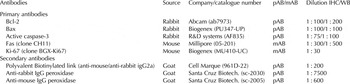
For IHC, the streptavidin–biotin immunoperoxidase method was performed as described by Andreotti et al. (Reference Andreotti, Pereyra, Baravalle, Renna, Ortega, Calvinho and Dallard2014). Briefly, sections were dewaxed, hydrated and subjected to microwave pretreatment for antigen retrieval. Endogenous peroxidase activity was blocked with 1% H2O2 in methanol, and non-specific binding was blocked with 10% normal goat serum (Sigma-Aldrich Corp., St. Louis, MO). Tissue sections were incubated with the primary antibodies (Table 1) for 18 h at 4 °C. Slides were washed with PBS and incubated with the biotinylated second antibody for 30 min at 25 °C (Table 1). Detection was by a streptavidin-peroxidase solution (CytoScan HRP Detection System, Cell Marque), with 3·3-diaminobenzidine (Liquid DAB-Plus Substrate Kit; Zymed, San Francisco, CA, USA) as chromogen. The sections were then counterstained with Mayer haematoxylin, dehydrated, and mounted. Negative control sections in which the primary antibody was replaced by non-immune rabbit serum were included. To exclude the possibility that endogenous peroxidase activity was unsuccessfully blocked, some sections were incubated with DAB reagent alone. Serial sections of bovine mammary tissue similarly processed and with known reactivity for the antigen were used as positive controls in each assay to normalise the image analysis.
Antibodies, source and commercially purchased reagents used

pAB, polyclonal antibody; mAB, monoclonal antibody; IHC, immunohistochemistry; WB, Western blot
Antigen/antibody specificity
To test specificity of the primary antibodies that detect apoptotic-related proteins (Bcl-2, Bax, Fas and active caspase-3), bovine mammary tissue extracts were separated in SDS-polyacrylamide gels (15% resolving gel). Proteins were transferred onto nitrocellulose membranes (Amersham, Buckinghamshire, UK), blocked for 1 h 30 min in 5% nonfat milk in TBS containing 0·05% Tween-20 (Sigma-Aldrich Corp., St. Louis, MO) and then incubated overnight at 4 °C with specific primary antibodies. Following washing, membranes were treated for 1 h 30 min with secondary peroxidase-conjugated antibody (Table 1). Immunopositive bands were visualised with a chemiluminescent detection kit (ECL-Plus; GE-Amersham, Buckinghamshire, UK). Ki-67 specificity has been previously tested in bovine tissue in previous studies (Dallard et al. Reference Dallard, Baravalle, Andreotti, Ortega, Neder and Calvinho2011).
TUNEL assay
The epithelial and stromal apoptotic cells were visualised using a terminal deoxynucleotidyl transferase (TdT)-mediated X-dUTP nick end labelling (TUNEL) assay. A commercial kit (ApopTag Plus Peroxidase, Chemicon International, USA) was used following previous descriptions (Dallard et al. Reference Dallard, Baravalle, Ortega, Ruffino, Heffel and Calvinho2008). Briefly, after deparaffinisation and hydration, slides were incubated with 20 µg/ml of proteinase K (Dako North America Inc., Carpinteria CA, USA) for 15 min at 25 °C. Tissue sections were quenched in 3% H2O2 in PBS, incubated in equilibration buffer for 10 min, and then with terminal deoxynucleotidyl transferase (TdT) for 60 min at 37 °C. Sections were washed with stop wash buffer and then incubated with antidigoxigenin-peroxidase for 30 min at 25 °C. Tissue sections were washed in PBS and then incubated with DAB for colour development. Sections were washed, counterstained with Mayer haematoxylin, dehydrated and mounted.
Image analysis
Image analysis was performed using the Image Pro-Plus 3·0·1 system (Media Cybernetics, Silver Spring, MA). Images were digitised with an Olympus C5060 digital camera mounted on a conventional light microscope (Olympus BH-2; Olympus Co., Tokyo, Japan) using objective magnification of 40×. Image resolution was set to 1200 × 1600 pixels. Each pixel of the image corresponded to 0·13 µm2 at the respective magnification and each field represented a tissue area of 0·031 mm2. The system captured each image and automatically corrected for background. This prevented differential readings due to different lighting conditions.
Immunohistochemistry and TUNEL-positive cells quantification
The area labelled by each antibody reaction (for Bcl-2, Bax, Fas and active caspase-3) was quantified as a percentage of total explored area, using a segmentation colour method previously described (Andreotti et al. Reference Andreotti, Pereyra, Baravalle, Renna, Ortega, Calvinho and Dallard2014). The darkest from the main colour (brown) of the immunolabelled area was identified and assigned to ‘black’ in the black-and-white mask generated by the software, definitively separating it from the rest of the colours in the picture. These values were verified and normalised, with the controls carried across 2 or 3 runs of IHC, and the same region (verified by image comparison) was used for calibration. The black percentage area was calculated for at least 50 images for each slide with parenchymal and stromal structures.
Quantification of TUNEL-positive cells in the parenchyma (epithelium) and stroma of mammary gland was measured as described previously (Dallard et al. Reference Dallard, Baravalle, Ortega, Ruffino, Heffel and Calvinho2008). Cells were classified as unlabelled epithelial, unlabelled stromal, labelled epithelial or labelled stromal cells. Cells were counted as epithelial if were part of the mammary parenchyma, which included epithelial and myoepithelial cells. All connective tissue cells were classified as stromal, including fibroblasts, adipocytes and endothelial cells. Labelled brown nuclei were readily visible and a cell was classified as labelled when the nuclear staining was at least twice as intense as the background. Quantification of epithelial and stromal cells labelled with anti-Ki-67 protein was performed in a similar manner as for TUNEL. A minimum of 1000 cells in at least 20 microscopic fields at 40× magnification were counted in each section. Epithelial and stromal cells showing an intense nuclear staining were quantified and expressed as a percentage of the total number of cells counted per section. The balance between proliferating and apoptotic cells was estimated using the ratio between percentages of Ki-67 immunopositive cells and percentages of TUNEL positive cells following previous descriptions (Dallard et al. Reference Dallard, Baravalle, Andreotti, Ortega, Neder and Calvinho2011).
Statistical analysis
A statistical software package (SPSS 11·0 for Windows; SPSS, Inc., Chicago, IL) was used to perform statistical analysis. Data were analysed using the general linear model (GLM) procedure. The model tested for main effects of infection status, time and the interaction between the infection status*time. Individual means were compared by t-test. The level of significance was set at P < 0·05. Data are presented as means of three mammary gland zones obtained from the same mammary quarter ± sem (standard error of the mean).
Results
Immunohistochemistry for Bcl-2, Bax, Fas, and active caspase-3
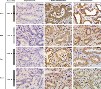
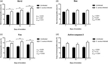
The positive immunohistochemical reaction with each protein (Bcl-2, Bax, Fas and active caspase-3) was detected by brown cytoplasmic staining and evaluated by the immunopositive area. Representative patterns of immunostaining of Bcl-2, Bax, Fas and active caspase-3 in S. aureus-infected and uninfected quarters are shown in Fig. 1 and a quantitative analysis of the expression of these proteins from immunohistochemical analysis is shown in Fig. 2.
Representative images of Bcl-2, Bax, Fas and active caspase-3 (Casp3) immunostaining in S. aureus-infected quarters and uninfected controls at 14 d of involution are shown in the right panels. Verification of antibody specificity by Western blot analysis of mammary tissue homogenates and negative controls for immunostaining are shown in the left two panels, respectively. Magnification is the same for all panels (400×) and is indicated by 25 µm bars.

Relative expression (measured as percentage of immunopositive area) of (a) Bcl-2, (b) Bax, (c) Fas and (d) active caspase-3 in S. aureus-infected quarters and uninfected controls at 7, 14 and 21 d of involution. Values represent the mean ± sem. Asterisks represent statistically significant differences between S. aureus-infected and uninfected quarters at each sampling time (***P < 0·001). The overall effects of infection status (IS), time (T) and their interaction are also shown.

Immunostaining for Bcl-2 protein was observed in all tissue sections, ranging from weak to intense, and was mainly associated with the mammary parenchyma structures. Positive reaction was observed around the nuclei and in the cytoplasm of epithelial and stromal cells. The patterns of localisation for Bcl-2 in mammary tissue were similar in both S. aureus-infected than uninfected quarters (Fig. 1). Protein expression of Bcl-2 was higher in S. aureus-infected than uninfected quarters (P < 0·001) and was affected by time of sampling (P < 0·001; Fig. 2a). There was a significant interaction between infectious status and time of sampling (P = 0·01; Fig. 2a), being percentage of immunopositive area for Bcl-2 higher in S. aureus-infected than in uninfected quarters at 7, 14 and 21 d of involution (P < 0·001; Fig. 2a).
Expression of Bax protein was primarily associated with the epithelium of the alveoli and ducts of the mammary parenchyma (Fig. 1). Protein expression of Bax was higher in S. aureus-infected than uninfected quarters during all evaluated times (P < 0·001) and was not affected by time of sampling (P > 0·05; Fig. 2b). There was no interaction between infectious status and time of sampling (P > 0·05; Fig. 2b).
Immunostaining for Fas protein was observed in all tissue sections, ranging from weak to intense, and was associated with structures of the mammary parenchyma and stroma (Fig. 1). Positive reaction was in the cytoplasm of epithelial and stromal cells. Protein expression of Fas was higher in S. aureus-infected than uninfected quarters (P < 0·001) and was affected by time of sampling (P < 0·001; Fig. 2c). There was a significant interaction between infectious status and time of sampling (P < 0·001; Fig. 2c), being percentage of immunopositive area for Fas higher in S. aureus-infected than in uninfected quarters at 7, 14 and 21 d of involution (P < 0·001; Fig. 2c).
Immunostaining for active caspase-3 was observed in the cellular nucleus and cytoplasm of epithelial and stromal cells. The patterns of localisation for active caspase-3 in mammary tissue were similar in both S. aureus-infected than uninfected quarters (Fig. 1). Protein expression of active caspase-3 was higher in S. aureus-infected than uninfected quarters during al evaluated times (P < 0·001) and was not affected by time of sampling (P > 0·05; Fig. 2d). There was no interaction between infectious status and time of sampling (P > 0·05; Fig. 2d).
Antibody specificity
Western blot recognition of Bcl-2, Bax, Fas, active caspase-3 in mammary homogenates from S. aureus-infected quarters is summarised in Fig. 1. Western blot analysis revealed positive bands of appropriate sizes for each of the proteins studied. Antibodies against the proteins detected a single band at 25 kDa corresponding to Bcl-2 mature form; a single band at 21 kDa for Bax; a single band at 43 kDa for Fas; and a single band at 17 kDa for active caspase-3.
Detection of apoptosis in situ (TUNEL)
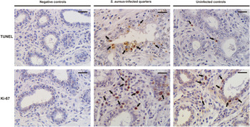
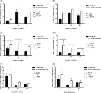
Apoptotic cells were found both in alveolar epithelium and mammary stroma, distributed as single cells rather than clusters. Apoptotic events were evident as TUNEL-positive cells juxtaposed with healthy cells (Fig. 3). Percentage of apoptotic cells in mammary parenchyma and stroma are summarised in Fig. 4. Percentage of apoptosis in parenchymal cells were affected by the infection status (P < 0·05); being higher in S. aureus-infected quarters compared with uninfected quarters during all evaluated times. Moreover, percentage of apoptosis in parenchymal cells were affected by time of sampling (P < 0·001), showing the highest percentage of apoptotic cells at 14 d of involution both in uninfected and S. aureus-infected quarters. No interaction between infection status and time of sampling was detected (P > 0·05; Fig. 4a).
Representative images of apoptosis detection using TUNEL assay and Ki-67 immunostaining in S. aureus-infected quarters and uninfected controls at 14 d of involution. Black arrows indicate TUNEL and Ki-67 immunopositive cells. Negative controls for immunostaining are shown in the left panel. Magnification is the same for all panels (400×) and is indicated by 25 µm bars.

Percentage of apoptotic cells detected using TUNEL assay, percentage of proliferating cells by Ki-67 immunostaining and proliferation/apoptosis ratio in S. aureus-infected quarters and uninfected controls at different times of involution. (a) Percentage of TUNEL positive cells in mammary parenchyma and (b) stroma. (c) Percentage of Ki-67 positive cells in mammary parenchyma and (d) stroma. (e) Ratio between Ki-67 positive cells/TUNEL positive cells in mammary parenchyma and (f) stroma. Values represent the mean ± sem. Asterisks represent statistically significant differences between S. aureus-infected and uninfected quarters at each sampling time (*P < 0·05; **P < 0·01; ***P < 0·001). The overall effects of infection status (IS), time (T) and their interaction are also shown.

Percentage of apoptosis in stromal cells were higher in S. aureus-infected quarters compared with uninfected quarters during all evaluated times (P < 0·001). Moreover, percentage of apoptosis in stromal cells was affected by time of sampling (P < 0·001), showing the highest percentage of apoptotic cells at 14 d of involution both in uninfected and S. aureus-infected quarters. There was no interaction between infectious status and time of sampling (P > 0·05; Fig. 4b).
Cellular proliferation
Nuclei labelled for Ki-67 were evident in both parenchyma and stroma of mammary gland (Fig. 3). Percentage of proliferating cells in mammary parenchyma and stroma are summarised in Fig. 4. Regarding parenchymal cells, the percentage of Ki-67 labelled cells was higher in S. aureus-infected than uninfected quarters (P < 0·001) and was affected by time of sampling (P = 0·01, Fig. 3c). There was a significant interaction between infectious status and time of sampling (P = 0·01; Fig. 3c). Percentage of proliferating cells were higher in S. aureus-infected than in uninfected quarters at 7 (P = 0·01), 14 (P < 0·001) and 21 d of involution (P = 0·001, Fig. 3c).
Regarding stromal cells, the percentage of Ki-67 labelled cells were higher in S. aureus-infected than uninfected quarters (P < 0·001) and were affected by time of sampling (P < 0·05; Fig. 3d). There was a significant interaction between infectious status and time of sampling (P < 0·05; Fig. 3d). Percentage of proliferating cells were higher in S. aureus-infected than in uninfected quarters at 7 (P < 0·01), 14 (P = 0·001) and 21 d of involution (P < 0·05; Fig. 3d).
Proliferating to apoptotic cells index (Ki-67/TUNEL)
The ratio between percentage of Ki-67 immunopositive cells and percentage of TUNEL positive cells are shown in Fig. 4e, f. Regarding parenchymal cells, the proliferation/apoptosis ratio was affected by the time of sampling, showing at 7 d of involution the highest ratio both in S. aureus-infected and uninfected quarters (P < 0·001). In parenchymal cells, the proliferation/apoptosis ratio was not affected by IMI (P < 0·05, Fig. 4e) and there was no interaction between infectious status and time of sampling (P > 0·05 Fig. 4e).
Regarding stromal cells, the proliferation/apoptosis ratio was affected by the time of sampling showing at 7 d of involution the highest ratio both in S. aureus-infected and uninfected quarters (P < 0·001). Moreover, in stromal cells, the proliferation/apoptosis ratio was affected by infection presence, being higher in S. aureus-infected than uninfected quarters during all evaluated times (P = 0·01). No interaction between infection status and time of sampling was detected (P > 0·05; Fig. 4f).
Discussion
In the present study, the effect of chronic S. aureus IMI on apoptosis, selected apoptosis regulatory proteins expression, cell proliferation and proliferation-apoptosis ratio in bovine mammary glands during active involution was assessed. Variations in the expression of pro and anti-apoptotic proteins in bovine mammary gland and the exact role of genes that regulate apoptosis during IMI has not been fully explored (Dallard et al. Reference Dallard, Baravalle, Ortega, Ruffino, Heffel and Calvinho2008; Zhao & Lacasse, Reference Zhao and Lacasse2008). In addition, little information is available regarding signalling events involved in mammary cells apoptosis in vitro after S. aureus infection (Hu et al. Reference Hu, Cui, Tao, Xiu, Wang and Wang2014) and cell death mechanisms in S. aureus chronically infected mammary glands during active involution. In this regard, in previous studies from our laboratory using a lipopolysaccharide (LPS)-based biological response modifier or placebo (vehicle alone) infused in S. aureus chronically infected and uninfected bovine mammary glands during involution, we observed an increase in percentages of immune expression for Bax protein in parenchymal and stromal cells in S. aureus chronically infected placebo-treated quarters compared with uninfected placebo-treated quarters at 7, 14 and 21 d of mammary involution (Dallard et al. Reference Dallard, Baravalle, Ortega, Ruffino, Heffel and Calvinho2008). We conducted the present study in order to substantiate previous observations on placebo-treated quarters. In accord with earlier findings (Dallard et al. Reference Dallard, Baravalle, Ortega, Ruffino, Heffel and Calvinho2008) a significant effect of S. aureus IMI on percentages of immunostaining for Bax and Bcl-2 was observed, showing both proteins higher expression in S. aureus-infected than uninfected quarters at all involution stages studied. Moreover, for Bcl-2, a significant effect of time was observed, showing higher immunostaining percentage S. aureus-infected quarters compared with uninfected quarters at 7, 14 and 21 d with a progressive increase as involution progressed. These results indicate an over-expression of both proteins during active involution in quarters chronically infected with S. aureus compared with uninfected quarters, implying an increase in apoptotic cell death through the pro-apoptotic protein Bax, as well as apoptosis inhibition by progressive increase in anti-apoptotic protein Bcl-2. This finding suggests a host attempt to control the effects induced by IMI to maintain integrity of tissue affected by the inflammatory reaction. Although significance of increased apoptosis during S. aureus IMI at active involution is not fully understood, early studies have demonstrated that epithelial cells apoptosis induction upon infection with S. aureus may result in breakdown of the epithelial barrier that can favour bacterial access to deep tissues without stimulating bactericidal activities, while simultaneously being provided with a protective barrier against exogenous host immune defences and/or antibiotics (Bayles et al. Reference Bayles, Wesson, Liou, Fox, Bohach and Trumble1998).
Fas antigen is a member of the tumour necrosis factor receptor superfamily and can mediate apoptotic cell death in various cells types (Kikuchi et al. Reference Kikuchi, Kuribayashi and Imajoh-Ohmi2014). Song et al. (Reference Song, Sapi, Brown, Nilsen, Tartaro, Kacinski, Craft, Naftolin and Mor2000) carried out a study in healthy mice mammary glands and reported high levels of both Fas and FasL proteins and caspase-3 at day 1 after weaning, coinciding with appearance of apoptotic cells in ducts and glands. These authors suggested that Fas-FasL interaction could play an important role in mice mammary tissue normal remodelling. In the present study, Fas protein immunostaining was visualised in parenchymal cells cytoplasm and to a lesser extent in stromal cells both in S. aureus-infected and uninfected quarters. The localisation pattern observed coincided with that reported by Song et al. (Reference Song, Sapi, Brown, Nilsen, Tartaro, Kacinski, Craft, Naftolin and Mor2000) in mice mammary glands. Hu et al. (Reference Hu, Cui, Tao, Xiu, Wang and Wang2014) demonstrated that S. aureus induced apoptosis in pBMECs in a time- and dose-dependent manner via the Fas-FADD death receptor and subsequent activation of caspase-3 and caspase-8. In the present study Fas expression in mammary tissue was higher in S. aureus-infected than uninfected quarters at 7, 14 and 21 d of involution, suggesting an important role of this receptor in S. aureus chronically infected bovine mammary glands.
Caspase-3 belongs to a family of highly conserved cysteine proteases that mediate the course of apoptotic cell suicide. Activation of caspase cascades triggers apoptosis in certain bacteria-infected cells (Krzymińska et al. Reference Krzymińska, Szczuka and Kaznowski2012). As mentioned above, Hu et al. (Reference Hu, Cui, Tao, Xiu, Wang and Wang2014) demonstrated that induction of pBMEC apoptosis upon infection with S. aureus was associated with activation of caspase-3 and caspase-8. These results are in accord with previous studies in a bovine mammary epithelial cell line (MAC-T) where S. aureus infection also induced activation of caspase-3 and caspase-8 (Wesson et al. Reference Wesson, Deringer, Liou, Bayles, Bohach and Trumble2000). In the present study we found a significant effect of S. aureus IMI on percentages of immunostaining for active caspase-3 in mammary tissue, as indicated by higher expression in S. aureus-infected than uninfected quarters in all involution stages studied. These results are in accord with Dallard et al. (Reference Dallard, Baravalle, Ortega, Ruffino, Heffel and Calvinho2008), who found an increase of active caspase-3 immunostaining in epithelial and stromal cells of mammary gland chronically infected with S. aureus compared with uninfected glands during active involution.
Regarding quantification of apoptotic cells by TUNEL assay, in the present study, percentage of apoptosis in parenchyma and stroma were significantly affected by IMI, being higher in S. aureus-infected than uninfected quarters during all evaluated times. Dallard et al. (Reference Dallard, Baravalle, Ortega, Ruffino, Heffel and Calvinho2008) observed similar results in mammary glands chronically infected with S. aureus during active involution using the same apoptosis detection system, demonstrating an increase in apoptosis percentages in S. aureus-infected quarters compared with uninfected in parenchymal and stromal cells. These results provide evidence of increased apoptotic activity in response to S. aureus chronic infection.
During mammary gland remodelling which includes extensive cell death, proliferation and differentiation of mammary cells, a dynamic balance between mitosis and apoptosis occurs (Capuco et al. Reference Capuco, Akers and Smith1997). In this study, percentages of parenchymal and stromal cells proliferation evaluated through Ki-67 nuclear staining were higher in S. aureus-infected than uninfected quarters in all involution periods evaluated. In S. aureus-infected quarters, the highest parenchymal cells proliferation percentages were observed at day 14, whereas in stromal cells the highest percentages were observed at day 7 and gradually decreased towards 21 d of involution. Consistent with results obtained in this study, Dallard et al. (Reference Dallard, Ruffino, Heffel and Calvinho2007, Reference Dallard, Baravalle, Ortega, Ruffino, Heffel and Calvinho2008) showed that cell proliferation, measured by Ki-67 immunostaining, was increased both in epithelial and stromal cells in S. aureus-chronically infected quarters compared with uninfected quarters during bovine mammary gland early involution. These findings are connected to previous observations of increased protein expression of TGF-β isoforms in S. aureus chronically infected glands during active involution (Andreotti et al. Reference Andreotti, Pereyra, Baravalle, Renna, Ortega, Calvinho and Dallard2014) and taken together may represent a mechanism to counteract and reduce tissue damage caused by chronic inflammatory reaction and contribute to cellular repair.
In order to evaluate the effect of S. aureus IMI on the proliferation/apoptosis index, Ki-67/TUNEL ratio was estimated. In parenchymal cells, no effect of IMI on the proliferation/apoptosis index was observed. However, in stromal cells, the proliferation/apoptosis index was significantly affected by IMI, showing higher ratio in S. aureus-infected than uninfected quarters during all evaluated times. These results showed that although proliferation and apoptosis processes were increased during mammary gland involution in the presence of chronic S. aureus-infection, the highest cell turnover occurs on mammary stromal compartment in an attempt to ameliorate tissue damage caused by chronic infection. Whereas bacterial toxins, or the pro-inflammatory mediators, may cause cell death by apoptosis, increased stromal cell proliferation may compensate for cell loss during inflammation. These results are connected to earlier observation of Andreotti et al. (Reference Andreotti, Pereyra, Baravalle, Renna, Ortega, Calvinho and Dallard2014) who demonstrated both increased protein expression of collagen I and percentage of tissue area composed by stroma in chronic S. aureus-infected mammary glands during involution, which was associated with repair mechanisms directed to limit the scope of inflammation and injury to the host (Andreotti et al. Reference Andreotti, Pereyra, Baravalle, Renna, Ortega, Calvinho and Dallard2014).
Conclusion
The higher expression of apoptosis-related proteins and increment in percentages of parenchymal and stromal apoptotic cells observed in S. aureus-infected compared with uninfected quarters, reinforces the theory that increased apoptosis of epithelial and stromal cells might favour the persistence of S. aureus in bovine mammary gland. Moreover, the intense tissue damage caused by chronic infection stimulates cell turnover and promotes a shift in the proliferation/apoptosis index to favour proliferation of stromal mammary cells. This imbalance could induce aberrant involution with permanence of non-secretory tissue and increase of stromal components.
This work was supported by Argentine National Agency for the Promotion of Science and Technology (ANPCyT) (PICT 0872/2010) and National Institute of Agricultural Technology (INTA – AESA 52:203992).