Introduction
Neurocognitive dysfunction has consistently been demonstrated in schizophrenia (Rund, Reference Rund1998) and to some extent in bipolar disorder (Bora, Yucel, & Pantelis, Reference Bora, Yucel and Pantelis2009) and is recognized as a possible intermediate phenotype and predictor of functional outcome in both disorders (Green, Reference Green2006). These findings are paralleled by reports of cortical brain tissue loss. In schizophrenia, there is substantial evidence for widespread cortical structure abnormalities such as cortical volume reduction as measured with magnetic resonance imaging (MRI) (Arnone et al., Reference Arnone, Cavanagh, Gerber, Lawrie, Ebmeier and McIntosh2009; Ellison-Wright & Bullmore, Reference Ellison-Wright and Bullmore2010; Glahn et al., Reference Glahn, Laird, Ellison-Wright, Thelen, Robinson, Lancaster and Fox2008). For bipolar disorder, results have been more heterogeneous, but recent meta-analyses of studies on bipolar disorder patients have summarized that cortical gray matter reduction in the anterior cingulate and fronto-insular regions are characteristic features (Bora, Fornito, Yucel, & Pantelis, Reference Bora, Fornito, Yucel and Pantelis2010; Ellison-Wright & Bullmore, Reference Ellison-Wright and Bullmore2010). Taken together with the relationships between specific cortical brain structures and neurocognitive function found in MRI-based studies of healthy adults (Dickerson et al., Reference Dickerson, Fenstermacher, Salat, Wolk, Maguire, Desikan and Fischl2008; Fornito et al., Reference Fornito, Yucel, Wood, Stuart, Buchanan, Proffitt and Pantelis2004; Gur et al., Reference Gur, Cowell, Turetsky, Gallacher, Cannon, Bilker and Gur1998; Sanfilipo et al., Reference Sanfilipo, Lafargue, Rusinek, Arena, Loneragan, Lautin and Wolkin2002; Walhovd et al., Reference Walhovd, Fjell, Dale, Fischl, Quinn, Makris and Reinvang2006) as well as dysfunction in individuals with focal brain lesions (Stuss, Floden, Alexander, Levine, & Katz, Reference Stuss, Floden, Alexander, Levine and Katz2001; Stuss, Bisschop, et al., Reference Stuss, Bisschop, Alexander, Levine, Katz and Izukawa2001), these findings suggest that cortical structural integrity may be important for cognitive performance in schizophrenia and bipolar disorder. In patients with schizophrenia, performance on specific neurocognitive tests has been related to regional brain cortical volumes (Antonova et al., Reference Antonova, Kumari, Morris, Halari, Anilkumar, Mehrotra and Sharma2005; Bonilha et al., Reference Bonilha, Molnar, Horner, Anderson, Forster, George and Nahas2008; Matsui et al., Reference Matsui, Suzuki, Zhou, Takahashi, Kawasaki, Yuuki and Kurachi2008; Minatogawa-Chang et al., Reference Minatogawa-Chang, Schaufelberger, Ayres, Duran, Gutt, Murray and Busatto2009; Rusch et al., Reference Rusch, Spoletini, Wilke, Bria, Di Paola, Di Iulio and Spalletta2007; Wolf, Hose, Frasch, Walter, & Vasic, Reference Wolf, Hose, Frasch, Walter and Vasic2008). The most consistent findings are positive relationships between frontal lobe volumes and executive functioning and with verbal memory. Only a few studies have investigated structure/function relationships in bipolar disorder (Bearden, Hoffman, & Cannon, Reference Bearden, Hoffman and Cannon2001). In one study, subregional volumes of the anterior cingulate were differently related to executive functioning in bipolar patients and healthy controls (Zimmerman, DelBello, Getz, Shear, & Strakowski, Reference Zimmerman, DelBello, Getz, Shear and Strakowski2006). However, volume is the product of thickness and surface area, and the ability to separately study these two cortical characteristics enhances the opportunity to reveal even more specific relationships between cortical anatomy and cognitive abilities. Moreover, it has been proposed that cortical thickness and surface area are products of two well-differentiated ontogenetic processes and that the two parameters can separately be affected by genetic defects or extrinsic factors (Rakic, Reference Rakic1988). Indeed, it has been shown that separate genetic processes determine the development of cortical thickness and surface area (Panizzon et al., Reference Panizzon, Fennema-Notestine, Eyler, Jernigan, Prom-Wormley, Neale and Kremen2009) and suggested that surface area may be determined earlier in neurodevelopment and be less affected by environmental factors than cortical thickness (Rakic, Reference Rakic1988; Habets, Marcelis, Gronenschild, Drukker, & Os, Reference Habets, Marcelis, Gronenschild, Drukker and van Os2010). How abnormal brain structure as found in schizophrenia or bipolar disorder relates to the impaired cognition is not known, but abnormal regional neuronal organization or density (Fornito, Yucel, & Pantelis, Reference Fornito, Yucel and Pantelis2009; Selemon, Reference Selemon2001) has been proposed as factors. Cortical thickness and surface area measures may represent different underlying cellular organization (Rakic, Reference Rakic1988) and hence relate differently to neurocognitive test scores. Previous MRI studies have demonstrated fronto-temporal cortical thinning in schizophrenia (Nesvag et al., Reference Nesvag, Lawyer, Varnas, Fjell, Walhovd, Frigessi and Agartz2008; Crespo-Facorro et al., Reference Crespo-Facorro, Roiz-Santianez, Perez-Iglesias, Rodriguez-Sanchez, Mata, Tordesillas-Gutierrez and Vazquez-Barquero2011; Kuperberg et al., Reference Kuperberg, Broome, McGuire, David, Eddy, Ozawa and Fischl2003). One first-episode schizophrenia (FES) study reported regional cortical surface area reduction in patients, while there were no differences in cortical thickness between patients and healthy controls (Gutierrez-Galve et al., Reference Gutierrez-Galve, Wheeler-Kingshott, Altmann, Price, Chu, Leeson and Ron2010).
We have previously, in an independent sample of healthy participants and patients with chronic schizophrenia demonstrated that cortical thickness in localized frontal, temporal, and occipital regions was positively related to verbal learning, verbal IQ, and executive functioning (Hartberg et al., Reference Hartberg, Lawyer, Nyman, Jonsson, Haukvik, Saetre and Agartz2010). The relationships between temporal and occipital regions with verbal IQ were disrupted in schizophrenia. In a recent study, the investigators reported a weak correlation between total and parietal cortical thickness and measures of attention in FES patients (Crespo-Facorro et al., Reference Crespo-Facorro, Roiz-Santianez, Perez-Iglesias, Rodriguez-Sanchez, Mata, Tordesillas-Gutierrez and Vazquez-Barquero2011). However, Crespo-Facorro et al. only investigated relationships within the patient group and used only lobar measurements. In contrast, Gutierrez-Galve et al. (Reference Gutierrez-Galve, Wheeler-Kingshott, Altmann, Price, Chu, Leeson and Ron2010) found regional cortical surface area to be related to general cognitive function (IQ) and working memory, while there were no relationships between cortical thickness and cognitive performance (Gutierrez-Galve et al., Reference Gutierrez-Galve, Wheeler-Kingshott, Altmann, Price, Chu, Leeson and Ron2010).
Finally, the large-scale epidemiological study by Lichtenstein et al. (Reference Lichtenstein, Yip, Bjork, Pawitan, Cannon, Sullivan and Hultman2009) found shared and distinct genetic liability for schizophrenia and bipolar disorder. Association studies have proposed the same candidate genes in both disorders (Chumakov et al., Reference Chumakov, Blumenfeld, Guerassimenko, Cavarec, Palicio, Abderrahim and Cohen2002; Hattori et al., Reference Hattori, Liu, Badner, Bonner, Christian, Maheshwari and Gershon2003; Kunugi et al., Reference Kunugi, Vallada, Sham, Hoda, Arranz, Li and Collier1997; Maier, Hofgen, Zobel, & Rietschel, Reference Maier, Hofgen, Zobel and Rietschel2005; Neves-Pereira et al., Reference Neves-Pereira, Mundo, Muglia, King, Macciardi and Kennedy2002; Rosa et al., Reference Rosa, Cuesta, Fatjo-Vilas, Peralta, Zarzuela and Fananas2006; Shifman et al., Reference Shifman, Bronstein, Sternfeld, Pisante, Weizman, Reznik and Darvasi2004; Williams et al., Reference Williams, Norton, Dwyer, Moskvina, Nikolov, Carroll and O'Donovan2010), suggesting that some of the pathophysiology may be shared. Also, comparisons of cognitive performance between schizophrenia and bipolar disorder demonstrate largely similar neurocognitive profiles between the disorders (Barch, Reference Barch2009; Jabben, Arts, van Os, & Krabbendam, Reference Jabben, Arts, van Os and Krabbendam2010) but the magnitude of impairment is greater in schizophrenia, suggesting that the differences between the patient groups are quantitative rather than qualitative. The similarities in neurocognitive profiles provide further support for the hypothesis of common underlying brain pathology (Lewandowski, Cohen, & Ongur, Reference Lewandowski, Cohen and Ongur2011). Also, our research group recently demonstrated that cortical thinning in the frontal lobe appears to be common to schizophrenia and bipolar disorder type 1 (Rimol et al., Reference Rimol, Hartberg, Nesvag, Fennema-Notestine, Hagler, Pung and Agartz2010). However, there is stronger evidence for an early neurodevelopmental component in the pathology of schizophrenia, than in bipolar disorder (Murray et al., Reference Murray, Sham, van Os, Zanelli, Cannon and McDonald2004), and differences between cortical thickness and surface area relationships with cognition may reflect differences in timing of adverse risk factors between the disorders. Only a few studies have compared cortical structure/function relationships across schizophrenia and bipolar disorder patients to determine illness-specificity. Minatogawa-Chang et al. (Reference Minatogawa-Chang, Schaufelberger, Ayres, Duran, Gutt, Murray and Busatto2009) found the middle frontal gyrus volume to be related to a composite cognitive test score in a first-episode psychosis group; however, subdividing into schizophrenic and affective psychosis did not yield illness-specific relationships.
Taken together, although frontal and temporal brain cortical abnormalities are suggestive of cognitive dysfunction in both disorders, findings from studies addressing structure/function relationships in schizophrenia are inconsistent, and there is a paucity of studies on bipolar disorder. Thus, it is still unclear if and how cortical structure is related to neurocognition in schizophrenia and bipolar disorder, and if these relationships are different or similar between the disorders. The objective of the present study was to investigate how cortical thickness and surface area were related to cognitive performance in a large sample (n = 430) of patients with schizophrenia or bipolar spectrum disorders, and healthy control participants. The three diagnostic groups were investigated combined for general relationships between brain structure and cognitive function as well as separately for group-specific relationships. We also aimed at clarifying differences in structure/function relationships between schizophrenia and bipolar disorder, which to our knowledge, has not been done using both cortical thickness and surface area before.
Based on findings in the previous literature, we hypothesized that (1) relationships between cortical thickness and surface area in cortical regions and specific neurocognitive functions in patients and healthy controls would show similarities, but that some of the relationships would be distinctly specific to either patient group; and (2) cortical thickness and cortical surface area would relate differently to neurocognitive test scores.
Method
Participants
All participants were recruited between 2003 and 2009 as part of an ongoing study on psychotic disorders, the Thematically Organized Psychosis (TOP) Research Study (Rimol et al., Reference Rimol, Hartberg, Nesvag, Fennema-Notestine, Hagler, Pung and Agartz2010) in Norway. Inpatients and outpatients were referred from psychiatric units from four major hospitals in the greater Oslo area. To ensure a representative control group, the healthy controls were randomly selected from statistical records from the same catchment area as the patient groups, and contacted by letter inviting them to participate.
All participants gave informed consent to participation, and the study has been approved by the Regional Committee for Medical Research Ethics and the Norwegian Data Inspectorate.
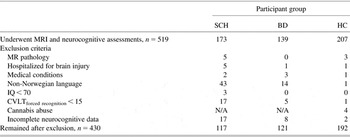
All participants who had undergone both MRI scanning and neuropsychological testing were included in the current study, but were excluded if they met the following criteria (Table 1): a history of hospitalized head injury, neurological disorder, IQ < 70 points, and age outside the range of 18–65 years. To ensure valid neurocognitive test performance, all participants had to have Norwegian as their first language or have received their compulsory schooling in Norway and had to score 15 or above in the forced recognition trial in the California Verbal Learning Test (CVLT)-II (Delis, Kramer, Kaplan, & Ober, Reference Delis, Kramer, Kaplan and Ober2004), which is a measure of adequate test effort. All MRI scans were evaluated by a neuroradiologist, and excluded if significant pathology was present, that is, lesions >1 cm in diameter or other gross pathology judged not to be within normal variation. The exclusion criteria for MRI pathology were equal for patients and controls.
Overview of the number of participants in each participant group who were excluded according to the listed criteria

Note. In total, 89 participants were excluded according to the criteria; 59 participants were excluded for one criterion, 10 participants were excluded for two criteria, and 20 participants were excluded for three criteria. SCH, schizophrenia-spectrum patients; BD, bipolar-spectrum disorder patients; HC healthy control participants.
We included 117 schizophrenia and 121 bipolar spectrum disorder patients, and 192 healthy controls in the present study. The schizophrenia spectrum included patients with schizophrenia (n = 94), schizophreniform disorder (n = 7) and schizoaffective disorder (n = 16), the bipolar disorder group included bipolar disorder type 1 (n = 76) and type 2 (n = 45). In the following, we refer to schizophrenia spectrum as “schizophrenia” and bipolar spectrum disorder as “bipolar disorder.”
The healthy control sample was given an interview to evaluate severe mental disorder symptoms and investigated with the Primary Care Evaluation of Mental Disorders (PRIME-MD) (Spitzer et al., Reference Spitzer, Williams, Kroenke, Linzer, deGruy, Hahn and Johnson1994). Controls were excluded if they had a drug abuse/dependency the last 3 months or had used drugs within the last 2 weeks, if they or any of their first-degree relatives had a life-time history of a severe psychiatric disorder, or if they had a history of medical problems thought to interfere with brain function.
Clinical Assessment
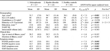
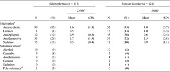
Trained physicians and clinical psychologists performed the clinical assessments. Diagnosis was based on the Structured Clinical Interview for DSM-IV Axis I disorders (SCID-I A-E) (First, Reference First2002). Current positive and negative symptoms were rated using the Positive and Negative Syndrome Scale (PANSS) (Kay, Fiszbein, & Opler, Reference Kay, Fiszbein and Opler1987). Psychosocial functioning was assessed with the Global Assessment of Functioning (GAF) scale, split version. Diagnostic interrater reliability was found to be satisfactory, with overall agreement for DSM-IV diagnostic categories of 82% with κ = 0.77 (95% CI: 0.60–0.94), and with intraclass coefficients = 0.73 and 0.86, respectively, for the PANSS positive and negative subscales and the GAF scales (Engh et al., Reference Engh, Friis, Birkenaes, Jonsdottir, Klungsoyr, Ringen and Andreassen2010). Demographic and clinical data are displayed in Table 2. Missing data are described in the footnote of Table 2. Data on current medication were derived from interviews and medical records. To obtain a clinically representative sample, all patients were included regardless of alcohol or illicit drug use, but abuse/dependency was diagnosed (SCID-E) if present (Table 3).
Demographics and clinical data

ANOVA = univariate analysis of variance; NART = National Adult Reading Test – Norwegian version; IQ = Intelligent Quotient; PANSS = Positive and Negative Syndrome Scale; GAF = Global Assessment of Functioning.
Note. Number of missing data: Handedness; schizophrenia: 5, bipolar disorder: 15; education; schizophrenia: 1; NART IQ; schizophrenia: 11, bipolar disorder: 2; age of onset and illness duration; schizophrenia: 1, bipolar disorder: 6.
aTukey post hoc tests.
bAge was defined as age at MRI scanning. The age range (years) for the subject groups: Schizophrenia 18–52, bipolar disorder 19–65, healthy controls 18–57.
cHandedness was determined by hand preference when writing.
dYears of education were registered as years of schooling as reported by the subjects during the interview.
eAge at onset was defined as age at first contact with the mental health service due to a primary symptom. The age at onset range (years) for the patient groups: Schizophrenia 15–48 and bipolar disorder 11–59. Duration of illness was defined as number of years between age at onset and age at MRI scanning. Median (Interquartile range) age at onset: schizophrenia: 25.5 (9.0), bipolar disorder: 26.0 (16.8); Median (Interquartile range) illness duration: schizophrenia: 2.7 (7.8), bipolar disorder: 4.0 (7.8).
Overview of medication and substance abuse in the patient groups

DDD = Defined Daily Doses; SD = Standard Deviation.
aInformation on medication dosages were converted into DDD for standardization purposes according to the WHO guidelines (http://www.whocc.no/atcddd).
bEight (7%) patients with schizophrenia and 4 (3%) patients with bipolar disorder received typical antipsychotic medication and 84 (72%) patients with schizophrenia and 46 (38%) patients with bipolar disorder received atypical antipsychotic medication, while 7 (6%) patients with schizophrenia and 1 (1%) patient with bipolar disorder received both. Thirteen (11%) patients with schizophrenia and 18 (15%) patients with bipolar disorder received no psychopharmacological medication.
cDiagnoses of current or partial remission of abuse or dependency. None of the patients in the current study were currently abusing or had a dependency on hallucinogens or opiates.
dPoly-substance dependency refers to behavior of repeated use, qualifying for a diagnosis of dependency of three groups of substances, but no single substance predominated.
Neurocognitive Assessment
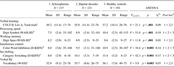
Trained psychologists performed the neurocognitive assessments. A comprehensive test battery was administered in a fixed order. Premorbid IQ was estimated using the National Adult Reading Test – Norwegian version (Sundet & Vaskinn, Reference Sundet and Vaskinn2008). For the present study, neurocognitive tests were included from the full test-battery if (1) they or near identical tests had previously shown a relationship with brain cortical thickness in an independent study of schizophrenia (Hartberg et al., Reference Hartberg, Lawyer, Nyman, Jonsson, Haukvik, Saetre and Agartz2010), or (2) had previously been found to differ between schizophrenia and bipolar disorder patients (Simonsen et al., Reference Simonsen, Sundet, Vaskinn, Birkenaes, Engh, Faerden and Andreassen2011). In total, six measures were selected (Table 4): Verbal learning was tested with the Total recall trial score (list A1–A5) from the California Verbal Learning Test, Second edition (CVLT-II) (Delis et al., Reference Delis, Kramer, Kaplan and Ober2004). Processing speed was assessed with the Digit Symbol subtest from the Wechsler Adult Intelligence Scale, Third edition (WAIS-III) (Wechsler, Reference Wechsler2003). Working memory was assessed with the Digit Span subtest (sum of forward and backward trials) from the WAIS-III (Wechsler, Reference Wechsler2003). This is a broad working memory measure and also measures aspects of attention. Interference control was tested with the Inhibition subtest from the Color-Word Interference Test (Delis-Kaplan Executive Function Scale, D-KEFS) (Delis, Kaplan, & Kramer, 2005). Set-shifting was measured with the Category Switching subtest (correct scores) from the Verbal Fluency Test (D-KEFS) (Delis et al., Reference Delis, Kaplan and Kramer2005). Both interference control and set-shifting are considered to be aspects of executive functioning. Verbal IQ was estimated with the Vocabulary subtest from the Wechsler Abbreviated Scale of Intelligence (WASI) (Wechsler, Reference Wechsler2007).
Results of neurocognitive comparisons

ANOVA = analysis of variance; CVLT-II = California Verbal Learning Test – Revised; WAIS-III = Wechsler Adult Intelligence Scale III Revision; D-KEFS = Delis-Kaplan Executive Functioning System; WASI = Wechsler Abbreviated Scale of Intelligence.
Note. Standardized scores are shown and used in the analyses.
aEta square effect size.
bTukey's post-hoc test.
cT-scores.
dS-scores.
Manual-based, age- and sex-adjusted scaled scores [(T-scores: 50 ± 10) or (S-scores: 10 ± 3)] are reported in the neurocognitive group comparisons. For T- and S-scores, higher scores equal better performance on all measures. Raw scores were used in the main analyses of brain structure/function relationships. For raw scores, higher scores equal better performance on all measures except for the Color-Word Interference Test.
Brain Imaging
MR image acquisition
All participants underwent MRI scanning on a 1.5T Siemens Magnetom Sonata scanner (Siemens Medical Solutions, Erlangen, Germany) equipped with a standard head coil. After a conventional three-plane localizer, two sagittal T1-weighted magnetization prepared rapid gradient echo (MPRAGE) volumes were acquired with the Siemens tfl3d1_ns pulse sequence (TE = 3.93 ms, TR = 2730 ms, TI = 1000 ms, flip angle = 7°; FOV = 24 cm, voxel size = 1.33 × 0.94 × 1 mm3, number of partitions = 160). Acquisition parameters were optimized for increased gray/white matter image contrast. There was no scanner upgrade during the study period.
MR image processing
The image files in DICOM format were transferred to a Linux workstation for morphometric analysis. Images were corrected for non-linear warping caused by gradient coil non-linearities, using tools developed through the Morphometry Biomedical Informatics Research Network (mBIRN), and the two T1-weighted images were rigid body registered to each other and averaged together to increase the signal to noise ratio. The FreeSurfer software was used for cortical surface reconstruction followed by a procedure to obtain a representation of the gray/white matter boundary and the pial surface (Dale, Fischl, & Sereno, Reference Dale, Fischl and Sereno1999; Fischl & Dale, Reference Fischl and Dale2000). Manual corrections of topological defects were performed blinded to group identity. Cortical thickness was calculated as the average distance between the gray/white matter boundary and the pial surface within each ROI. Surface area was calculated as the sum of the areas of each tessellation falling within a given ROI; this was done in each subject's native space. ICV estimates were obtained from the automated procedure for brain volume measurements implemented in Freesurfer (Buckner et al., Reference Buckner, Head, Parker, Fotenos, Marcus, Morris and Snyder2004).
Selection of brain regions to be used in the analysis
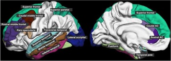
We selected 28 predefined cortical regions (Figure 1). These regions have shown to be abnormal in schizophrenia or bipolar disorder in previous MRI studies (Arnone et al., Reference Arnone, Cavanagh, Gerber, Lawrie, Ebmeier and McIntosh2009; Lyoo et al., Reference Lyoo, Sung, Dager, Friedman, Lee, Kim, Kim and Renshaw2006; Nesvag et al., Reference Nesvag, Lawyer, Varnas, Fjell, Walhovd, Frigessi and Agartz2008) and were comparable with those used in the similar study on cortical thickness/area and cognition in FES patients (Gutierrez-Galve et al., Reference Gutierrez-Galve, Wheeler-Kingshott, Altmann, Price, Chu, Leeson and Ron2010). Since cortical thickness/function relationships may be lateralized (Hartberg et al., Reference Hartberg, Lawyer, Nyman, Jonsson, Haukvik, Saetre and Agartz2010), the left and right hemispheres were investigated separately.
AC, Anterior cingulate. The software enables surface division into 34 regions-of-interest (ROIs) in each hemisphere, which are neuroanatomically labeled (Desikan et al., Reference Desikan, Segonne, Fischl, Quinn, Dickerson, Blacker and Killiany2006; Fischl et al., Reference Fischl, van der Kouwe, Destrieux, Halgren, Segonne, Salat and Dale2004). We selected 28 predefined cortical regions within all brain lobes, but predominantly from the frontal and temporal lobes. The figure shows lateral and midsagittal views of the selected parcellations.

Statistical Analysis
All analyses were performed in the statistical software SPSS 16.0. All analyses were two-tailed.
Group differences in demographic, clinical, neurocognitive, and brain structure measurements
Demographic, clinical, and standardized neuropsychological variables were compared across groups using Student's t tests, χ2 analyses and analyses of variance (ANOVAs), followed by Tukey's post hoc tests. Neuropsychological test comparisons for a subject sample partly overlapping with the present have been published earlier (Simonsen et al., Reference Simonsen, Sundet, Vaskinn, Birkenaes, Engh, Faerden and Andreassen2011), but were repeated for this particular sample with standardized T- and S-scores. Group comparisons of cortical thickness and surface area measurements from each of the predefined brain regions were analyzed using ANCOVAs, with age and sex as covariates.
Structure/function relationships by regression analysis
The main analyses of relationships between cortical measurements and neurocognitive function were conducted as follows. First, multiple stepwise linear regression analyses were performed for the combined sample and separately for each of the three subject groups (groupwise analyses). Each cortical measure was entered as the dependent variable. Age, sex (and diagnosis for the combined group analyses), and all six neurocognitive test scores (raw scores) were entered as independent variables. All analyses were corrected for age and sex differences.
Group differences in correlations between cortical thickness/area and neurocognition
Next, to test for differences in relationships, partial correlation coefficients from the group-wise regression analyses, selected at alpha = 0.01, were contrasted with the corresponding correlation coefficients from the other groups in a pairwise manner using Fisher z transformations. Sixteen regions were selected on the basis of the separate group analyses for pairwise between-group contrasts of correlation coefficients based on this criterion. Bonferroni correction was applied to both the main analyses and to the pairwise contrast analyses. Separate analyses were performed for the left and right hemisphere and for thickness and area. We performed 56 regression analyses for each of the three groups and for the combined sample. Thus, the resulting alpha level was 0.05/14 = 0.0036 for the main analyses and 0.05/3 = 0.017 for the pairwise contrast analyses.
Confounding factors
Possible confounding factors were investigated with ethnicity as a covariate for all groups and PANSS total positive and total negative symptom scores, and duration of illness and antipsychotic DDD (both logarithmic transformed) as covariates for the patient groups. Only patients who were prescribed medication were included.
Subgroup analysis
All analyses were repeated with bipolar disorder type 1 separately, and for the narrow schizophrenia group (including schizophrenia and schizophreniform disorder), and excluding patients with current cannabis abuse. Since 82% of the bipolar I disorder patients and 18% of the bipolar II disorder patients had experienced psychotic symptoms, history of psychosis was included as a covariate in the analyses of the bipolar disorder group.
Results
Demographic and Clinical Characteristics
The three diagnostic groups differed on demographic variables with regard to age, sex, and ethnicity (Table 2). The schizophrenia group had lower mean age and more males than the two other groups. The groups also differed on symptom ratings. There were no group differences in handedness, education, premorbid IQ, intracranial volume, age at onset, or duration of illness.
Group Differences in Neurocognitive Performance
The groups differed on all neuropsychological measures (Table 4). The largest effect sizes were observed for processing speed and attention-demanding set-shifting. The schizophrenia group performed worse than the bipolar group and healthy controls on all test scores, while the bipolar group performed worse than healthy controls on the processing speed, interference control and set-shifting tasks.
Group Differences in Cortical Thickness and Area
Thickness reduction was found in 11 frontal and temporal regions in both patients groups compared with healthy controls (Table S1): the bilateral caudal and rostral middle frontal regions; pars opercularis; superior frontal regions; the right fusiform, middle, and superior temporal regions; in two regions in schizophrenia compared with bipolar disorder; the left caudal anterior cingulate; and the right middle temporal region. Cortical thickness was reduced in the left superior temporal and the left lateral occipital regions in schizophrenia patients relative to healthy controls. The cortical thickness results parallel those reported by Rimol et al. (Reference Rimol, Hartberg, Nesvag, Fennema-Notestine, Hagler, Pung and Agartz2010). Cortical surface area was smaller in four regions in schizophrenia relative to bipolar disorder and healthy controls (Table S2); the right caudal and rostral middle frontal and fusiform regions and the left middle temporal region. Surface area was larger in the left superior frontal region and the right temporal pole in bipolar disorder relative to healthy controls and schizophrenia patients.
Relationships Between Cortical Thickness/Area and Neurocognition
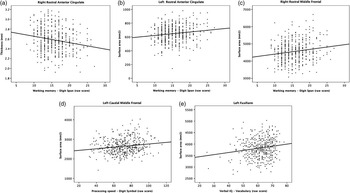
In the combined group, there was one significant cortical thickness relationship and four significant surface area relationships with neurocognitive performance (Figure 2). Increased cortical thickness in the right rostral anterior cingulate was related to poorer working memory performance. A larger surface area was related to better neurocognitive performance for the relationships between left rostral anterior cingulate and right rostral middle frontal regions with working memory performance, between left caudal middle frontal region and processing speed, and between left fusiform region and verbal IQ.
Relationships in the combined sample. Scatter plots showing significant relationships between cortical thickness (mm) and cortical surface area (mm2) with neurocognitive performance, adjusted for age and sex in the combined sample. (a) Larger cortical thickness in the right rostral anterior cingulate related to poorer working memory performance (B = 0.17; P = .0004); (b) larger cortical surface area in the left rostral anterior cingulate related to better working memory performance (B = 0.14; P = .003); (c) larger cortical surface area in the right rostral middle frontal region related to better working memory performance (B = 0.14; P = .001); (d) larger cortical surface area in the left caudal middle frontal region related to better processing speed performance (B = 0.15; P = .003); (e) larger cortical surface area in the left fusiform region related to better verbal IQ (B = 0.14; P = .002).

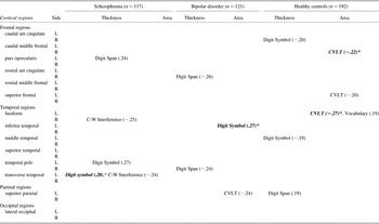
The results (p < .01) for the groupwise analyses are displayed in Table 5. In the schizophrenia group, larger cortical thickness in the left transverse temporal region was significantly related to better processing speed performance. In the bipolar disorder group, larger cortical surface area in the left inferior temporal region was significantly related to better processing speed performance, whereas in the healthy control group, larger cortical surface area in the right caudal middle frontal and left fusiform regions was significantly related to poorer verbal learning. There were no overlapping relationships between surface area and thickness.
Relationships (p < .01) between cortical thickness and surface area and neurocognitive test scores in each participant group

Note. Test (Domain): Digit Span (Working memory); Digit Symbol (Processing speed); C-W Interference (Interference control); CVLT California Verbal Learning test (Verbal learning); Vocabulary (Verbal IQ). Partial correlation coefficients are shown for p values < .01.
*Significant after Bonferroni correction for multiple comparisons.
Group Differences in Correlations Between Cortical Thickness/Area and Neurocognition
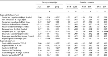
Results for the group comparisons are displayed in Table 6. Eight of the contrasts were statistically significant. There were three significantly different correlations between schizophrenia and bipolar disorder: between cortical thickness in the left pars opercularis and working memory; the left transverse temporal region and processing speed; and the right temporal pole and working memory. The latter correlation also differentiated the bipolar disorder patients from the healthy control group. The schizophrenia group displayed positive correlations in all three regions, while the bipolar group correlations were negative. There were four significantly different correlations between schizophrenia and healthy controls in temporal lobe regions: between cortical thickness in the left middle and transverse temporal regions and the left temporal pole and processing speed; and between surface area in the left fusiform region and verbal learning. All four correlations were positive in the schizophrenia group, while they were all negative in the healthy control group. There were no overlapping relationships between surface area and thickness.
Pairwise contrasts of correlations (p < .01) between cortical thickness or area and neurocognitive tests

CTR = Healthy control subjects; SCH = Schizophrenia; BD = Bipolar disorder; r = partial correlation coefficient; Z = Fisher's z value; ant = anterior; caud = caudal; lh = left hemisphere; rh = right hemisphere; CVLT = California Verbal Learning test (Verbal learning).
*Groupwise correlations significant at alpha level = 0.01. Bold indicates significant group differences (Bonferroni corrected).
Confounding Factors and Subgroup Analyses
The patterns of relationships in the narrow schizophrenia and bipolar disorder type 1 groups were essentially the same as in the schizophrenia and bipolar spectrum groups, respectively. Overall, the strength of relationships remained the same after controlling for ethnicity, duration of illness, symptom scores, history of psychosis, and antipsychotic medication, and when excluding patients with current cannabis abuse.
Discussion
This is the first study to investigate relationships between regional cortical thickness/area and neurocognitive performance and directly compare between patients with schizophrenia and bipolar disorder investigated with the same MRI scanner, post-processing methodology, and neurocognitive test battery. Contrary to our expectations, we detected only a limited number of significant cortical structure/function relationships; five relationships were significant in the combined group of participants, that is, were found to be the same in all three groups. In keeping with our expectations, however, there were significant group differences in relationships; three specific relationships between regional cortical thickness/function were different in schizophrenia compared with bipolar disorder, of which the negative relationship between cortical thickness in the right temporal pole and working memory was specific to bipolar disorder, and the positive relationship between cortical thickness in the left transverse temporal region and processing speed was specific to schizophrenia. Also, we found that cortical thickness and surface area related differently to neurocognitive functioning.
From our previous report on cortical thickness relationships with cognitive performance in chronic schizophrenia (Hartberg et al., Reference Hartberg, Lawyer, Nyman, Jonsson, Haukvik, Saetre and Agartz2010), we expected more widespread cortical thickness relationships in the present study. The patients who participated in the present study had been ill for a relatively short period of time (Table 2) as compared to other chronic patient cohorts, and in this regard were similar to the participants in two studies on FES patients (Crespo-Facorro et al., Reference Crespo-Facorro, Roiz-Santianez, Perez-Iglesias, Rodriguez-Sanchez, Mata, Tordesillas-Gutierrez and Vazquez-Barquero2011; Gutierrez-Galve et al., Reference Gutierrez-Galve, Wheeler-Kingshott, Altmann, Price, Chu, Leeson and Ron2010). Both FES studies reported sparse results for relationships between cortical structure and specific neurocognitive domains. Also, the effect sizes for group differences in cortical thickness and neurocognitive performance were smaller in the present study (Table S1, Table 4) than in those with chronic patients (Heinrichs & Zakzanis, Reference Heinrichs and Zakzanis1998; Nesvag et al., Reference Nesvag, Lawyer, Varnas, Fjell, Walhovd, Frigessi and Agartz2008), which might have limited our ability to detect subtle relationships. Furthermore, comparisons between younger and older adults have revealed stronger and positive relationships in the older participants (Zimmerman et al., Reference Zimmerman, Brickman, Paul, Grieve, Tate, Gunstad and Gordon2006) regardless of having a schizophrenia diagnosis or not (Premkumar, Kumari, Corr, Fannon, & Sharma, Reference Premkumar, Kumari, Corr, Fannon and Sharma2008), suggesting that structure/function relationships generally become more pronounced with higher age. Unfortunately, the age distribution in the present study, which was skewed toward younger age, did not allow for testing of such a hypothesis. Furthermore, there may be non-linear effects of age or a dynamic age-dependent interplay between structure and function, which makes the relationships change over time and difficult to detect in cross-sectional studies. An example of this would be the demonstrated progression of gray matter loss concurrent with neurocognitive improvements in the first years after illness onset in schizophrenia (Zipparo et al., Reference Zipparo, Whitford, Redoblado Hodge, Lucas, Farrow, Brennan and Harris2008), which render complex patterns of structure/function relationships, and both negative and positive relationships may result from this.
Although few, the observed common significant relationships between all three groups suggest that there exist general relationships that are not disrupted by having a diagnosis of either schizophrenia or bipolar disorder. For the relationship between cortical thickness in the right rostral anterior cingulate (AC) and working memory, the correlation was strongest in the bipolar disorder group, which is in accordance with the negative, but non-significant correlation between rostral AC gray matter volumes and measures of working memory that was previously demonstrated in bipolar disorder patients and healthy controls (Zimmerman et al., Reference Zimmerman, Brickman, Paul, Grieve, Tate, Gunstad and Gordon2006). The rostral AC has been related to emotional processes (Devinsky, Morrell, & Vogt, Reference Devinsky, Morrell and Vogt1995) known to be of importance in bipolar disorder, as well as implicated in circuitries disrupted in schizophrenia (Benes, Reference Benes2010). The present results suggest that structural integrity in this region is important for working memory performance, regardless of group status. The results for frontal surface area relationships in the combined group are supported by similar findings in FES patients (Gutierrez-Galve et al., Reference Gutierrez-Galve, Wheeler-Kingshott, Altmann, Price, Chu, Leeson and Ron2010).
Although the present correlations between regional cortical thickness and working memory and processing speed are of small/medium effect sizes (Cohen, Reference Cohen1988), they are, as shown in the pair wise contrast analyses, in opposite directions in the schizophrenia and bipolar disorder patients, which suggest different patterns of relationships. Consistent with this, post-mortem studies demonstrate cellular differences in some, but not all regions of the frontal lobe between schizophrenia and bipolar patient groups (Selemon & Rajkowska, Reference Selemon and Rajkowska2003). The fact that we found only weak relationships even in a large sample of participants may suggest that brain structure and function are weakly related in these disorders.
In accordance with our second hypothesis, there were different relationships between neurocognition and cortical thickness compared with cortical surface area. Furthermore, there were more differences in relationships between participant groups for thickness than for area. Cortical thickness demonstrates variable regional heritability (Kremen et al., Reference Kremen, Prom-Wormley, Panizzon, Eyler, Fischl, Neale and Fennema-Notestine2010) and may be sensitive to environmental influences such as drug use (Habets et al., Reference Habets, Marcelis, Gronenschild, Drukker and van Os2010), and illness-related factors (Goldman et al., Reference Goldman, Pezawas, Mattay, Fischl, Verchinski, Chen and Meyer-Lindenberg2009). We, therefore, corrected for cannabis abuse, illness duration, and medication use in the analyses, and our interpretation is that the present differences in cortical thickness relationships between subject groups reflect pathophysiological processes inherent to the disorders and not environmental factors. Although environmental effects on cortical surface area development cannot be excluded (White, Andreasen, & Nopoulos, Reference White, Andreasen and Nopoulos2002), surface area is proposed to be mainly determined during embryonic and neonatal life without undergoing major subsequent changes (Rakic, Reference Rakic1988), and as such represents a window for investigating early neurodevelopmental disturbances. Reduced regional surface area (Table S2) and the disrupted relationship with verbal learning in schizophrenia, but not in bipolar disorder, support theories of more severe neurodevelopmental disturbances in schizophrenia than in bipolar disorder.
We have previously demonstrated that a positive relationship between cortical thickness in the right fusiform (temporo-occipital) region and Verbal IQ in healthy controls were disrupted in chronic schizophrenia (Hartberg et al., Reference Hartberg, Lawyer, Nyman, Jonsson, Haukvik, Saetre and Agartz2010), which suggested that this region might be of special importance in the disorder. However, in the present study cortical area in the left fusiform region was significantly correlated with verbal learning in the healthy control group and differentiated between healthy control group and the schizophrenia patients. The temporo-occipital region is important for word recognition and is activated during word reading tasks in fMRI studies (Brem et al., Reference Brem, Bucher, Halder, Summers, Dietrich, Martin and Brandeis2006), with a left lateralization activation pattern. The present results add to the evidence that these regions have been specifically excluded from the normal functioning network underlying word processing in schizophrenia.
There were a few limitations to this study. The group differences in age and sex may have affected the results, although they were corrected for in all statistical analyses. Also, there were group differences in ethnicity, which could have affected the present results, although co-varying for ethnicity did not change the main findings. To give an account of the pattern of relationships in each group, separate group analyses were conducted which resulted in a large number of statistical tests and the results must be interpreted with some caution. However, the threshold for selection of groupwise relationships for further group comparisons are in accordance with the threshold used by other researchers in the field (Antonova et al., Reference Antonova, Kumari, Morris, Halari, Anilkumar, Mehrotra and Sharma2005; Crespo-Facorro et al., Reference Crespo-Facorro, Roiz-Santianez, Perez-Iglesias, Rodriguez-Sanchez, Mata, Tordesillas-Gutierrez and Vazquez-Barquero2011). Furthermore, a conservative multiple comparison control was applied to the final results. In the present study, the combined digit span score was used as a measure of working memory. However, the digit span forward and backward tasks may measure discrete functions (Lezak, Reference Lezak1995) differentially associated with the diathesis for schizophrenia (Conklin, Curtis, Katsanis, & Iacono, Reference Conklin, Curtis, Katsanis and Iacono2000). Separate analyses of the backward task score were not performed due to limited range of scores and low variability. The combined and the backward scores were highly correlated (r = 0.8). Thus, we feel confident that we have revealed relevant relationships with a broad measure of working memory.
Use of medication can affect cortical structure (Navari & Dazzan, Reference Navari and Dazzan2009). However, corrections for current dose of antipsychotic medication did not affect the results. Only 17 participants were prescribed lithium, and the effect on cortical structure could not be investigated.
In conclusion, we find significant relationships between regional cortical thickness/surface area and neurocognitive test scores, of which some are common to all groups and others are unique to schizophrenia or bipolar disorder. The results add to the knowledge on how cortical structure is important to specific cognitive functioning in general and how relationships may specifically be altered in schizophrenia and bipolar disorder. Different patterns of relationships between schizophrenia and bipolar disorder suggest differences in underlying pathophysiology, which could be related to differences in neurodevelopmental processes, but whether these patterns are consistent should be tested in independent samples. Future longitudinal studies are needed to determine how structure and function interrelate over time.
Acknowledgments
We thank patients and controls for their participation in the study. We also thank Merete Øibakken, Martin Furan, Petr S. Bjerkan, Thomas Bjella, and Eivind Bakken, for technical and administrative assistance, Oslo University Hospital and the University of Oslo to support the Thematically Organized Psychosis (TOP) Research Study group. The study was supported by grants from the Research Council of Norway (grant number 190311/V50); Eastern and Western Norway Health Authority (grant number 2004-123) and South-Eastern Norway Regional Health Authority (grant numbers 2008-011, 2009-037, 2009-038).
FINANCIAL DISCLOSURES
I.M. has received Speaker's honorarium from Janssen, AstraZeneca and Lundbeck. R.N. has received Speaker's honorarium from Janssen and AstraZeneca. A.M.D. is a founder and holds equity in CorTechs Labs, and also serves on the Scientific Advisory Board. The terms of this arrangement have been reviewed and approved by the University of California, San Diego, in accordance with its conflict of interest policies. O.A.A. has received Speaker's honorarium from AstraZeneca, Janssen, BMS, and GSK. All other authors report no conflict of interest.
Supplementary materials
To review these additional data and analyses, please access the online-only supplementary Tables 1 and 2. Please visit journals.cambridge.org/INS, then click on the link “Supplementary Materials” at this article.