Highlights
-
• CMB/cSS occurred in 4.7% of memory clinic patients.
-
• WMH burden and education not CMB/cSS burden best explained cross-sectional cognition.
-
• ∼23% met Boston v2.0 probable CAA, highlighting diagnostic and safety implications.
Background
As global populations age, cognitive impairment is increasingly burdening societies and families. Although Alzheimer’s disease (AD) still accounts for most dementia diagnoses in older adults, there has been increasing focus on vascular contributions to cognitive disorders. Reference Livingston, Huntley and Sommerlad1 Neuroimaging innovations allow for the detection of several vascular conditions, including cerebral microbleeds (CMBs) and cortical superficial siderosis (cSS). Microbleeds (MBs) are small, chronic bleeding lesions that appear as hypointense foci on blood-sensitive MRI, reflecting hemosiderin-laden macrophages in the brain parenchyma. Reference Charidimou and Werring2 cSS is characterized by the deposition of blood breakdown products in the cortical sulci along the convex surfaces of the cerebral hemispheres, typically presenting with a distinctive low-signal-intensity rim on MRI. Reference Charidimou, Linn and Vernooij3 There are several possible causes of these lesions. Hypertensive arteriopathy, for example, is commonly involved in deep or mixed MB distributions, while cerebral amyloid angiopathy (CAA) – the pathological deposition of amyloid-β in the vessel walls of small- to medium-sized cerebral vessels – is more often associated with lobar MBs and cSS. Reference Poels, Vernooij and Ikram4,Reference Na, Park and Kim5 This amyloid-mediated fragility plays a significant role in the development of hemorrhagic lesions in older adults. Reference Charidimou, Boulouis and Xiong6,Reference Lummel, Wollenweber and Demaerel7 The current literature has shown that MBs are associated with poorer cognitive performance across various types of dementia, including AD, dementia with Lewy bodies and Parkinson’s disease dementia. Reference Chin, Holper, Loveland, Churilov, Yassi and Watson8 Lobar-located MBs are related to more deficits in executive function, processing speed and memory, while disseminated cSS may raise the risk of recurrent hemorrhages and further cognitive deterioration. Reference Akoudad, Wolters and Viswanathan9–Reference Zhou, Liu, Yan and Jin11 In AD, brain microhemorrhages such as MBs may play a crucial role in disease pathophysiology by potentially bridging two prominent theories of AD neuropathogenesis – the amyloid cascade hypothesis and the vascular hypothesis. These microhemorrhages might influence clinical progression and carry therapeutic implications. Reference Cordonnier and van der Flier12 However, it remains unclear which vascular-associated variable are associated with MBs and cSS. Another important biomarker associated with cognitive impairment – and a key feature of cerebral small vessel disease (CSVD) – are white matter hyperintensities (WMH), which appear as hyperintense regions on T2-weighted or FLAIR images. Reference Wardlaw, Smith and Dichgans13 Their burden is most frequently assessed by the Fazekas scale, a semi-qualitative scale for scoring lesion burden, although alternative rating methods and automated volumetric techniques have been developed. Reference Pradeep, Raghavan and Przybelski14 WMH burden is strongly associated with vascular-associated variable and consistently linked to poorer cognitive performance, emphasizing the influence of cerebrovascular pathology on the dementia trajectory beyond neurodegeneration. Reference Wardlaw, Smith and Dichgans13,Reference Kynast, Lampe and Luck15 Although previous studies have linked CMBs with age and hypertension, most of this work has focused on stroke populations or small, mixed cohorts. In contrast, our study draws on a large, real-world memory clinic population with comprehensive cognitive assessments. This allows us to explore how hemorrhagic markers like CMBs and cSS relate to white matter changes and cognitive performance across different types of cognitive impairment, offering a more clinically meaningful perspective.
Materials and methods
Patient selection
We conducted a retrospective comparative study, at the University Health Network (UHN) in Toronto, Ontario, Canada, between 2014 and 2022, wherein a total of 2923 patients with cognitive impairment who underwent a dementia protocol MRI were screened. We grouped patients by what was seen on their MRI. Those with ≥1 hemorrhagic marker a CMB or cSS identified on susceptibility-sensitive sequences (gradient-recalled echo [GRE] or susceptibility-weighted imaging [SWI]) were classified as cases. Patients without CMBs or cSS on the same MRI protocol served as the comparison group. From the CMB/cSS-negative pool, we sampled at an approximately 2:1 ratio to cases; after routine MRI/clinical quality control, the final totals were 137 cases and 278 controls (∼2:1). This study was approved by the University Health Network Research Ethics Board (CAPCR ID #22-5431). As this was a retrospective chart review, informed consent was waived by the REB. An overview of the screening and selection process is shown in Figure 1, which details the total sample screened and the identification of each group.
Patient selection flow diagram. Flowchart illustrating the selection process of study participants. A total of 2923 patients with cognitive impairment who underwent a dementia protocol MRI at the University Health Network (2014–2022) were screened based on inclusion and exclusion criteria. Patients with microbleeds (MBs) and/or cortical superficial siderosis (cSS) on GRE/SWI sequences were identified (n = 137), and a comparison group of patients without MB/cSS findings was selected using a 2:1 matching ratio (n = 278), resulting in a final sample of 415 participants. GRE = gradient-recalled echo; SWI = susceptibility-weighted imaging; WMH = white matter hyperintensities; CMBs = cerebral microbleeds; CAA = cerebral amyloid angiopathy.

The inclusion criteria for both cohorts were (1) patients with cognitive complaints or impairment, including subjective cognitive impairment, mild cognitive impairment or dementia and (2) patients who underwent a brain MRI dementia protocol. The exclusion criteria for both groups were as follows: (1) patients with acute stroke and significant traumatic brain injury (TBI), defined as evidence of diffuse axonal injury, brain contusions or subdural hematoma on imaging; we include the non-significant TBI; (2) patients with other imaging changes or diseases that could sufficiently explain cognitive impairment; and (3) patients with a known history of brain macro-hemorrhages, including intracerebral hemorrhage (ICH) and subarachnoid hemorrhage (SAH), were also excluded. An overview of the screening and selection process is shown in Figure 1.
Brain MRI and SVD markers
All patients underwent brain MRI as part of a standardized dementia protocol on either 1.5T or 3T scanners (GE or Siemens). Susceptibility-sensitive sequences included either GRE or SWI, with minimum slice thickness of 2 mm. Among the 137 patients with MBs and/or cSS, 22 were imaged using GRE and 115 with SWI. Among the 278 control subjects, 109 underwent GRE and 169 underwent SWI. MRI reads were obtained retrospectively from clinical radiology reports.
CMBs: Presence was coded when a report described ≥1 CMB. When a numeric total was provided, it was recorded as the CMB count; if only qualitative terms (“few” and “multiple”) were used, cases contributed to presence/absence analyses but were coded missing for count-based analyses. CMB topography was categorized as lobar, deep or mixed per the report. We did not sub-classify lobar CMBs by individual lobes (e.g., frontal and parietal) because regional labeling was inconsistently documented across reports and scanners.
-
cSS: Coded present/absent. Extent (focal vs disseminated) was not consistently documented and was therefore not analyzed.
-
WMH: When reported, WMH burden used the Fazekas score (0–3); if Fazekas was not stated, WMH was not imputed. For some summaries, WMH was dichotomized low (0–1) versus high (2–3).
-
Acquisition covariates: To partially account for detection variability, multivariable models adjusted for sequence (GRE vs SWI) and field strength (1.5T vs 3T).
Clinical variables
Information on vascular and medical comorbidities was obtained from patient charts and included hypertension, diabetes, and hyperlipidemia. Hypertension was coded present if a documented diagnosis and/or active antihypertensive therapy was recorded at the index visit; otherwise, absent. (Standardized longitudinal control metrics ambulatory/home BP, time-in-range were not consistently available and were not modeled.)
Hyperlipidemia was determined by elevated levels of cholesterol and/or triglycerides or the use of lipid-lowering
substance exposures: Smoking and alcohol were coded present versus absent because dose measures (pack-years; units/week) were inconsistently documented in charts. Recreational substance use (including cannabis) was variably recorded in a generic “substance use” field and was therefore analyzed as present/absent without substance-specific breakdown.
Medications: Antithrombotics were defined as antiplatelet and/or anticoagulant use at the time of MRI (yes/no).
Cognitive assessment
Cognitive function was assessed using the Mini-Mental State Examination (MMSE) or the Montreal Cognitive Assessment (MoCA) within 1 to 2 years of the MRI scan. When multiple assessments were available, we selected the assessment closest to the MRI to minimize interval bias. The MMSE examines multiple cognitive domains including orientation, registration, attention and calculation, recall and language, with a maximum score of 30 points. The MoCA assesses cognitive domains such as visuospatial/executive functions, naming, memory, attention, language, abstraction and orientation, with a total score of 30 points. A score of 26 or above is considered normal cognition. An additional point was added for individuals with less than 12 years of education. For patients assessed using MMSE, raw scores were converted into z-scores based on published normative data adjusted for age and education. The same was done for MoCA scores. To ensure comparability, analyses used standardized z-scores, and MMSE and MoCA were not pooled as raw scores.
Statistical analysis
We treated this as a cross-sectional study and report associations, not causal effects. Continuous variables are summarized as mean ± SD when approximately normal, or median [IQR] when skewed; categorical variables are summarized as n (%). Group differences were tested with independent-samples t tests or Mann–Whitney U tests for continuous data, and χ 2 or Fisher’s exact tests for categorical data (two-sided α = 0.05). We used multivariable logistic regression to estimate adjusted odds of having any hemorrhagic marker (CMB and/or cSS) and multivariable linear regression to examine cross-sectional associations between imaging markers (CMBs/cSS and WMH/Fazekas) and global cognitive z-scores. Covariates were prespecified for clinical relevance: age, sex, education (modeled with flexible polynomial terms), hypertension, psychiatric history, smoking, remote TBI/concussion, WMH (Fazekas) and MRI acquisition factors (GRE vs SWI; 1.5T vs 3T). In secondary analyses, we evaluated CMB burden as a count and by categories (0, 1, 2–4, ≥5) and assessed topography (lobar/deep/mixed); cSS was analyzed as present/absent due to limited reporting of extent. Analyses were conducted in R (v4.3.1).
Results
Prevalence and subgroup characteristics
One hundred and thirty-seven cases were identified as having MBs and cSS yielding a prevalence of 4.7% (137 out of 2923). Within the cases with hemorrhagic markers, 103 patients (75.0%) had CMBs, only 9 patients (6.6%) had cSS and 25 patients (18.4%) had both CMBs and cSS. Thirty-one patients (22.6%) met Boston Criteria version 2.0 criteria for probable CAA, defined as age ≥50 and the presence of two or more strictly lobar hemorrhagic lesions (i.e., lobar MBs or cSS), without evidence of deep hemorrhages or other explanations.
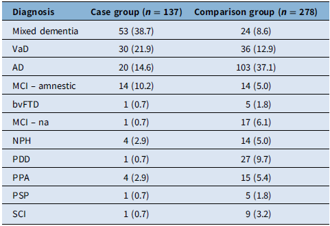
Patients in the hemorrhagic case group were older than those in the comparison group. The hemorrhagic case group also had a higher prevalence of hypertension (p < 0.001), and there were more people with TBI/concussion. There were no statistically significant differences between the two groups in terms of sex (male/female), diabetes, hyperlipidemia, smoking and alcohol use (p > 0.05). The Fazekas scale scores were significantly higher in the hemorrhagic case group. The detailed characteristics are presented in Table 1. Baseline clinical diagnoses are summarized in Table 2. Dementia was the most common stage in both groups, with mixed/vascular etiologies more frequent among cases (e.g., mixed 38.7% and vascular dementia [VaD] 21.9%) and AD more frequent among controls (37.1%).
Baseline characteristics of the study population

Table 1. Long description
A table comparing baseline characteristics of two study groups. The table has 12 rows and 5 columns. The columns are labeled ‘Characteristic’, ‘Case group (n = 137)’, ‘Comparison group (n = 278)’, and ‘p-Value’. The characteristics include Age (SD), Sex (M/F), Hypertension (%), Diabetes (%), Hyperlipidemia (%), Smoking (%), Alcohol use (%), TBI/concussion (%), Antithrombotic medications (%), and Fazekas scale, mean (SD). Each row provides specific values for each characteristic in both groups and the corresponding p-value. Notable trends include higher percentages of hypertension and TBI/concussion in the case group, and significant differences in the Fazekas scale scores between the two groups.
SD = standard deviation; M/F = male/female; TBI = traumatic brain injury; Fazekas scale = a visual rating scale for white matter hyperintensities.
TBI/concussion denotes remote, resolved mild concussion (>12 months) without persistent cognitive sequelae; moderate–severe or recent symptomatic TBI was excluded.
Clinical diagnoses of the study population

Table 2. Long description
A table comparing clinical diagnoses between case and comparison groups. The table has 11 rows and 3 columns. The columns are labeled ‘Diagnosis’, ‘Case group (n = 137)’, and ‘Comparison group (n = 278)’. The row labels include various diagnoses such as Mixed dementia, VaD, AD, MCI - amnestic, bvFTD, MCI - na, NPH, PDD, PPA, PSP, and SCI. Each row lists the number and percentage of cases and comparisons for each diagnosis. For example, Mixed dementia has 53 cases (38.7 percent) and 24 comparisons (8.6 percent). VaD has 30 cases (21.9 percent) and 36 comparisons (12.9 percent). AD has 20 cases (14.6 percent) and 103 comparisons (37.1 percent). The table shows that Mixed dementia and VaD are more prevalent in the case group, while AD is more prevalent in the comparison group.
AD = Alzheimer’s disease; bvFTD = behavioral variant frontotemporal dementia; MCI = mild cognitive impairment (amnestic and non-amnestic); NPH = normal pressure hydrocephalus; PPA = primary progressive aphasia; PSP = progressive supranuclear palsy; SCI = subjective cognitive impairment; VaD = vascular dementia.
Percentages are column-wise within dementia cases. DLB was not separately coded at baseline in this dataset; Lewy body spectrum is represented here by PDD where recorded.
The multivariable analysis of the associated variable for MBs and cSS
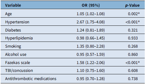
In adjusted logistic regression models (age, sex, education, hypertension, psychiatric history, smoking, remote TBI/concussion, Fazekas and MRI sequence/field strength), older age, hypertension and higher WMH burden (Fazekas) were associated with the presence of MBs/cSS (e.g., age OR 1.05, 95% CI 1.02–1.08; hypertension OR 2.67, 95% CI 1.75–4.08; Fazekas OR 1.58, 95% CI 1.22–2.06). Education, smoking, remote TBI/concussion and antithrombotic use (antiplatelet/anticoagulant) were not independently associated. Full estimates appear in Table 3. Associations are interpreted as cross-sectional correlates, not causal effects.
Multivariate logistic regression model showing odds of association between predictor variables and cerebral MBs

Table 3. Long description
The table presents the results of a logistic multivariate model analyzing the association of different variables with the presence of cerebral MBs. It includes three columns: Variable, OR (95%), and p-Value. The variables listed are Age, Hypertension, Diabetes, Hyperlipidemia, Smoking, Alcohol use, Fazekas scale, TBI/concussion, and Antithrombotic medications. Each row provides the odds ratio (OR) with a 95% confidence interval and the corresponding p-value for each variable. Notable findings include significant associations for Age (OR 1.05, p-Value 0.002), Hypertension (OR 2.67, p-Value <0.001), and Fazekas scale (OR 1.58, p-Value <0.001). Other variables such as Diabetes, Hyperlipidemia, Smoking, Alcohol use, TBI/concussion, and Antithrombotic medications show non-significant associations with p-values greater than 0.05.
OR = odds ratio; TBI = traumatic brain injury; MBs = microbleeds.
*p < 0.05 was considered statistically significant.
Cognitive assessment
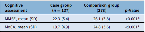
Unadjusted MMSE/MoCA scores were lower in the MBs/cSS group (see Table 4). In fully adjusted linear models using global cognitive z-scores, education showed the strongest association with cognition, whereas the presence of MBs/cSS was not independently associated after accounting for covariates (including WMH).
Cognitive function of the study population

Table 4. Long description
A table comparing cognitive assessment scores between two groups. The table has four rows and three columns. The columns are labeled ‘Cognitive assessment’, ‘Case group (n = 137)’, and ‘Comparison group (278)’. The rows are labeled ‘MMSE, mean (SD)’ and ‘MoCA, mean (SD)’. The values in the table are as follows: Row 1: MMSE, mean (SD), 22.3 (5.4), 26.1 (3.8). Row 2: MoCA, mean (SD), 19.7 (4.9), 24.8 (3.6). The p-Value for both comparisons is less than 0.001.
MMSE = Mini-Mental State Examination; MoCA = Montreal Cognitive Assessment; SD = standard deviation.
*p < 0.05 was considered statistically significant.
Impact of MB location and other factors on cognitive scores
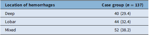
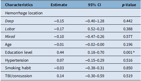
The distribution of MB locations is summarized in Table 5. In adjusted linear models (Table 6), MB location (deep, lobar, mixed) was not associated with global cognitive z-scores (all p > 0.38). Age was not independently associated, whereas higher education correlated with better cognitive performance (estimate 0.44; 95% CI 0.18–0.70; p < 0.001). Overall model fit was modest (R 2 = 0.08; adjusted R 2 = 0.04), consistent with unmeasured contributors to cognition in this cross-sectional cohort.
Location of MBs

Table 5. Long description
The table presents the distribution of hemorrhage locations in a case group of 137 individuals. It has two columns: Location of hemorrhages and Case group (n = 137). The table includes three rows with the following data: Row 1: Deep, 40 (29.4); Row 2: Lobar, 44 (32.4); Row 3: Mixed, 52 (38.2). The table shows the number and percentage of cases for each location of hemorrhages.
MBs = microbleeds; n = number of participants.
Linear regression model of the effect of MB location on z-standardized cognitive scores

Table 6. Long description
A table with four columns and nine rows. The columns are labeled Characteristics, Estimate, 95% CI, and p-Value. The rows list different characteristics and their corresponding estimates, confidence intervals, and p-values. The characteristics include Hemorrhage location with subcategories Deep, Lobar, and Mixed, Age, Education level, Hypertension, Smoking habit, and TBI/concussion. Each row provides specific values for these characteristics.
MBs = microbleeds; CI = confidence interval; TBI = traumatic brain injury.
*p < 0.05 was considered statistically significant.
Sex-stratified analysis of the effect on cognitive z-scores of having MBs in initial brain MRI at baseline assessment
Linear regression models were conducted in females (n = 192) and males (n = 176) to examine whether the association between vascular-associated variable, MBs/cSS (case comparison) and cognitive performance (standardized z-scores) differed by sex. In both groups, higher education was significantly associated with better cognitive scores (females: 0.45, 95% CI: 0.06–0.83, p = 0.024; males 0.42, 95% CI: 0.05–0.79, p = 0.027). In contrast, the presence of MBs/cSS, as well as age, hypertension, cigarette smoking, TBI/concussion and Fazekas score terms, were not significant predictors in either sex. Model fit was modest (females: R 2 = 9.9%, adjusted R 2= 3.3%; males: R 2 = 12.9%, adjusted R 2 = 6.0%). A formal MBs/cSS×sex interaction term was not significant, indicating no evidence that the MBs/cSS–cognition relationship differs by sex. Taken together, regression coefficients were similar between sexes, suggesting comparable effects of these vascular factors on cognitive performance in females and males.
Cognitive z-scores by WMH burden
A WMH burden-stratified analysis was conducted to evaluate whether the association of MBs/cSS and other vascular-associated variable with cognitive performance are modified by WMH severity. Patients were divided into two groups, low WMH (Fazekas 0–1) and high WMH (Fazekas 2–3). In the group with low WMH, higher education positively affects cognitive z- scores (0.51, 95% CI: 0.16–0.86, p = 0.005). By contrast, MBs/cSS (estimate = 0.13, 95% CI: −0.25 to 0.52, p = 0.501), as well as age, hypertension, psychiatric disorders, cigarette smoking and TBI/concussion, did not show a notable effect on cognition under relatively low WMH burden. While, in the high WMH group, none of the tested factors – including MBs/cSS (estimate = 0.15, 95% CI: −0.21 to 0.50, p = 0.414), age, education or other vascular-associated variable significantly predicted cognitive z-scores. Patients in this group, on average, displayed lower cognitive performance overall, suggesting that when WMH burden is advanced, it may overshadow any additional influences from MBs/cSS or other associated variable. These results suggest that, in individuals with a relatively low WMH burden, higher education is a protective factor for cognitive impairment. Given the cross-sectional design and smaller strata, power to detect modest interactions may be limited; nonetheless, education remained the most consistent correlate of cognitive performance across WMH strata.
Discussion
This study aimed to investigate the prevalence, associated variable and cognitive impact of CMBs and cSS. Out of 2923 patients who were screened for cognitive impairment and underwent dementia protocol MRI, 137 (approximately 4.7%) were identified as having MBs and/or cSS. The prevalence of MBs and/or cSS in our cohort (4.7%) is lower than that reported in some recent studies, where rates of CMBs have been observed in approximately 20–25% of older adults. Reference Vuong, Morrill and Graff-Radford31 This discrepancy likely reflects differences in study design and population characteristics. Population- based and longitudinal cohorts, such as the ARIC-Neurocognitive Study, often include older individuals with varying degrees of vascular risk and may utilize highly sensitive imaging protocols, leading to higher detection rates. In contrast, our study was conducted in a memory clinic-based cohort with specific referral patterns and inclusion criteria, which may influence the observed prevalence. Additionally, differences in MRI acquisition techniques (e.g., SWI vs GRE), field strength and criteria for defining MBs may further contribute to variability across studies. These factors highlight the importance of interpreting prevalence estimates within the context of study methodology and population.
This study is among the limited number of investigations examining CMBs and cSS in a large memory clinic-based cohort as most prior studies have focused on population-based samples or specific disease groups rather than real-world memory clinic populations. Reference Poels, Vernooij and Ikram4,Reference Chin, Holper, Loveland, Churilov, Yassi and Watson8 By applying Boston Criteria v2.0 and stratifying by WMH severity, we provide a more granular view of hemorrhagic lesion patterns and their cognitive correlates, beyond what has been reported in stroke-focused or population-based studies. Reference Poels, Vernooij and Ikram4,Reference Chin, Holper, Loveland, Churilov, Yassi and Watson8,Reference Hussein, Shawqi and Bahbah16
Notably, many novel dementia therapies – especially those targeting amyloid – exclude patients with a higher burden of hemorrhagic markers – such as ≥4 CMBs or the presence of cSS – due to the increased risk of ICH. Reference van Dyck, Swanson and Aisen17 In our study, MBs were defined as the presence of at least one lesion, and therefore the proportion of patients meeting stricter exclusion thresholds is likely smaller. Nonetheless, approximately 5% of our cohort exhibited hemorrhagic markers, suggesting that a subset of patients may be ineligible for such therapies depending on specific criteria. In comparing those with MBs and SS, our results demonstrated that patients in the hemorrhagic case group were significantly older and had a higher prevalence of hypertension compared to the comparison group. These findings are consistent with previous studies that have identified age and hypertension as major associated variable for CMBs. Hypertension is a well-established risk factor for CMBs, particularly in older populations, Reference Poels, Vernooij and Ikram4,Reference Greenberg, Vernooij and Cordonnier19 although prior studies have explored this relationship in different contexts and populations. Reference Xia, Wang and Yang18 These patterns support a shared small-vessel substrate and highlight that therapy eligibility and safety require joint review of hemorrhagic and ischemic SVD features. Although TBI/concussion occurred more frequently in our MB/cSS group (14.7% vs 2.5%, p < 0.001), it was not independently associated with MB/cSS after adjustment for age, hypertension and WMH (Fazekas). Notably, although remote, mild head injury is recognized as a potential contributor to MBs/cSS, in older populations, vascular-associated variable may play a more important associated variable. Reference Greenberg, Vernooij and Cordonnier19 Similarly, while antithrombotic use (antiplatelet/anticoagulant) was somewhat higher in the MB/cSS group (17.5% vs 10.8%, p ≈ 0.055), it too failed to emerge as an independent predictor after adjustment for other factors – consistent with reports that routine anticoagulation alone does not fully account for MB/cSS development, despite some studies citing an elevated MB risk in antithrombotic users. Reference Xu, Xu, Yi, Yi and Ma20 The absence of significant differences between the groups in terms of sex, diabetes, hyperlipidemia, smoking and alcohol use indicates that these factors may not play as pivotal a role in the CMBs and cSS as age and hypertension. This is corroborated by the Rotterdam Scan Study, which also found age and hypertension to be the most important associated variable for CMBs, while other factors like smoking had a less clear association. Reference Poels, Vernooij and Ikram4
The distribution of clinical diagnoses between the case and comparison groups highlighted notable differences. Mixed dementia was the most common diagnosis in the case group (38.7%), followed by VaD (21.9%) and AD (14.6%). Conversely, AD was the most prevalent diagnosis in the comparison group (37.1%), followed by VaD (12.9%) and PDD (9.7%). This distribution aligns with the understanding that CMBs are often associated with vascular pathologies and so mixed dementias. Reference Wang, Jiang and Suo21 Moreover, these findings are in line with previous research which indicates that CMBs are more frequently observed in patients with VaD and mixed dementia than in those with AD alone. Reference Boyano, Ramos and López-Alvarez22 A meta-analysis by Hussein et al. (2023) found that patients with VaD exhibit a higher prevalence of CMBs, supporting our findings that vascular factors are closely linked with CMBs. Reference Hussein, Shawqi and Bahbah16 The high prevalence of mixed dementia in our hemorrhagic case group suggests there may be a relationship between AD pathology and CVD that may promote MB formation. Vascular disease is increasingly recognized as an associated variable for AD, with chronic cerebral hypoperfusion and small vessel damage accelerating amyloid-beta accumulation and thereby contributing to mixed dementia. Reference Ii, Ishikawa and Matsuyama23 Supporting this, Fazekas scale scores were significantly higher in the hemorrhagic case group, consistent with prior research associating high Fazekas scores with MBs and highlighting the role of small vessel disease in MB pathogenesis. Reference Pradeep, Raghavan and Przybelski14,Reference Kynast, Lampe and Luck15 Moreover, advanced CVD has been implicated in more rapid progression of AD, underscoring the interplay between vascular and amyloid pathologies in cognitive decline and the occurrence of hemorrhagic markers. Reference Ii, Ishikawa and Matsuyama23 Notably, nearly one-quarter (24.8%) of the case group had cSS, highlighting its importance as a marker of cerebrovascular damage. Although hypertension and small vessel ischemic changes appear to be primary drivers of MBs/cSS in this sample, the potential role of CAA must also be recognized – particularly given that cSS often arises in the context of CAA. In our cohort, 31 (22.6%) of our hemorrhagic cases group met Boston Criteria for probable CAA, reinforcing its clinical relevance as a distinct pathological subgroup among patients with hemorrhagic markers. Na et al. Reference Na, Park and Kim5 reported that cSS serves as a strong indicator of vascular amyloid in cognitively impaired patients, reinforcing the link between cSS and amyloid pathology. Similarly, Charidimou et al Reference Charidimou, Linn and Vernooij3 have demonstrated that strictly lobar MBs and cSS are hallmark imaging findings in CAA. Despite these connections, many participants in our cohort presented with mixed or deep MBs alongside prevalent hypertension, making pure CAA less likely. Nonetheless, mixed vascular pathology cannot be ruled out; older patients may harbor both hypertensive arteriopathy and CAA. Reference Ii, Ishikawa and Matsuyama23 Our multivariable model identified age, hypertension and Fazekas scores as variables associated with MBs/cSS, consistent with literature implicating vascular burden in these markers. Reference Hussein, Shawqi and Bahbah16,Reference Xu, Xu, Yi, Yi and Ma20 Interestingly, smoking was not significantly associated with MB risk in our model. This aligns with findings from other studies, which found that lifestyle factors like smoking had a less pronounced impact on the development of CMBs when controlling for age and hypertension. Reference Mok and Kim24,Reference Chen, Liang and Yuan25 Similarly, neither TBI/concussion nor the use of antithrombotic medications showed a significant association with CMBs, albeit higher in the MB/cSS group. As clarified in Methods, “antithrombotics” included both antiplatelet and anticoagulant agents; dose, indication, duration and timing relative to MRI were not available, limiting more granular analyses. Cognitive assessments using MMSE and MoCA scores were lower in the hemorrhagic case group, consistent with greater global impairment. However, in adjusted analyses, education and WMH burden were the dominant correlates of performance, while MBs/cSS showed only modest independent associations – echoing prior work that vascular comorbidity and cognitive reserve shape global scores. Reference Akoudad, Wolters and Viswanathan9,Reference Li, Yuan and Yang10,Reference Emanuel, van Duinkerken and Wattjes26 The model explained a small fraction of variance (∼8%), suggesting additional influences, likely neurodegenerative processes, also contribute. Clinically, MBs/cSS remain important for etiologic context and safety considerations (e.g., antithrombotics and anti-amyloid therapies), even if their independent cross-sectional effect on global scores is limited. Regression analysis examining the impact of MB location on cognitive z-scores at baseline revealed no differences based on the location of hemorrhages (deep, lobar or mixed). However, education level was significantly associated with better cognitive performance, highlighting the protective effect of higher education. This protective effect of education is well -documented in the literature, often referred to as “cognitive reserve,” which allows individuals to maintain cognitive function despite brain pathology. Reference Bindoff, Summers, Hill, Alty and Vickers27,Reference Zahodne, Mayeda and Hohman28 Once again, since our model only explained 8% of the variance in cognitive scores, other factors – including neurodegenerative processes – may exert a stronger influence. This aligns with the multifactorial nature of cognitive decline, in which genetic predispositions, lifestyle and various comorbidities interact with vascular changes to shape outcomes. Reference Woodard, Sugarman and Nielson29 In sex-stratified analyses, the patterns were similar: education remained positively associated with cognition, whereas MBs/cSS, age, hypertension and WMH terms were not independently associated with z-scores. This mirrors prior population findings showing no meaningful interaction by sex for MB–cognition associations. Reference Poels, Vernooij and Ikram4,Reference Akoudad, Wolters and Viswanathan9,Reference Chung, Chou and Chen30 This is in keeping with results from the Rotterdam Scan Study, which also showed no significant interaction with sex for the impact of MBs on cognition. Reference Poels, Vernooij and Ikram4
Finally, in addition to identifying associated variable for MBs, we investigated whether these hemorrhagic markers and vascular-associated variable impact cognitive z-scores in the context of varying WMH burden. Subjects were classified into low Fazekas (0–1) and high Fazekas (2–3) groups. In the low WMH group, higher education was protective against cognitive decline, aligning with previous studies that indicated that greater educational attainment was associated with increased cognitive reserve and reduced susceptibility to mild vascular pathology. Reference Zahodne, Mayeda and Hohman28 However, MBs/cSS and other vascular-associated variable had no influence on cognition in this subgroup. In the high WMH group, none of the modeled predictors – including MBs/cSS – were associated with cognitive performance, and participants exhibited lower cognitive z-scores overall. This implies that when WMH burden is extensive, small vessel disease may be responsible for most of the cognitive decline, masking the contributions of MBs/cSS or of protective factors such as education. Reference Wardlaw, Smith and Dichgans13 Since WMH does not only reflect small vessel disease, they may also include changes from neurodegeneration, inflammation or demyelination, making it more difficult to identify which associated variables are the most relevant. Reference Kynast, Lampe and Luck15
Limitations
This retrospective, cross-sectional study has several limitations. First, the comparison group was randomly sampled rather than individually matched; despite multivariable adjustment, residual confounding is possible. Second, cognition was assessed within 12 months of MRI (nearest-in-time score used), so MRI–test interval variability may attenuate associations, and longitudinal change was not examined. Third, hypertension, smoking, alcohol and substance use were coded as present/absent (dose and control metrics – e.g., time-in-target BP, pack-years, units/week – were not uniformly available), which may underestimate graded effects. Fourth, imaging markers were abstracted from routine clinical reports without centralized, blinded re-reading; detection can vary by sequence and field strength (GRE vs SWI; 1.5T vs 3T), and cSS extent and lobe-specific CMB mapping were not systematically captured. Fifth, medication data for “antithrombotics” combined antiplatelets and anticoagulants without dose, indication or timing relative to MRI. Sixth, some vascular and genetic covariates (e.g., atrial fibrillation, kidney disease, OSA, APOE ϵ4) were incompletely available. Finally, findings derive from a memory clinic cohort and may not generalize to community samples. Overall, while these findings are supported by a relatively large clinical cohort, they should be interpreted as hypothesis-generating given the inherent limitations of the study design.
From a clinical perspective, therefore, our findings suggest that the presence of hemorrhagic markers (MBs and cSS) in a memory clinic population may identify a subgroup of patients with greater vascular burden and poorer cognitive performance. This may have implications for risk stratification and for informing clinical decision-making, particularly in contexts where hemorrhagic risk is relevant. However, given the heterogeneity of the cohort, incomplete data capture and the limited characterization of MB burden and location, these findings should be interpreted with caution. Prospective studies with standardized imaging protocols and detailed assessment of MB burden are needed to clarify potential dose–response relationships and to better define their role in clinical practice. These findings provide a basis for future hypothesis-driven investigations in this area.
Conclusion
In this real-world memory clinic cohort, CMBs and/or cSS were uncommon (∼5%) and co-occurred with older age, hypertension and greater WMH burden. After adjustment, education showed the strongest association with global cognitive performance, whereas the independent association of hemorrhagic markers with cognition was modest. These results support interpreting ischemic (WMH) and hemorrhagic (CMB/cSS) small-vessel features in parallel, and they underscore practical implications: optimize vascular-associated variable (particularly blood pressure) and review hemorrhagic markers carefully when considering antithrombotics/anticoagulants, acute stroke therapies and anti-amyloid treatment eligibility and ARIA risk. Because this study is cross-sectional, associations should not be taken as causal; prospective, longitudinal work with standardized exposure quantification and centralized imaging reads is needed to test whether CMB/cSS burden predicts subsequent cognitive decline.
Author contributions
HA: study concept and design; data collection; imaging review; data curation; drafting of the manuscript. JCVG: statistical analysis; methodology; data verification; critical revisions. PAL: imaging methodology; imaging ratings/verification; critical revisions. DFTW: patient recruitment; clinical characterization; critical revisions. MI: study design; interpretation; critical revisions. MCT: supervision; study concept; interpretation; critical revisions. All authors approved the final version and agreed to be accountable for all aspects of the work.
Funding statement
This research received no specific grant from any funding agency, commercial or not-for-profit sectors.
Competing interests
H.A. reports no disclosures relevant to the manuscript. J-C. Vargas-González reports no disclosures relevant to the manuscript. A.P reports no disclosures relevant to the manuscript. T.D reports no disclosures relevant to the manuscript.
M.C.T. has served as an advisor to Eisai, Novo Nordisk and Lilly. She has received in-kind support from Roche Diagnostics. She is a member of the Scientific Advisory Board for the Women’s Brain Project, PSP Canada and Brain Injury Canada. She is an investigator in clinical trials for Avanex, Biogen, Janssen, Merck, Novo Nordisk, Passage Bio and UCB. She is supported by the Marion and Gerald Soloway Chair in Brain Injury and Concussion Research. M.I. is a paid consultant to BioArctic AB and Eisai Pharmaceuticals.