




No CrossRef data available.
Article contents
Neuropsychological results and neuropathological findings at autopsy in a case of mild traumatic brain injury
Published online by Cambridge University Press: 01 September 2004
Abstract
Autopsy studies were undertaken in a 47-year-old college-educated male patient who, 7 months prior to an unexpected death, had sustained a mild traumatic brain injury (TBI) as manifested by brief loss of consciousness and an initial Glasgow Coma Scale score of 14. The patient died from cardiac arrest secondary to an undiagnosed and unknown arteriosclerotic cardiovascular disease as assessed by the coroner's office at the time of autopsy. Gross inspection of the brain at autopsy was normal; however, microscopic analysis demonstrated what were considered trauma findings of hemosiderin-laden macrophages in the perivascular space and macrophages in the white matter, particularly the section taken from the frontal lobe. The patient had partially returned to work at the time of death, but had encountered problems with diminished cognitive performance in his work as an appraiser. Neuropsychological studies were generally within normal limits although several tests of either speed of processing or short-term memory showed lower than expected performance. This case demonstrates the presence of subtle neuropathological changes in the brain of a patient who sustained a mild TBI and was still symptomatic for the residual effects of the injury 7 months post injury when he unexpectedly died. (JINS, 2004, 10, 794–806.)
Information
- Type
- CASE STUDY
- Information
- Journal of the International Neuropsychological Society , Volume 10 , Issue 5 , September 2004 , pp. 794 - 806
- Copyright
- 2004 The International Neuropsychological Society
INTRODUCTION
Most of our knowledge concerning the neuropathological features of traumatic brain injury (TBI) either come from clinically relevant animal models of experimental brain injury or from human post-mortem studies typically involving patients who die shortly after or within days of their injuries. There are some studies where patients have survived for longer periods of time, but these are typically individuals with severe deficits and/or coma (Adams et al., 2000; Ommaya et al., 2002; Strich, 1956, 1961), where the cause of death is either attributable to the brain injury or the combination of brain or systemic injuries. As would be expected, the majority of these neuropathological reports involve patients with moderate to severe TBI. An exception is Blumbergs et al. (1994) report involving patients with mild TBI who died not from the head injury but from complications of systemic injuries or other causes. Even though these patients had Glasgow Coma Scores (GCS; Teasdale & Jennett, 1974) in the mild range (i.e., 14–15) remarkably all were found to have axonal injury. Unfortunately, in the Blumbergs et al. (1994) study only 1 subject survived more than 4 weeks; that subject was 80 years old, no specific assessment for neurobehavioral sequelae was obtained or reported, and no neuropsychological testing was obtained.
The issue of residual neural damage in mild TBI has been controversial (Alexander, 1995), but considerable data are accumulating indicating enduring pathophysiological effects associated with mild TBI. For example, Garnett et al. (2000a, 2000b) and Brooks et al. (2000) recently demonstrated abnormal magnetic resonance spectroscopy (MRS) findings with normal structural imaging, even in cases meeting criteria for mild TBI (i.e., GCS ≥ 13). Magnetization transfer and MR signal intensity studies have similarly demonstrated abnormalities in otherwise normal appearing MR scans from individuals with mild TBI (Goetz et al., 2004; Hofman et al., 2002; Kumar et al., 2003; McGowan et al., 2000; Sinson et al., 2001; Thatcher et al., 1997). Lewine et al. (1999) in a group of mild TBI subjects, also with normal structural MR imaging findings, demonstrated significant magnetoencephalographic (MEG) abnormalities and studies using diffusion tensor MR imaging have shown white matter abnormalities following mild TBI (Arfanakis et al., 2002). Sequential quantitative MR imaging has even demonstrated progressive degenerative changes in mild TBI (MacKenzie et al., 2002).
The likelihood of significant sequelae from mild TBI and their apparent occult presentation to any macroscopic inspection also comes from the prevalence and incidence studies of dementia and neuropsychiatric outcome. For example, several studies (Guo et al., 2000; O'Meara et al., 1997; Plassman et al., 2000; Schofield et al., 1997), although not all (Mehta et al., 1999), have demonstrated an association between a history of mild TBI in individuals who presumably had fully “recovered” and later in life developed dementia (Guo et al., 2000; O'Meara et al., 1997; Plassman et al., 2000; Schofield et al., 1997). These studies support the presence of a permanent neuropathologic basis to mild TBI, even though clinical “recovery” has occurred. With regard to neuropsychiatric outcome, even patients with mild TBI have a significant increase in depression (Fann et al., 2004; Holsinger et al., 2002; Jorge et al., 2004).
Recently, we evaluated a 47-year-old patient who sustained a well-documented mild TBI and seemed to be on his way to a good recovery when he died unexpectedly at 7 months post injury. Because of the unexplained nature of his sudden death, a mandatory autopsy took place and although an exhaustive neuropathological investigation was not undertaken, gross neuropathological inspection of the brain was made at autopsy along with several tissue specimens examined microscopically.
Importantly, neuropsychological testing and behavioral information was obtained approximately 6 months after injury along with neuroimaging data shortly before the patient died. This patient had returned to work on a limited basis and was attempting to resume a normal life once again. Accordingly, post-injury behavioral, neuroimaging, and neuropsychological data were available to compare to the autopsy and neuropathological data.
CASE STUDY
History of Accident
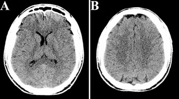
At age 47 this otherwise healthy college educated male real estate appraiser was involved in a motor vehicle accident (MVA) when the patient's vehicle struck a large load of lumber that had come loose and across the center divide from an oncoming flat-bed semi-truck hauling lumber. The patient, as the only occupant, was the seat-belted driver traveling at freeway speed (65 mph) when, without warning, the head-on collision occurred. The collision demolished the vehicle and because of the lumber debris and other traffic factors at the time of impact, it was estimated that 1–3 min transpired before the patient was attended to by individuals at the scene of the accident. According to police reports an eyewitness considered him to be “unconscious.” Actual duration of loss of consciousness (LOC) is unknown, but when emergency medical personnel arrived on the scene, approximately 15 min post injury, his GCS was rated at 14 due to confusion. He had to be extricated from the damaged vehicle (approximately 45 min), after which he was helicopter evacuated to a Trauma I hospital. During transit his GCS was rated a 15 with a similar rating given on ER admission. The impact of lumber through the windshield, produced what the emergency medical responders recorded as “massive bleeding,” especially from one laceration extending approximately 10 cm from the forehead posteriorly. He also complained of severe neck pain. After admission to the ER, CT imaging of the brain was interpreted to be within normal limits (see Figure 1). However, cervical CT imaging demonstrated grade I C4/5 subluxation with fractures of the left C3/4 and C4/5 posterior lateral elements. Clinically, neurosurgical evaluation found him to be neurologically normal except for significant loss of left deltoid strength, left C5 sensation and diminished left triceps reflex. Neurosurgically, he was successfully treated with an anterior fusion at C3/4 and C4/5.

Day-of-injury CT scan that was interpreted as within normal limits. There is slight asymmetry of the anterior horn but this was not considered to be clinically significant. Interhemispheric fissure was at midline and there is appropriate white gray matter differentiation with no edema or blood byproducts noted (scan is in radiological perspective where left is on the viewer's right).
Injury severity classification
GCS was never reported lower than 14 and then recorded as 15 when assessed both initially and later in the ER; in addition, he exhibited no significant anterograde amnesia—all consistent with mild TBI. While some classification schemes of brain injury would potentially categorize this as “complicated mild,” or even mild–moderate because of sustaining a cervical fracture (Alexander, 1995; Varney & Roberts, 1999), he was never clinically assessed as having anything more than mild injury. Furthermore, as indicated above, he was hospitalized for only four days and that was based solely on the fact that he had undergone neurosurgical repair for the cervical fracture. Accordingly, for the purposes of this case study, for the reason given above, the patient was considered to have sustained a mild TBI.
Course of recovery
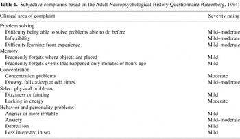
The patient had what would be described as a routine course of recovery. He was hospitalized for only 4 days. He convalesced at home and underwent physical therapy for approximately 3 months. Within 1 month post injury he attempted to return to work, but found it difficult to focus and sustain mental effort. Nonetheless, he gradually increased his workload but never succeeded in reaching his previous level of function prior to his untimely death. His subjective complaints are summarized in Table 1. Subjectively, throughout this post-injury period, he found that his endurance was diminished and that the appraisal work was more difficult. He reported taking longer to do his work with diminished accuracy and “no stamina.” He also reported that before the accident he could conceptualize, visualize and anticipate an appraisal project from “beginning to end” and that he could no longer perform these tasks like he once did. He complained of not being “creative” and lacking “enthusiasm” for his work, like he once did. His wife described him as being “slower” in his ability to process and problem solve. Also, coworkers in his office reported that the amount and accuracy of his work had diminished. These were the factors that actually precipitated the reason for the neuropsychological consultation. During this post-injury period he had numerous follow-up medical visits including examinations by the neurosurgeon, plastic surgeon, family physician, and a physiatrist who was managing his treatment. All performed standard physical exams including cardiac and blood pressure studies with no systemic abnormalities detected. When released by the neurosurgeon he resumed his physical conditioning program without incident or complication. Accordingly, it is unlikely that any of the above mentioned symptoms were due to the underlying cardiovascular disease that would soon take his life, although they cannot be ruled out. Prior to the accident he had no significant health concerns, was completely asymptomatic for any of the problems identified in Table 1, and had never been diagnosed with a pre-existing medical problem that could explain these complaints. All symptoms listed in Table 1 had their onset after this injury.
Subjective complaints based on the Adult Neuropsychological History Questionnaire (Greenberg, 1994)

Premorbid functioning
Vocationally, this patient was very successful in his line of business and had achieved considerable recognition by holding several important state association positions in his professional field. He had a bachelor's degree in finance. School records indicated that he graduated from high school with a 2.2 grade point average (on a 4.0 scale) and maintained approximately a B average in college. His high school records also indicated that on some unspecified intelligence testing he obtained an “IQ score of 109.” The patient was in excellent health at the time of the accident. Prior to the accident he was a mountain hiker, runner and biker and competitively sparred in karate. In college he was a varsity wrestler and in high school played several competitive sports. He had no known history of heart disease prior to his unexpected death and there was no family history of cardiovascular or neurological disease or disorder. Routine pre-injury physical exams were negative although minimally elevated blood pressure was noted, but never thought significant enough to treat. There was no prior head injury, other than incidental contact during competitive sport activities and no history of loss of consciousness for any reason. He was on no medication at the time of the accident and likewise was on no medication at the time of neuropsychological assessment. Nonetheless, it could not be ruled out that he had sustained a prior mild head injury from contact sports he competed in as a youth. He was not obese and his weight was appropriate for his height and age; he engaged in routine physical fitness before and after the accident and had resumed a vigorous exercise program.
Neuropsychological consultation
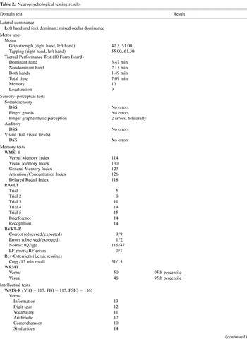
Due to the aforementioned symptoms, the patient was referred for neuropsychological consultation by his treating physiatrist. Results of neuropsychological testing are summarized in Table 2. For the most part, neuropsychological studies, including tests of memory were performed within normal limits. On intellectual testing with the Wechsler Adult Intelligence Scale–Revised (WAIS–R; Wechsler, 1981) he obtained a Full Scale IQ score of 116 with no difference between verbal and performance abilities. On the Wechsler Memory Scale–Revised (WMS–R; Wechsler, 1987) his General Memory Index was 123, and all other indices were within a standard deviation. He made only one error on the Benton Visual Retention Test (BVRT; Benton, 1992), no errors on the verbal component of the Warrington Recognition Memory Test (WRMT; Warrington, 1984) and only two errors on the visual component. On the Paced Auditory Serial Addition Test (PASAT, Lezak, 1995) he made only four errors. Academic performance on the Wide Range Achievement Test–Revision 3 (WRAT–3, Jastak & Wilkinson, 1993) ranged from the 53rd to the 73rd percentile. He displayed superior word fluency as reflected by the mean word fluency on the FAS of 26 and only one item not named on the Boston Naming Test (BNT; Goodglass & Kaplan, 1987). Visual–perceptual function was normal (28/30) on the Hooper Visual Organization Test (HVOT; Hooper, 1958) and normal (36/36) on the Raven Coloured Progressive Matrices test (Raven's CPM; Raven, 1947). The only hint of slightly sub-par performance came on the Digit Symbol subtest on the WAIS–R (scaled score = 9), Trial 1 (5/15) of the Rey Auditory Verbal Learning Test (RAVLT; Rey, 1964) and 15-min recall (13/36) of the Rey-Osterrieth Complex Figure Test (ROCFT; Rey, 1964).
Neuropsychological testing results

Table 2 continued

Self-reported neurobehavioral findings
Table 1 outlines the Adult Neuropsychological History Questionnaire (Greenberg, 1994) of the subjective complaints of the patient that developed after the injury. These are the common complaints associated with post-concussive syndrome as outlined in the Diagnostic and Statistical Manual of Mental Disorders–Fourth Edition (DSM–IV; American Psychiatric Association, 1994, pp. 704–706). Table 2 summarizes the various symptom and self-report inventories. The Symptom Checklist–90–R (SCL–90–R; Derogatis, 1996) results demonstrated T scores of approximately 60 (i.e., 1 SD above the norm) on the somatic and depression scales. His Beck Anxiety and Depression Inventories (BAI and BDI, Beck, 1978, 1990) yielded scores of 5 and 9 (within normal limits) and a short-form of the Minnesota Multiphasic Personality Inventory (MMPI; Faschingbauer, 1974) yielded a T score of 60 on the depression subscale. Taken together these findings indicated his subjective symptoms represented an endorsement of mild symptomatology mostly in terms of cognitive, motivation and drive rather than affective problems at the time of neuropsychological consultation, although there was some endorsement of the latter.
Neuroimaging studies
On admission to the ER the patient underwent CT imaging of the head and the findings were negative (see Figure 1). No other cranial imaging was done acutely. As stated above, CT imaging of the neck demonstrated cervical fractures. Approximately 6 months post injury, as part of the comprehensive work-up for his ongoing symptoms, MR and single photon emission computed tomography (SPECT) studies were undertaken, using 99mTc-HMPAO radionuclide. Unfortunately, on the day neuroimaging studies were performed, technical problems were encountered during MR imaging and the procedure had to be abandoned, with no clinically interpretable images obtained. SPECT imaging was accomplished on that day, with studies reportedly within normal limits. Regrettably, the patient died 11 days later, just 3 days prior to when the MR scan had been rescheduled.
Death
In an attempt to regain his physical stamina, the patient had resumed exercising on a regular basis since about 1 month following the accident. Seven months to the day post injury, during a semi-strenuous morning jog, he collapsed and died. Because of the unexplained death occurring on public property, a full autopsy was ordered. The Office of the Medical Examiner, State of Utah, Department of Health performed the autopsy. The final pathological diagnosis for the cause of death was “ischemic heart disease” due to “arteriosclerotic cardiovascular disease.”
Pathological examination of the brain
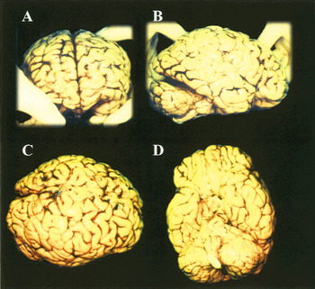
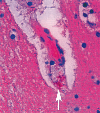
Gross anatomic inspection of the brain and spinal cord was described as normal. Brain weight was assessed at 1,510 g. The outward appearance of the brain was normal and no residual contusions or surface abnormalities were detected (see Figure 2). The brain was routinely sectioned and inspected and no gross abnormalities were identified. Two sections of the brain were taken for microscopic analysis—one frontal lobe, the other temporal lobe at the level of the hippocampus. These sections were examined microscopically where “hemosiderin-laden macrophages” (see Figure 3) were observed in perivascular spaces along with “scattered macrophages in the white matter.” More were reported in the section taken from the frontal lobe than the temporal lobe where the pathologist described the findings as a “sprinkling of lymphocytes.” In the sections viewed, neurons appeared intact. The one section with the hippocampus was specifically examined for pyramidal cell loss, which was not observed. The spinal cord was also sectioned at several levels and no abnormalities, including no hemosiderin or macrophages, were found at any level. One histological slide was decolorized and re-stained with Elastic Van Gieson (EVG) to determine the status of the elastic lamina of vessels. The EVG stain revealed no abnormalities, such as reduplication and thickening common in hypertensive individuals, further supporting the conclusions that the abnormal neuropathological findings were trauma related.

Post-mortem gross brain at the time of autopsy demonstrating no abnormalities. Positioning of the brain was based on routine placement by the pathologist.

Histological findings of hemosiderin (arrow) in a perivascular space indicating prior hemorrhagic lesion, most likely trauma induced, according to the neuropathologist's conclusions.
DISCUSSION
In this case study of mild TBI gross brain inspection revealed no visible pathological findings. As shown in Figure 2, no old surface contusions, regions of focal atrophy or any other visible abnormality was present, confirming the generally normal gross appearance expected in mild TBI. However, at the histological level, presence of hemosiderin-laden macrophages in perivascular spaces, scattered macrophages and lymphocytes in the white matter were identified. The neuropathologist's final diagnosis was “cerebral white matter with increased perivascular hemosiderin laden macrophages” that was attributed to the prior trauma. Appearance of lymphocytes in the white matter was more prevalent in the section taken from the frontal cortex, but this has to be viewed cautiously as only select sections were reviewed as part of the coroner directed neuropathological examination and histological assessment was not based on a systematic sectioning of the brain. Nonetheless, Scheid et al. (2003) using a T2-weighted gradient-echo sequence at 3 Tesla field strength found more than double the signal abnormality consistent with prior microbleeds in the frontal region, than the temporal lobe in an MR imaging study of 66 chronic TBI patients. Although the findings of the current case study presented herein were nonspecific, and no further microscopic or ultrastructural details were available, these abnormalities were consistent with injury to brain parenchyma with concomitant immunologic and phagocytotic response to injury (Grady et al., 2003; Soares et al., 1995; Tracey, 2002). The board-certified neuropathologist who performed the examination concluded that most likely these were trauma-based findings in the brain of this individual who had sustained a mild TBI. No similar pathology was noted in spinal cord sections, even at the level where the fracture occurred.
In their review of the cellular mechanisms in brain injury, McIntosh et al. (1998) suggest that presence of macrophages may be part of progressive tissue necrosis, presumably because they release cytotoxic molecules (including oxygen free radicals and inflammatory cytokines) mediating the local inflammatory response to brain injury and to the phagocytosis of debris from dying cells. Macrophages may also be present as part of reparative responses to brain injury as well (Silver & Miller, 2004). Experimental models of brain injury that routinely demonstrate macrophages at perilesional sites (Schwab et al., 2001; Vela et al., 2002) have even shown that the degree of macrophage presence positively correlates with the severity of damage. Also, animal models have shown that where no gross detection of hemorrhaging has been identified, hemosiderin staining may be evident histologically (Adelson et al., 2001). Furthermore, the microvasculature of the brain is also susceptible to shear injury and microhemorrhaging (Dietrich et al., 1994; Scheid et al., 2003). Accordingly, presence of hemosiderin is likely a marker of parenchymal injury as well (Oehmichen et al., 2003; Raghupathi & Margulies, 2002).
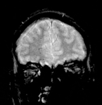
By their very nature, case study reports have to be viewed cautiously and conservatively. This patient did die from what was assessed by the coroner's office as “arteriosclerotic cardiovascular disease,” even though he had never been assessed for having, or treated for, any cardiovascular condition prior to his death. Presence of occult cardiovascular disease at the time of vigorous exercise could have been the trigger for sudden death (see Albert et al., 2000). How his cardiovascular condition may have contributed to the histological findings is not known since, in a post-mortem case study such as this, there is no post-hoc method to control for these factors. On the other hand, given the nature of the injury, the timing of his symptoms and the relevant histopathological findings consistent with trauma, the likely explanation is that these findings were traumatically related. Further credence for this conclusion comes from another recent case, presented in Figure 4, with very similar clinical history (i.e., mild TBI, GCS = 14), where the gradient recalled echo MR scan (Besenski, 2002; Fazekas et al., 1999; Preul et al., 2003) over 1 yr post injury demonstrated numerous small hemosiderin deposits. Histological studies of brain injury have shown disseminated hemosiderin deposits within brain parenchyma (Oehmichen et al., 2003). Other neuroimaging studies have clearly demonstrated the increased frequency of hemosiderin deposits in frontal over temporal regions in TBI (Scheid et al., 2003). However, hemosiderin deposits of the size seen in the patient in this case study would have been under the threshold of detection by MR imaging (Wardlaw & Statham, 2000). Immunological response and presence of macrophages is indicative of a response to injury (Adelson et al., 2001; Folkerts et al., 1998; Grady et al., 2003; Medana & Esiri, 2003; Stoll et al., 1989) and it was concluded by the neuropathologist that these neuropathological findings were attributable to the trauma in question.

Frontal coronal view of MRI (gradient recalled echo) in a separate patient, but with similar history of mild TBI. At the time of this injury, the patient was a passenger in a vehicle involved in a rollover accident, following rear impact. He had apparent brief LOC and paramedic rated his GCS as 14 at the scene, but 15 on arrival to the ER. Note the scattered hemosiderin deposits (black signal) scattered throughout the frontal region bilaterally, but more so in the right frontal (scan is in radiological perspective). The patient was 16 years of age at the time of injury, 17 years old at the time of imaging and without any history of prior head trauma or any other risk factor for microhemorrhaging. He was discharged from the hospital on the day of injury following several hours of observation where his mental status did not change. He had similar subjective symptoms and complaints as the patient who is the focus of the case study, independently verified by high school teachers who knew him before and after the injury.
Neuropsychologically, this patient's studies were broadly within normal limits, yet his subjective complaints, confirmed both by his spouse and coworkers, of changes in cognitive abilities, personality and temperament were consistent with mild TBI. If a liberal interpretation of his neuropsychological studies is undertaken, comparing to either his reported pre-morbid IQ of 109 or his post-injury WAIS–R Full Scale IQ of 116, there were only three tasks where the patient performed below expectations: a speed of processing task (Digit Symbol) and two memory tasks (Trial 1 RAVLT performance and 15-min recall of the ROCFT). These slightly diminished performances levels would be fitting with the patient's subjective complaint that some of his cognitive abilities were “off.” However, these are also subtle cognitive changes that occur in individuals diagnosed with depression and/or post-traumatic stress disorder (Brandes et al., 2002).
Even if the above mentioned tests are detecting subtle abnormalities in cognitive status of this patient, the remainder of his neuropsychological studies would have to be considered entirely within normal limits, despite his subjective cognitive and neurobehavioral complaints. The ecological validity of neuropsychological testing has limitations (Silver, 2000), where the controlled environment of the laboratory does not necessarily match the cognitive challenges of the outside world. In this case, the neuropsychological studies provide minimal insight into the residual neurobehavioral effects of this head injury, but the patient's presenting complaints and alteration in job performance are most fitting with the behavioral indicators of what DSM–IV labels a post-concussive syndrome (pp. 704–706). Neuropsychologically, the patient's memory was generally within normal limits. His complaints centered more on high-level executive problems, drive and motivation. Interestingly, recent research in the area of multiple sclerosis has implicated axonal injury as a factor in fatigue (Tartaglia et al., 2004). Accordingly, the connection between white matter burden and fatigue may be associated with the white matter damage in TBI.
Unfortunately, the brain was not preserved for more complete analysis as the findings were based on a clinical autopsy and what is reported here is based entirely on the final coroner's report, where the neuropathological analysis was performed by a board certified neuropathologist. These findings are not specific, but the presence of hemosiderin laden macrophages in the white matter clearly indicate perturbation of brain parenchyma or blood vessels and the likelihood of at least some microhemorrhaging secondary to trauma (see Davis & Robertson, 1985; Graham et al., 2002; Malamud & Hirano, 1973; Yokota et al., 2002). Lastly, with regard to hemosiderin deposits, it should be pointed out that they can occur spontaneously and for a variety of non-traumatic reasons (Imaizumi et al., 2004; Johnston & Marx, 2003; Wong et al., 2003).
It should be noted that had the patient lived, litigation would have likely occurred. However, the patient was clinically referred by his treating physiatrist; he was following a rigorous physical retraining and reconditioning program in an attempt to recover and had returned to work. As indicated above, the neuropsychological test performance was generally within normal limits and all validity and measures of effort were performed normally as well. Accordingly, the issue of potential litigation had he lived, was not thought to have affected his performance on neuropsychological tests. Also, it is not known what role his latent cardiovascular status may have played with respect to his subjective complaints.
This study has definite implications for cases of mild TBI. As indicated in the introduction, there is compelling evidence that presence of prior head injury, even mild, constitutes a vulnerability factor for later in life dementia (Guo et al., 2000; Plassman et al., 2000) and long-term neuropsychiatric sequela (Fann et al., 2004; Holsinger et al., 2002; Jorge et al., 2004). Obviously since in most cases of mild TBI patients resume approximate levels of premorbid ability, the increased risk of dementia implies underlying latent neural damage. In most cases of mild TBI conventional neuroimaging studies are negative, as they were in this case. However, recent studies have clearly demonstrated quantitative, MR signal, and biochemical markers of injury to the white matter in mild TBI where seemingly conventional imaging detects no abnormality (Bigler, 2001, 2002; Brooks et al., 2000; Garnett et al., 2000a, 2000b; McGowan et al., 2000; Sinson et al., 2001; Thatcher et al., 1997; Zhang et al., 2003). Furthermore, MacKenzie et al. (2002) in a longitudinal study have demonstrated degenerative changes that accompany even mild TBI. This case histologically demonstrates markers of injury present in the white matter below conventional methods of detection using contemporary neuroimaging methods, more so in the frontal region. Furthermore, behaviorally the patient presented with classic post-concussive symptomatology that had persisted until his unexpected death 7 months post injury. Slowed neural transmission or disrupted integration of neural pathways due to white matter injury (Adams et al., 2000; Gennarelli et al., 1998; Mathias et al., 2004; Povlishock & Christman, 1995; Smith & Meaney, 2000), particularly in the frontal region, could certainly be at the basis of the patient's complaints following this mild TBI.
In summary, the post-mortem findings in a case of mild TBI (brief LOC; initial GCS of 14) are presented that documents neuropathology under the threshold of detection by CT or SPECT imaging and even direct visual inspection of the gross brain. Markers of previous trauma were identified in the white matter under microscopic analysis. At a minimum these findings demonstrated the likelihood of perturbation of white matter, and presence of microhemorrhagic lesions in mild TBI, in both frontal and temporal lobe regions. The connection between sustaining a mild brain injury, the persistence of neurobehavioral symptoms and problems in this patient, most of which were not observed in standardized neuropsychological testing, and the histopathology of white matter injury is made.
ACKNOWLEDGMENTS
Parts of the ongoing traumatic brain injury (TBI) research have been supported by funds from the Ira Fulton Foundation. The reviews of Tracy McIntosh, Ph.D., of earlier revisions of this manuscript are gratefully acknowledged as is the manuscript assistance of Jo Ann Petrie.
References
REFERENCES
Adams, J.H.,
Graham, D.I., &
Jennett, B.
(2000).
The neuropathology of the vegetative state after an acute brain
insult.
Brain,
123,
1327–1338.CrossRefGoogle Scholar
Adelson, P.D.,
Jenkins, L.W.,
Hamilton, R.L.,
Robichaud, P.,
Tran, M.P., &
Kochanek, P.M.
(2001).
Histopathologic response of the immature rat to diffuse traumatic
brain injury.
Journal of Neurotrauma,
18,
967–977.CrossRefGoogle Scholar
Albert, C.M.,
Mittleman, M.A.,
Chae, C.U.,
Lee, I.M.,
Hennekens, C.H., &
Manson, J.E.
(2000).
Triggering of sudden death from cardiac causes by vigorous
exertion.
New England Journal of Medicine,
343,
1355–1361.CrossRefGoogle Scholar
Alexander, M.P.
(1995).
Mild traumatic brain injury: Pathophysiology, natural history, and
clinical management.
Neurology,
45,
1253–1260.CrossRefGoogle Scholar
American Psychiatric Association.
(1994).
Diagnostic and statistical manual of mental disorders
(4th ed.).
Washington, DC:
Author.
Arfanakis, K.,
Haughton, V.M.,
Carew, J.D.,
Rogers, B.P.,
Dempsey, R.J., &
Meyerand, M.E.
(2002).
Diffusion tensor MR imaging in diffuse axonal injury.
American Journal of Neuroradiology,
23,
794–802.Google Scholar
Beck, A.T.
(1978).
Beck Depression Inventory manual.
San Antonio, TX:
The Psychological Corporation.
Beck, A.T.
(1990).
Beck Anxiety Inventory.
San Antonio, TX:
The Psychological Corporation.
Benton, A.L.
(1992).
Benton Visual Retention Test–5th Edition.
New York:
The Psychological Corporation.
Besenski, N.
(2002).
Traumatic injuries: Imaging of head injuries.
European Radiology,
12,
1237–1252.CrossRefGoogle Scholar
Bigler, E.D.
(2001).
Quantitative magnetic resonance imaging in traumatic brain injury.
Journal of Head Trauma Rehabilitation,
16,
1–21.CrossRefGoogle Scholar
Bigler, E.D.,
Anderson, C.V., &
Blatter, D.D.
(2002).
Temporal lobe morphology in normal aging and traumatic brain
injury.
AJNR American Journal of Neuroradiology,
23,
255–266.Google Scholar
Blumbergs, P.C.,
Scott, G.,
Manavis, J.,
Wainwright, H.,
Simpson, D.A., &
McLean, A.J.
(1994).
Staining of amyloid precursor protein to study axonal damage in
mild head injury.
Lancet,
344,
1055–1056.CrossRefGoogle Scholar
Brandes, D.,
Ben-Schachar, G.,
Gilboa, A.,
Bonne, O.,
Freedman, S., &
Shalev, A.Y.
(2002).
PTSD symptoms and cognitive performance in recent trauma survivors.
Psychiatry Research,
110,
231–238.CrossRefGoogle Scholar
Brooks, W.M.,
Stidley, C.A.,
Petropoulos, H.,
Jung, R.E.,
Weers, D.C.,
Friedman, S.D.,
Barlow, M.A.,
Sibbitt, W.L., &
Yeo, R.A.
(2000).
Metabolic and cognitive response to human traumatic brain injury: A
quantitative proton magnetic resonance study.
Journal of Neurotrauma,
17,
629–640.CrossRefGoogle Scholar
Davis, R.L. &
Robertson, D.M.
(1985).
Textbook of neuropathology.
Baltimore:
Williams & Wilkins.
Derogatis, L.R.
(1996).
Symptom Checklist–90–R.
Minneapolis, MN:
National Computer Systems, Inc.
Dietrich, W.D.,
Alonso, O., &
Halley, M.
(1994).
Early microvascular and neuronal consequences of traumatic brain
injury: A light and electron microscopic study in rats.
Journal of Neurotrauma,
11,
289–301.CrossRefGoogle Scholar
Fann, J.R.,
Burington, B.,
Leonetti, A.,
Jaffe, K.,
Katon, W.J., &
Thompson, R.S.
(2004).
Psychiatric illness following traumatic brain injury in an adult
health maintenance organization population.
Archives of General Psychiatry,
61,
53–61.CrossRefGoogle Scholar
Faschingbauer, T.R.
(1974).
A 166-item written short form of the group MMPI: The FAM.
Journal of Consulting and Clinical Psychology,
42,
645–655.CrossRefGoogle Scholar
Fazekas, F.,
Kleinert, R.,
Roob, G.,
Kleinert, G.,
Kapeller, P.,
Schmidt, R., &
Hartung, H.P.
(1999).
Histopathologic analysis of foci of signal loss on gradient-echo
T2*-weighted MR images in patients with spontaneous intracerebral
hemorrhage: Evidence of microangiopathy-related microbleeds.
American Journal of Neuroradiology,
20,
637–642.Google Scholar
Folkerts, M.M.,
Berman, R.F.,
Muizelaar, J.P., &
Rafols, J.A.
(1998).
Disruption of MAP-2 immunostaining in rat hippocampus after
traumatic brain injury.
Journal of Neurotrauma,
15,
349–363.CrossRefGoogle Scholar
Garnett, M.R.,
Blamire, A.M.,
Corkill, R.G.,
Cadoux-Hudson, T.A.D.,
Rajagopalan, B., &
Styles, P.
(2000a).
Early proton magnetic resonance spectroscopy in normal-appearing
brain correlates with outcome in patients following traumatic brain
injury.
Brain,
123,
2046–2054.Google Scholar
Garnett, M.R.,
Blamire, A.M.,
Rajagopalan, B.,
Styles, P., &
Cadoux-Hudson, T.A.D.
(2000b).
Evidence for cellular damage in normal-appearing white matter
correlates with injury severity in patients following traumatic brain
injury: A magnetic resonance spectroscopy study.
Brain,
123,
1403–1409.Google Scholar
Gennarelli, T.A.,
Thibault, L.E., &
Graham, D.I.
(1998).
Diffuse axonal injury: An important form of traumatic brain damage.
Neuroscientist,
4,
202–215.CrossRefGoogle Scholar
Goetz, P.,
Blamire, A.,
Rajagopalan, B.,
Cadoux-Hudson, T.,
Young, D., &
Styles, P.
(2004).
Increase in apparent diffusion coefficient in normal appearing
white matter following human traumatic brain injury correlates with
injury severity.
Journal of Neurotrauma,
21,
645–654.CrossRefGoogle Scholar
Goodglass, H. &
Kaplan, E.
(1987).
The assessment of aphasia and related disorders
(2nd ed.).
Philadelphia:
Lea & Febiger.
Grady, M.S.,
Charleston, J.S.,
Maris, D.,
Witgen, B.M., &
Lifshitz, J.
(2003).
Neuronal and glial cell number in the hippocampus after
experimental traumatic brain injury: Analysis by stereological
estimation.
Journal of Neurotrauma,
20,
929–941.CrossRefGoogle Scholar
Graham, D.I.,
Gennarelli, T.A., &
McIntosh, T.K.
(2002).
Trauma. In
P.L. Lantos (Ed.),
Greenfield's neuropathology, pp.
823–895.
New York:
Hodder Arnold.
Greenberg, G.D.
(1994).
Adult neuropsychological history.
Worthington, OH:
IDS Publishing Corporation.
Guo, Z.,
Cupples, L.A.,
Kurz, A.,
Auerbach, S.H.,
Volicer, L.,
Chui, H.,
Green, R.C.,
Sadovnick, AD.,
Duara, R.,
DeCarli, C.,
Johnson, K.,
Go, R.C.,
Growdon, J.H.,
Haines, J.L.,
Kukull, W.A., &
Farrer, L.A.
(2000).
Head injury and the risk of AD in the MIRAGE study.
Neurology,
54,
1316–1323.CrossRefGoogle Scholar
Hofman, P.A.M.,
Verhey, F.R.J.,
Wilmink, J.T.,
Rozendaal, N., &
Jolles, J.
(2002).
Brain lesions in patients visiting a memory clinic with
postconcussional sequelae after mild to moderate brain injury.
Journal of Neuropsychiatry and Clinical Neurosciences,
14,
176–184.CrossRefGoogle Scholar
Holsinger, T.,
Steffens, D.C.,
Phillips, C.,
Helms, M.J.,
Havlik, R.J.,
Breitner, J.C.,
Guralnik, J.M., &
Plassman, B.L.
(2002).
Head injury in early adulthood and the lifetime risk of depression.
Archives of General Psychiatry,
59,
17–22.CrossRefGoogle Scholar
Hooper, H.E.
(1958).
The Hooper Visual Organization Test manual.
Beverley Hills, CA:
Western Psychological Services.
Imaizumi, T.,
Horita, Y.,
Chiba, M.,
Hashimoto, Y.,
Honma, T., &
Niwa, J.
(2004).
Dot-like hemosiderin spots on gradient echo T2-weighted magnetic
resonance imaging are associated with past history of small vessel
disease in patients with intracerebral hemorrhage.
Journal of Neuroimaging,
14,
251–257.CrossRefGoogle Scholar
Jastak, S. &
Wilkinson, G.
(1993).
The Wide Range Achievement Test–Revision 3.
Wilmington, DE:
Jastak Associates.
Johnston, K.C. &
Marx, W.F., Jr.
(2003).
Microhemorrhages on gradient echo MRI.
Neurology,
60,
518.CrossRefGoogle Scholar
Jorge, R.E.,
Robinson, R.G.,
Moser, D.,
Tateno, A.,
Crespo-Facorro, B., &
Arndt, S.
(2004).
Major depression following traumatic brain injury.
Archives of General Psychiatry,
61,
42–50.CrossRefGoogle Scholar
Kumar, R.,
Gupta, R.K.,
Husain, M.,
Vatsal, D.K.,
Chawla, S.,
Rathore, R.K., &
Pradhan, S.
(2003).
Magnetization transfer MR imaging in patients with posttraumatic
epilepsy.
AJNR American Journal of Neuroradiology,
24,
218–224.Google Scholar
Lewine, J.D.,
Davis, J.T.,
Sloan, J.H.,
Kodituwakku, P.W., &
Orrison, W.W.
(1999).
Neuromagnetic assessment of pathophysiologic brain activity induced
by minor head trauma.
American Journal of Neuroradiology,
20,
857–866.Google Scholar
Lezak, M.D.
(1995).
Neuropsychological assessment
(3rd ed.).
New York:
Oxford University Press.
MacKenzie, J.D.,
Siddiqi, F.,
Babb, J.S.,
Bagley, L.J.,
Mannon, L.J.,
Sinson, G.P., &
Grossman, R.I.
(2002).
Brain atrophy in mild or moderate traumatic brain injury: A
longitudinal quantitative analysis.
American Journal of Neuroradiology,
23,
1509–1515.Google Scholar
Malamud, N. &
Hirano, A.
(1973).
Atlas of neuropathology
(2nd ed.).
Berkley, CA:
University of California Press.
Mathias, J.L.,
Beall, J.A., &
Bigler, E.D.
(2004).
Neuropsychological and information processing deficits following
mild traumatic brain injury.
Journal of the International Neuropsychological Society,
10,
286–297.Google Scholar
McGowan, J.C.,
Yang, J.H.,
Plotkin, R.C.,
Grossman, R.I.,
Umile, E.M.,
Cecil, K.M., &
Bagley, L.J.
(2000).
Magnetization transfer imaging in the detection of injury
associated with mild head trauma.
American Journal of Neuroradiology,
21,
875–880.Google Scholar
McIntosh, T.K.,
Saatman, K.E.,
Raghupathi, R.,
Graham, D.I.,
Smith, D.H.,
Lee, V.M.-Y., &
Trojanowski, J.Q.
(1998).
The molecular and cellular sequelae of experimental traumatic brain
injury: Pathogenetic mechanisms.
Neuropathology and Applied Neurobiology,
24,
251–267.CrossRefGoogle Scholar
Medana, I.M. &
Esiri, M.M.
(2003).
Axonal damage: A key predictor of outcome in human CNS diseases.
Brain,
126,
515–530.CrossRefGoogle Scholar
Mehta, K.M.,
Ott, A.,
Kalmijn, S.,
Slooter, A.J.C.,
Van Duijn, C.M.,
Hoffman, A., &
Breteler, M.M.B.
(1999).
Head trauma and risk of dementia and Alzheimer's disease: The
Rotterdam Study.
Neurology,
53,
1959–1962.CrossRefGoogle Scholar
Oehmichen, M.,
Walter, T.,
Meissner, C., &
Friedrich, H.-J.
(2003).
Time course of cortical hemorrhages after closed traumatic brain
injury: Statistical analysis of posttraumatic histomorphological
alterations.
Journal of Neurotrauma,
20,
87–103.CrossRefGoogle Scholar
O'Meara, E.S.,
Kukull, W.A.,
Sheppard, L.,
Bowen, J.D.,
McCormick, W.C.,
Teri, L.,
Pfanschmidt, M.,
Thompson, J.D.,
Schellenberg, G.D., &
Larson, E.B.
(1997).
Head injury and risk of Alzheimer's disease by Apolipoprotein
E genotype.
American Journal of Epidemiology,
146,
373–384.CrossRefGoogle Scholar
Ommaya, A.K.,
Goldsmith, W., &
Thibault, L.
(2002).
Biomechanics and neuropathology of adult and paediatric head
injury.
British Journal of Neurosurgery,
16,
220–242.CrossRefGoogle Scholar
Plassman, B.L.,
Havlik, R.J.,
Steffens, D.C.,
Helms, M.J.,
Newman, T.N.,
Drosdick, D.,
Phillips, C.,
Gau, B.A.,
Welsh-Bohmer, K.A.,
Burke, J.R.,
Guralnik, J.M., &
Breitner, J.C.S.
(2000).
Documented head injury in early adulthood and risk of
Alzheimer's disease and other dementias.
Neurology,
55,
1158–1166.CrossRefGoogle Scholar
Povlishock, J. &
Christman, C.
(1995).
The pathobiology of traumatically induced axonal injury in animals
and humans: A review of current thoughts.
Journal of Neurotrauma,
12,
555–564.CrossRefGoogle Scholar
Preul, C.,
Kuhn, B.,
Lang, E.W.,
Mehdorn, H.M.,
Heller, M., &
Link, J.
(2003).
Differentiation of cerebral tumors using multi-section echo planar
MR perfusion imaging.
European Journal of Radiology,
48,
244–251.CrossRefGoogle Scholar
Pritchard, D.A.
(1998).
Tests of neuropsychological malingering (version 2.0).
New York:
CRC Press, LLC.
Raghupathi, R. &
Margulies, S.S.
(2002).
Traumatic axonal injury after closed head injury in the neonatal
pig.
Journal of Neurotrauma,
19,
843–853.CrossRefGoogle Scholar
Raven, J.C.
(1947).
Colored Progressive Matrices Sets A, Ab, B.
London:
H.K. Lewis.
Rey, A.
(1964).
L'examen clinique en psychologie [The clinical examination
in psychology].
Paris:
Presse Universitaire de France.
Scheid, R.,
Preul, C.,
Gruber, O.,
Wiggins, C., &
von Cramon, D.Y.
(2003).
Diffuse axonal injury associated with chronic traumatic brain
injury: Evidence from T2*-weighted gradient-echo imaging at 3 T.
American Journal of Neuroradiology,
24,
1049–1056.Google Scholar
Schofield, P.W.,
Tang, M.,
Marder, K.,
Bell, K.,
Dooneief, G.,
Chun, M.,
Sano, M.,
Stern, Y., &
Mayeux, R.
(1997).
Alzheimer's disease after remote head injury: An incidence
study.
Journal of Neurology, Neurosurgery, and Psychiatry,
62,
119–124.CrossRefGoogle Scholar
Schwab, J.M.,
Seid, K., &
Schluesener, H.J.
(2001).
Traumatic brain injury induces prolonged accumulation of
cyclooxygenase-1 expressing microglia/brain macrophages in
rats.
Journal of Neurotrauma,
18,
881–890.CrossRefGoogle Scholar
Silver, C.H.
(2000).
Ecological validity of neuropsychological assessment in childhood
traumatic brain injury.
Journal of Head Trauma Rehabilitation,
15,
973–988.CrossRefGoogle Scholar
Silver, J. &
Miller, J.H.
(2004).
Regeneration beyond the glial scar.
Neuroscience,
5,
146–156.CrossRefGoogle Scholar
Sinson, G.P.,
Bagley, L.J.,
Cecil, K.M.,
Torchia, M.,
McGowan, J.C.,
Lenkinski, R.E.,
McIntosh, T.K., &
Grossman, R.I.
(2001).
Magnetization transfer imaging and proton MR spectroscopy in the
evaluation of axonal injury: Correlation with clinical outcome after
traumatic brain injury.
American Journal of Neuroradiology,
22,
143–151.Google Scholar
Smith, D.H. &
Meaney, D.F.
(2000).
Axonal damage in traumatic brain injury.
Neuroscientist,
6,
483–495.CrossRefGoogle Scholar
Soares, H.D.,
Hicks, R.R.,
Smith, D., &
McIntosh, T.K.
(1995).
Inflammatory leukocytic recruitment and diffuse neuronal
degeneration are separate pathological processes resulting from
traumatic brain injury.
Journal of Neuroscience,
15,
8223–8233.CrossRefGoogle Scholar
Stoll, G.,
Trapp, B.D., &
Griffin, J.W.
(1989).
Macrophage function during Wallerian degeneration of rat optic
nerve: Clearance of degenerating myelin and Ia expression.
Journal of Neuroscience,
9,
2327–2335.CrossRefGoogle Scholar
Strich, S.J.
(1956).
Diffuse degeneration of the cerebral white matter in severe
dementia following head injury.
Journal of Neurology, Neurosurgery, and Psychiatry,
19,
163–185.CrossRefGoogle Scholar
Strich, S.J.
(1961).
Shearing of nerve fibres as a cause of brain damage due to head
injury. A pathological study of twenty cases.
Lancet,
2,
443–448.Google Scholar
Tartaglia, M.C.,
Narayanan, S.,
Francis, S.J.,
Santos, A.C.,
De Stefano, N.,
Lapierre, Y., &
Arnold, D.L.
(2004).
The relationship between diffuse axonal damage and fatigue in
multiple sclerosis.
Archives of Neurology,
61,
201–207.CrossRefGoogle Scholar
Teasdale, G. &
Jennett, B.
(1974).
Assessment of coma and impaired consciousness: A practical scale.
Lancet,
2,
81–84.CrossRefGoogle Scholar
Thatcher, R.W.,
Camacho, M.,
Salazar, A.,
Linden, C.,
Biver, C., &
Clarke, L.
(1997).
Quantitative MRI of the gray-white matter distribution in traumatic
brain injury.
Journal of Neurotrauma,
14,
1–14.Google Scholar
Varney, N.R. &
Roberts, R.J.
(1999).
The evaluation and treatment of mild traumatic brain injury.
Mahwah, NJ:
Lawrence Erlbaum Associates.
Vela, J.M.,
Yáñez, A.,
González, B., &
Castellano, B.
(2002).
Time course of proliferation and elimination of
microglia/macrophages in different neurodegenerative
conditions.
Journal of Neurotrauma,
19,
1503–1520.CrossRefGoogle Scholar
Wardlaw, J.M. &
Statham, P.F.
(2000).
How often is haemosiderin not visible on routine MRI following
traumatic intracerebral haemorrhage? Neuroradiology,
42,
81–84.Google Scholar
Warrington, E.K.
(1984).
Warrington Recognition Memory Test.
Windsor, UK:
NFER-Nelson.
Wechsler, D.
(1981).
Wechsler Adult Intelligence Scale–Revised (WAIS–R).
San Antonio, TX:
The Psychological Corporation.
Wechsler, D.
(1987).
Wechsler Memory Scale–Revised manual.
San Antonio, TX:
The Psychological Corporation.
Wong, K.S.,
Chan, Y.L.,
Liu, J.Y.,
Gao, S., &
Lam, W.W.
(2003).
Asymptomatic microbleeds as a risk factor for aspirin-associated
intracerebral hemorrhages.
Neurology,
60,
511–513.CrossRefGoogle Scholar
Yokota, H.,
Naoe, Y.,
Nakabayashi, M.,
Unemoto, K.,
Kushimoto, S.,
Kurokawa, A.,
Node, Y., &
Yamamoto, Y.
(2002).
Cerebral endothelial injury in severe head injury: The significance
of measurements of serum thrombomodulin and the von Willebrand
factor.
Journal of Neurotrauma,
19,
1007–1015.CrossRefGoogle Scholar
Zhang, L.,
Ravdin, L.D.,
Relkin, N.,
Zimmerman, R.D.,
Jordan, B.,
Lathan, W.E., &
Ulug, A.M.
(2003).
Increased diffusion in the brain of professional boxers: A
preclinical sign of traumatic brain injury? American Journal of Neuroradiology,
24,
52–57.Google Scholar
Day-of-injury CT scan that was interpreted as within normal limits. There is slight asymmetry of the anterior horn but this was not considered to be clinically significant. Interhemispheric fissure was at midline and there is appropriate white gray matter differentiation with no edema or blood byproducts noted (scan is in radiological perspective where left is on the viewer's right).
Subjective complaints based on the Adult Neuropsychological History Questionnaire (Greenberg, 1994)
Neuropsychological testing results
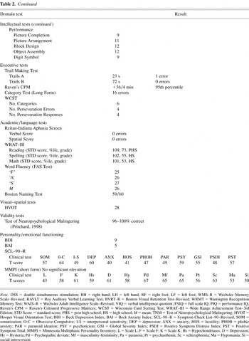
Table 2 continued
Post-mortem gross brain at the time of autopsy demonstrating no abnormalities. Positioning of the brain was based on routine placement by the pathologist.
Histological findings of hemosiderin (arrow) in a perivascular space indicating prior hemorrhagic lesion, most likely trauma induced, according to the neuropathologist's conclusions.
Frontal coronal view of MRI (gradient recalled echo) in a separate patient, but with similar history of mild TBI. At the time of this injury, the patient was a passenger in a vehicle involved in a rollover accident, following rear impact. He had apparent brief LOC and paramedic rated his GCS as 14 at the scene, but 15 on arrival to the ER. Note the scattered hemosiderin deposits (black signal) scattered throughout the frontal region bilaterally, but more so in the right frontal (scan is in radiological perspective). The patient was 16 years of age at the time of injury, 17 years old at the time of imaging and without any history of prior head trauma or any other risk factor for microhemorrhaging. He was discharged from the hospital on the day of injury following several hours of observation where his mental status did not change. He had similar subjective symptoms and complaints as the patient who is the focus of the case study, independently verified by high school teachers who knew him before and after the injury.
 You have
Access
- 108
- Cited by