Implications
Supplementing an immunomodulatory feed ingredient (Omnigen-AF; Phibro Animal Health, Teaneck, NJ, USA) to beef heifers did not modulate the physiological and leukocyte-mediated reactions elicited by bacterial lipopolysaccharide (LPS) administration. Heifers from this experiment were in adequate health conditions, whereas Omnigen-AF supplementation may strengthen innate immunity in immunosuppressed animals. Hence, Omnigen-AF supplementation did not provide additional benefits to innate immunocompetence of healthy beef cattle.
Introduction
The innate immune system is imperative for animal immunocompetence by eliciting immediate reactions to control or eliminate pathogenic infections (Abbas and Lichtman, Reference Abbas and Lichtman2007). These include leukocyte-mediated responses that initiate the attack against pathogens, but also regulate inflammatory and metabolic responses that affect productive and reproductive traits in livestock (Klasing and Korver, Reference Klasing and Korver1997; Cooke, Reference Cooke2017). Therefore, management alternatives to strengthen innate immunity are critical for welfare and productivity in livestock systems, such as beef cattle operations.
Nutritional status regulates the ability of the innate immune system to properly respond and control pathogenic infections (Carroll and Forsberg, Reference Carroll and Forsberg2007); hence, several nutritional strategies to enhance innate immunity in cattle have been investigated (Cooke, Reference Cooke2017). As example, Omnigen-AF (OMN) is a non-antibiotic feed ingredients with immunomodulatory properties associated with improved leukocyte function, surface L-selectin concentration, and phagocytosis of extracellular pathogens in dairy cattle (Wang et al., Reference Wang, Puntenney, Burton and Forsberg2009; Ryman et al., Reference Rodrigues, Cooke, Marques, Arispe, Keisler and Bohnert2013; Nace et al., 2014). Accordingly, Lippolis et al. (Reference Lippolis, Cooke, Schubach, Marques and Bohnert2017a) reported heightened innate immunity in newly weaned feedlot cattle supplemented with OMN. However, no research has fully established the leukocyte-mediated and overall immunomodulatory properties of OMN in beef cattle. One strategy to elicit the innate immune system and investigate such parameters is via bacterial LPS administration, and collection of whole blood for leukocyte mRNA expression (Düvel et al., Reference Düvel, Maaß, Heppelmann, Hussen, Koy, Piechotta, Sandra, Smith, Sheldon, Dieuzy-Labaye, Zieger and Schuberth2014; Rodrigues et al., Reference Perdew, Vanden Heuvel and Peters2015). Based on this rationale, we hypothesized that OMN supplementation will enhance innate immunity of beef cattle upon LPS administration, including leukocyte-mediated responses associated with early pathogenic control. Therefore, this experiment evaluated the effects of OMN supplementation on metabolic, inflammatory and whole blood mRNA responses of beef heifers administered bacterial LPS.
Material and methods
This experiment was conducted at the Oregon State University – Eastern Oregon Agricultural Research Center (Burns, OR, USA). All animals were cared for in accordance with acceptable practices and experimental protocols reviewed and approved by the Oregon State University, Institutional Animal Care and Use Committee (#4891).
Animals and treatments
Eight halter-trained, non-pregnant, non-lactating, nulliparous Angus×Hereford heifers were utilized (676±4 days of age). This animal model was chosen to focus on innate immune responses without the physiological influence of body development, pregnancy and lactation. During the experimental period, heifers were housed in an enclosed barn in individual pens (3×5 m) with ad libitum access to meadow foxtail (Alopecurus pratensis L.) hay, water and a granulated commercial vitamin+mineral mix (Cattleman’s Choice, Performix Nutrition Systems, Nampa, ID, USA) containing 14% Ca, 10% P, 16% NaCl, 1.5% Mg, 3200 mg/kg of Cu, 65 mg/kg of I, 900 mg/kg of Mn, 140 mg/kg of Se, 6000 mg/kg of Zn, 136 000 IU/kg of vitamin A, 13 000 IU/kg of vitamin D3 and 50 IU/kg of vitamin E.
Heifers were weighed for 3 consecutive days before the beginning of the experiment, and values were averaged as initial BW (473±8 kg). Heifers were ranked by initial BW into one of two groups with equivalent BW, and groups were assigned to crossover design containing two periods of 34 days each. Within each period, heifers from each group individually received (as-fed basis) 227 g/day of dried distillers grains (DDG) in addition to: (1) 56 g/heifer daily of OMN (n=4 heifers/period) or (2) no immunomodulatory feed supplementation (CON; n=4 heifers/period). Treatments were offered once daily at 0730 h before hay feeding. All heifers completely consumed their treatments within 20 min. The inclusion rate of OMN was according to the manufacturer’s recommendations for 2-year-old heifers and previous research in dairy cattle (Wang et al., Reference Wang, Puntenney, Burton and Forsberg2009; Ryman et al., Reference Rodrigues, Cooke, Marques, Arispe, Keisler and Bohnert2013; Brandão et al., Reference Brandão, Cooke, Corrá, Piccolo, Gennari, Leiva and Vasconcelos2016), whereas DDG was used as carrier for OMN to ensure adequate and complete treatment consumption.
Heifer BW was recorded on days 27 and 28 of each period, and averaged as pre-LPS BW (period 1=478±8 kg; period 2=486±7 kg). On day 28 (0800 h), heifers received an intravenous bolus dose of bacterial LPS (0.5 μg/kg of BW; Escherichia coli 0111:B4; Sigma-Aldrich, St. Louis, MO, USA). Bacterial LPS was dissolved into 5-ml of 0.9% saline immediately before administration, and infused using 10 ml sterile syringes (Monoject Covidien Animal Health; Mansfield, MA, USA) according to their pre-LPS BW. The 5-ml saline+LPS solution was infused into each heifer at 1 ml/s. Between periods, heifers were housed in a single paddock (30×25 m) with ad libitum access to meadow foxtail hay, water and the aforesaid granulated commercial vitamin+mineral mix for 10 days.
Sampling
Hay DM intake was evaluated daily from day 0 to 34 of each period by weighing and collecting samples of the offered and non-consumed hay at 0730 h, and drying samples for 96 h at 50°C in forced-air ovens for DM calculation. Samples of offered hay and DDG were collected twice within each period, pooled and analyzed for nutrient content by a commercial laboratory (Dairy One Forage Laboratory, Ithaca, NY, USA). Samples were analyzed by wet chemistry procedures for concentrations of CP, ADF and NDF, whereas net energy for maintenance (NEm) and gain (NEg) were calculated as in Lippolis et al. (Reference Lippolis, Cooke, Schubach, Marques and Bohnert2017a). Nutritional profile of hay and DDG were, respectively, (DM basis) 58% and 77% total digestible nutrients, 60% and 32% NDF, 43% and 19% ADF, 1.17 and 1.94 Mcal/kg of NEm, 0.61 and 1.27 Mcal/kg of NEg and 10.3% and 40.3% CP. Dietary TDN, NEm, NEg and CP concentrations were calculated according to hay and DDG intake, and nutritional profile of these ingredients.
On day 28 of each period (0700 h), a HOBO Water Temp Pro V2 data logger (Onset Company, Bourne, MA, USA) was inserted intravaginally in each heifer to record temperature at 10 min intervals. Each data logger was attached to a controlled internal drug-releasing device (CIDR, Zoetis Florham Park, NJ, USA) that did not contain hormone, and were removed from heifers 10 h after LPS administration. Blood samples were collected via jugular venipuncture at −1, 0, 0.5, 1, 1.5, 2, 2.5, 3, 4, 5, 6, 24, 48, 72, 96, 120 and 144 h relative to LPS administration into commercial blood collection tubes (Vacutainer, 10 ml; Becton Dickinson, Franklin Lakes, NJ, USA) containing freeze-dried sodium heparin for plasma collection. Blood samples were also collected via jugular venipuncture at 0, 0.5, 1, 1.5, 2 and 2.5 h relative to LPS administration into PAXgene tubes (BD Diagnostics, Sparks, MD, USA) for whole blood RNA extraction.
Blood laboratorial analyses
Plasma samples
After collection, blood samples were placed immediately on ice, centrifuged (2500×g for 30 min; 4°C) for plasma harvest, and stored at −80°C on the same day of collection. All plasma samples were analyzed for haptoglobin concentrations (Cooke and Arthington, Reference Cooke and Arthington2013). Plasma samples collected from −1 to 72 h relative to LPS administration were analyzed for concentrations of cortisol. Plasma samples collected from −1 to 6 h relative to LPS administration were analyzed for tumor necrosis factor alpha (TNFα) concentrations (bovine TNF-alpha ELISA kit no. ELB-TNFa-1; RayBiotech Inc., Norcross, GA, USA). This sampling design, in addition to the intravaginal temperature assessment previously described, was based on past research from our group to characterize the physiological, inflammatory and acute-phase reactions elicited by LPS administration (Rodrigues et al., Reference Perdew, Vanden Heuvel and Peters2015; Lippolis et al., Reference Lippolis, Cooke, Schumaher, Brandão, Silva, Schubach, Marques and Bohnert2017b). The intra- and interassay CV for haptoglobin and TNFα were, respectively, 2.9% and 11.4%, and 2.7% and 2.1%. Plasma cortisol was analyzed within a single assay, with an intra-assay CV of 4.1%.
PAXgene samples
Samples were collected within 2.5 h after LPS administration to characterize reactions associated with leukocyte recruitment and mobilization to interact with pathogens (Abbas and Lichtman, Reference Abbas and Lichtman2007), leading to a systemic increase in circulating concentrations of pro-inflammatory cytokines (Carroll et al., Reference Carroll, Reuter, Chase, Coleman, Riley, Spiers, Arthington and Galyean2009; Rodrigues et al., Reference Perdew, Vanden Heuvel and Peters2015; Lippolis et al., Reference Lippolis, Cooke, Schumaher, Brandão, Silva, Schubach, Marques and Bohnert2017b). Total RNA was extracted using the PAXgene Blood RNA Kit (Qiagen, Valencia, CA, USA), which included incubation at room temperature for 15 min in the presence of RNase-free DNase to remove contaminant genomic DNA. The use of PAXgene tubes (BD Diagnostics) and PAXgene Blood RNA Kit (Qiagen) has been previously used to obtain leukocyte mRNA from whole blood for gene expression analyses (Düvel et al., Reference Düvel, Maaß, Heppelmann, Hussen, Koy, Piechotta, Sandra, Smith, Sheldon, Dieuzy-Labaye, Zieger and Schuberth2014; Rodrigues et al., Reference Perdew, Vanden Heuvel and Peters2015). Quantity and quality of isolated RNA were assessed via UV absorbance (NanoDrop Lite; Thermo Fisher Scientific, Wilmington, DE, USA) at 260 nm and 260/280 nm ratio, respectively (Fleige and Pfaffl, Reference Fleige and Pfaffl2006). All samples had a 260/280 nm ratio between 1.8 and 2.0; hence, appropriate for cDNA synthesis (Fleige and Pfaffl, Reference Fleige and Pfaffl2006). Extracted RNA (120 ng) was reverse transcribed using the High Capacity cDNA Reverse Transcription Kit with random hexamers (Applied Biosystems, Foster City, CA, USA). Real-time RT-PCR was completed using the Fast SYBR Green Master Mix (Applied Biosystems) and specific primer sets (20 pM each; Table 1), with the StepOne Real-time PCR system (Applied Biosystems). Following incubation at 95°C for 10 min, 40 cycles of denaturation (95°C for 15 s) and annealing/synthesis (60°C for 2 min) were completed. Each RNA sample was analyzed in triplicate, and the absence of genomic contamination was verified by including a fourth reaction lacking exposure to the reverse transcriptase. At the end of each PCR, amplified products were subjected to a dissociation gradient (95°C for 15 s, 60°C for 30 s and 95°C for 15 s) to verify the amplification of a single product by denaturation at the anticipated temperature. A portion of the amplified products were purified with the QIAquick PCR purification kit (Qiagen) and sequenced at the Oregon State University – Center for Genome Research and Biocomputing to verify the specificity of amplification. All amplified products represented only the genes of interest.
Primer sequences, accession number and reference for all gene transcripts analyzed by real-time reverse-transcriptase PCRFootnote 1

1 Sources of primer sequences are described in Lippolis et al. (Reference Lippolis, Cooke, Schubach, Marques and Bohnert2017a). Responses from genes of interest were quantified based on threshold cycle (C T ) and normalized to the geometrical mean of C T values from β2-microglobulin and β-actin (Vandesompele et al., Reference Vandesompele, de Preter, Pattyn, Poppe, Van Roy, de Paepe and Speleman2002).
Responses were quantified based on the threshold cycle (C
T
), the number of PCR cycles required for target amplification to reach a pre-determined threshold. All C
T
responses from genes of interest were normalized to the geometrical mean of C
T
values from β2-microglobulin and β-actin (Vandesompele et al., Reference Vandesompele, de Preter, Pattyn, Poppe, Van Roy, de Paepe and Speleman2002) examined in the same sample and concurrently with target genes. Results are expressed as relative fold change (
 $$2^{{{\minus}\Delta \Delta C_{T} }} $$
), as described by Ocón-Grove et al. (2008). The CV for the geometrical mean of β2-microglobulin and β-actin C
T
values across all samples was 3.4%.
Statistical analysis
All variables were tested for normality with the Shapiro–Wilk test from the UNIVARIATE procedure of SAS (SAS Institute Inc., Cary, NC, USA), and results indicated W⩾0.81. Hence, non-transformed data were utilized for statistical analyses, as in previous studies with similar experimental design (Carroll et al., Reference Carroll, Reuter, Chase, Coleman, Riley, Spiers, Arthington and Galyean2009; Rodrigues et al., Reference Perdew, Vanden Heuvel and Peters2015; Lippolis et al., Reference Lippolis, Cooke, Schumaher, Brandão, Silva, Schubach, Marques and Bohnert2017b). All variables were analyzed using the MIXED procedure of SAS and Satterthwaite approximation to determine the denominator df for the tests of fixed effects. Heifer was considered the experimental unit and used as random variable for all analyses. Model statements contained the effects of treatment, time (day for hay DM intake or hour for temperature and blood variables), and the resultant interaction, in addition to period as an independent variable. All models also contained the sequence in which heifers were assigned to treatments, to account for potential carryover effects from period 1 to period 2, as an independent variable. The specified term for the repeated statement was day for hay DM intake or hour for blood and temperature variables, heifer (treatment×period) as subject, and the covariance structure used was first-order autoregressive based on the Akaike information criterion. Results are reported as least square means, and separated using LSD. Significance was set at P⩽0.05, and tendencies were determined if P>0.05 and ⩽0.10. Results are reported according to main treatment effects if no interactions were significant, or according to the highest-order interaction detected.
Results
The sequence effect was not significant (P⩾0.17; data not shown) across all variables. Period effects were significant (P⩽0.05) for hay DM intake (7.34 v. 7.72 kg/day for period 1 and 2, respectively; SEM=0.31), plasma haptoglobin (0.290 v. 0.387 mg/ml for period 1 and 2, respectively; SEM=0.026), and whole blood mRNA expression of interleukin 8 (11.2 v. 8.31 fold effect for periods 1 and 2, respectively; SEM=0.87) and TNFα (3.08 v. 2.44 fold effect for period 1 and 2, respectively; SEM=0.19), but not for the remaining variables (P⩾0.19; data not shown). Moreover, treatment×time interactions were not detected (P⩾0.37) for any of the variables evaluated herein; hence, treatment means are reported across sampling times.
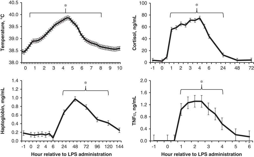
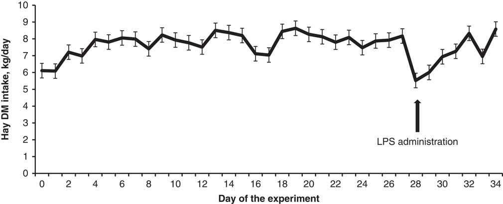
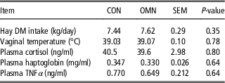
No treatment effects were detected (P⩾0.64) for intravaginal temperature and plasma concentrations of TNFα, cortisol and haptoglobin (Table 2). Time effects were detected (P<0.01) for all these variables (Figure 1), which promptly increased and peaked at 4.5, 2, 4 and 48 h, respectively, after LPS administration. No treatment difference was detected (P=0.35) for hay DM intake during the experiment (Table 2), which sharply decreased after LPS administration across treatments (day effect, P<0.01; Figure 2). Heifers gained (P=0.02) BW with the advance of the experiment (473, 478 and 486 kg of initial BW, pre-LPS BW in period 1, and pre-LPS BW in period 2, respectively; SEM=5), whereas BW gain was not compared among treatments as this experiment was not designed for such assessment.
Intravaginal temperature and plasma concentrations of cortisol, haptoglobin and tumor necrosis-alpha in beef heifers (n=16) administered an intravenous bolus dose (0.5 μg/kg of BW) of bacterial lipopolysaccharide (LPS) at 0 h. Time effects were detected for all variables (P<0.01), and values identified with an asterisk differ (P<0.05) from values obtained before LPS administration.

Hay DM intake in beef heifers (n=16) administered an intravenous bolus dose (0.5 μg/kg of BW) of bacterial lipopolysaccharide (LPS) on day 28. A day effect was detected (P<0.01), given that hay DM intake oscillated with the advance of the experiment, but sharply decreased after LPS administration.

Hay dry matter (DM) intake, vaginal temperature and plasma variables of beef heifers supplemented (OMN; n=8) or not (CON; n=8) with 56 g/day of Omnigen-AF (Phibro Animal Health, Teaneck, NJ, USA) from day 0 to day 34, and administered bacterial lipopolysaccharide on day 28 (0.5 μg/kg of BW)Footnote 1

TNFα=tumor necrosis factor alpha.
1 Results are reported according to main treatment effects and across days (DM intake) and hours (all other variables), given that treatment×time interactions were not detected (P⩾0.37).
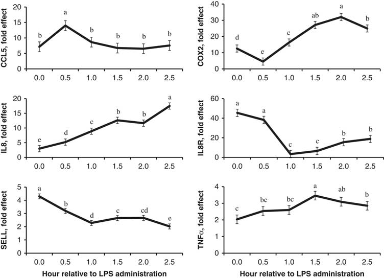
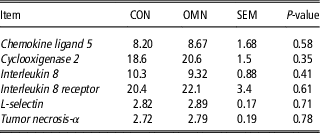
No treatment effects were detected (P⩾0.35) for whole blood mRNA expression of chemokine ligand 5, cyclooxygenase 2, interleukin 8, interleukin 8 receptor, L-selectin and TNFα (Table 3), whereas time effects were also detected for all these variables (P<0.01; Figure 3). Whole blood mRNA expression of chemokine ligand 5, TNFα and interleukin 8 were upregulated and peaked at 0.5, 1.5 and 2.5 h, respectively, after LPS administration. Administration of bacterial LPS initially downregulated cyclooxygenase 2 mRNA expression at 0.5 h, which was then upregulated and peaked 2 h after LPS administration. Whole blood mRNA expression of interleukin 8 receptor and L-selectin were downregulated and reached nadir levels by 1 and 2.5 h, respectively, after LPS administration.
Whole blood mRNA expression of chemokine ligand 5 (CCL5), cyclooxygenase 2 (COX2), interleukin 8 (IL8), IL8 receptor (IL8R), L-selectin (SELL) and tumor necrosis-alpha (TNFα) in beef heifers (n=16) administered an intravenous bolus dose (0.5 μg/kg of BW) of bacterial lipopolysaccharide (LPS) at 0 h. Time effects were detected for all genes (P<0.01); a,b,c,d,ehours with different superscript letters differ (P<0.05).

Whole blood mRNA expression from beef heifers supplemented (OMN; n=8) or not (CON; n=8) with 56 g/day of Omnigen-AF (Phibro Animal Health, Teaneck, NJ) from day 0 to day 34, and administered bacterial lipopolysaccharide on day 28 (0.5 μg/kg of BW)Footnote 1

1 Results are reported according to main treatment effects and across hours, given that treatment×hour interactions were not detected (P⩾0.44).
Discussion
The lack of sequence effects across all variables indicate that the order in which heifers were assigned to treatments had no impact on physiologic and innate immunity responses, corroborating that there were no carryover effects from period 1 to 2. Regarding the period effects detected herein, greater hay DM intake during period 2 should be primarily associated with increasing heifer BW from period 1 to 2 (National Academies of Sciences, Engineering, and Medicine (NASEM), Reference Nace, Nickerson, Kautz, Breidling, Wochele, Ely and Hurley2016). Plasma haptoglobin response to LPS administration was greater in period 2 whereas whole blood mRNA expression of interleukin 8 and TNFα were greater in period 1, which are conflicting outcomes based on the acute-phase nature of these reactions (Düvel et al., Reference Düvel, Maaß, Heppelmann, Hussen, Koy, Piechotta, Sandra, Smith, Sheldon, Dieuzy-Labaye, Zieger and Schuberth2014; Rodrigues et al., Reference Perdew, Vanden Heuvel and Peters2015). Nonetheless, all other variables responded similarly across periods, indicating that metabolic, inflammatory and whole blood mRNA responses to LPS administration were altogether similar between periods 1 and 2.
According to the manufacturer, OMN contains a mixture of active dried Saccharomyces cerevisiae, vitamins, minerals and other components (full formulation is proprietary) as described by Lippolis et al. (Reference Lippolis, Cooke, Schubach, Marques and Bohnert2017a). Yeast-based products contain pathogen-associated molecular patterns such as β-glucans that are recognized by the immune system, trigger innate immune responses by binding to toll-like receptors and prepare the immune system against pathogens (Heine and Ulmer, Reference Heine and Ulmer2005; Broadway et al., Reference Broadway, Carroll and Burdick Sanchez2015). Accordingly, supplementing OMN to periparturient dairy cows modulated leukocytes gene expression related to the inflammatory response, such as improved leukocyte function, surface L-selectin concentration and phagocytosis of extracellular pathogens during the transition period (Wang et al., Reference Wang, Puntenney, Burton and Forsberg2009; Ryman et al., Reference Rodrigues, Cooke, Marques, Arispe, Keisler and Bohnert2013; Nace et al., 2014). Brandão et al. (Reference Brandão, Cooke, Corrá, Piccolo, Gennari, Leiva and Vasconcelos2016) also reported improved milk production and innate immunity parameters in OMN-supplemented periparturient dairy cows.
In turn, published research examining OMN supplementation to beef cattle is still limited, particularly with focus on leukocyte-mediated responses associated with early pathogenic control. Lippolis et al. (Reference Lippolis, Cooke, Schubach, Marques and Bohnert2017a) reported that supplementing OMN to newly weaned beef steers upon arrival to the feedlot heightened, at least transiently, leukocyte-mediated responses including whole blood mRNA expression of interleukin-8 and TNFα, but did not improve immunocompetence against respiratory diseases, as well as BW gain, feed intake and feed efficiency during the initial 80 days in the feedlot. These authors, however, began OMN supplementation after steers were exposed to the immune challenges caused by weaning, auction, transportation, vaccination, commingling and feedlot entry (Cooke, Reference Cooke2017), whereas cattle should be adapted to OMN supplementation for several weeks before the stress or immune insult (Ryman et al., Reference Rodrigues, Cooke, Marques, Arispe, Keisler and Bohnert2013; Nace et al., 2014). Hence, the present experiment was designed to investigate physiological and leukocyte-mediated responses associated with early pathogenic control in beef cattle receiving OMN supplementation for 4 weeks. Moreover, LPS administration was selected to elicit innate immunity reactions that OMN supplementation are expected to modulate, based on Lippolis et al. (Reference Lippolis, Cooke, Schubach, Marques and Bohnert2017a) and previous dairy cattle research (Wang et al., Reference Wang, Puntenney, Burton and Forsberg2009; Ryman et al., Reference Rodrigues, Cooke, Marques, Arispe, Keisler and Bohnert2013; Nace et al., 2014). However, OMN supplementation to beef heifers did not impact any of the metabolic, inflammatory and whole blood mRNA responses elicited by LPS administration herein (Tables 2 and 3). Hay and DDG intake were also similar between OMN and CON heifers, whereas dietary TDN, NEm, NEg and CP across treatments were (DM basis) 58.5%, 1.19 Mcal/kg, 0.63 Mcal/kg and 11.1%, respectively. Hence, heifers from both treatments were in similar and adequate nutritional status to investigate the main hypothesis of this experiment, based on dietary nutrient profile and increasing BW with the advance of the experiment (NASEM, Reference Nace, Nickerson, Kautz, Breidling, Wochele, Ely and Hurley2016).
It should be noted that bacterial LPS administration decreased hay DM intake (Lippolis et al., Reference Lippolis, Cooke, Schumaher, Brandão, Silva, Schubach, Marques and Bohnert2017b; Figure 2) and provoked the expected increases in vaginal temperature, plasma cortisol and components of the acute-phase response such as plasma TNFα and haptoglobin (Cooke, Reference Cooke2017; Figure 1), which are key elements of the innate immune system for early pathogenic control (Abbas and Lichtman, Reference Abbas and Lichtman2007; Rodrigues et al., Reference Perdew, Vanden Heuvel and Peters2015; Lippolis et al., Reference Lippolis, Cooke, Schumaher, Brandão, Silva, Schubach, Marques and Bohnert2017b). Whole blood mRNA expression of innate immunity genes were also promptly impacted by bacterial LPS administration (Figure 3). As in Rodrigues et al. (Reference Perdew, Vanden Heuvel and Peters2015), whole blood mRNA expression of cyclooxygenase 2 and TNFα increased upon LPS administration, representing the synthesis of proinflammatory cytokines by leukocytes and subsequent increase in body temperature via cyclooxygenase 2 modulation (Carroll and Forsberg, Reference Carroll and Forsberg2007; Silva et al., Reference Ryman, Nickerson, Kautz, Hurley, Ely, Wang and Forsberg2008). Chemokine ligand 5 has a chemotactic activity by recruiting leukocytes into infection sites and inducing their proliferation and activation (Maghazachi et al., Reference Maghazachi, Al-Aoukaty and Schall1996), whereas interleukin 8 also induces neutrophil migration to infection sites and phagocytosis of pathogens (Bagglioni and Clark-Lewis, Reference Bagglioni and Clark-Lewis1992). Hence, the immediate increase in whole blood mRNA expression of these latter two genes upon LPS administration characterize critical innate immunity responses required for early leukocyte activity. Conversely, whole blood mRNA expression of interleukin 8 receptor and L-selectin were markedly decreased upon LPS administration (Figure 3). Downregulation of interleukin 8 receptor mRNA can be attributed to neutrophil-preserving mechanisms to avoid its overstimulation due to acute increase in interleukin 8 expression and synthesis by other leukocytes (Perdew et al., Reference Ocón-Grove, Cooke, Alvarez, Johnson, Ott and Ealy2006). L-selectin is an adhesion molecule that allows leukocytes such as neutrophils to roll along blood vessel walls surveying for signs of infection in peripheral tissues (Lawrence et al., 1991). Once infection is localized, L-selectin mRNA expression is downregulated and L-selectin molecules disengaged from cell surface, so neutrophils can transmigrate through the vascular endothelium and phagocytose pathogens (Burton et al., Reference Burton, Kehrli, Kapil and Horst1995; Weber et al., Reference Weber, Madsen, Smith, Ireland and Burton2001). Collectively, changes in whole blood mRNA expression of innate immunity genes reported herein are mostly novel in beef cattle, and provide unique insight on immediate leukocyte-based responses to control and eliminate pathogens such as bacterial LPS.
Contrary to our findings, Brandão et al. (Reference Brandão, Cooke, Corrá, Piccolo, Gennari, Leiva and Vasconcelos2016) administered LPS to lactating dairy cows consuming or not OMN throughout the transition period, and reported that OMN-supplemented cows had greater serum concentration of TNFα and haptoglobin compared with non-supplemented cohorts upon LPS administration. Yet, dairy cows utilized by Brandão et al. (Reference Brandão, Cooke, Corrá, Piccolo, Gennari, Leiva and Vasconcelos2016) were in early lactation when administered LPS, and still experiencing immunosuppression elicited by parturition and onset of lactation (Mallard et al., Reference Mallard, Dekkers, Ireland, Leslie, Sharif, Lacey Vankampen, Wagter and Wilkie1998; Drackley, Reference Drackley1999). Likewise, the majority of research reporting heightened innate immunity when OMN is supplemented utilized cattle under immunosuppressive conditions (Wang et al., Reference Wang, Puntenney, Burton and Forsberg2009; Nace et al., 2014; Lippolis et al., Reference Lippolis, Cooke, Schubach, Marques and Bohnert2017a). In this experiment, beef heifers were overtly healthy when administered treatments and the bacterial LPS, corroborated by the negligible concentrations of plasma TNFα, reduced plasma cortisol and haptoglobin concentrations, and normal intravaginal temperature before LPS administration (Merck and Co., Inc., Reference Silva, Gaivão, Leitão, Amaro, Lopes da Costa and Mateus1997; Figure 1). Hence, it can be speculated that innate immunocompetence of these heifers was optimal, and did not benefit from OMN to combat bacterial LPS as reported in immunosuppressed cattle. Perhaps OMN supplementation would enhance the physiological and leukocyte-mediated responses associated with early pathogenic control if bacterial LPS was administered to beef cattle under stressful and immunosuppressed conditions, such as after weaning, road-transport and feedlot entry (Cooke, Reference Cooke2017).
Conclusions
Supplementing OMN to immunocompetent beef heifers did not impact the physiological and leukocyte-mediated reactions elicited by bacterial LPS administration, including metabolic, inflammatory and whole blood mRNA responses. Moreover, LPS administration not only elicited the expected neuroendocrine and acute-phase reactions, but also regulated expression of innate immunity genes that provide novel insights on leukocyte-based responses in cattle. Therefore, OMN supplementation does not provide additional benefits to innate immunocompetence of healthy beef cattle administered bacterial LPS administration.
Acknowledgements
Dr Reinaldo Cooke is also affiliated as permanent professor to the Programa de Pós-Graduação em Zootecnia/São Paulo State University (UNESP), School of Veterinary Medicine and Animal Science, Botucatu 18168-000, Brazil.
Declaration of interest
No conflicts of interest to report.
Ethics statement
All animals were cared for in accordance with acceptable practices and experimental protocols reviewed and approved by the Oregon State University, Institutional Animal Care and Use Committee (#4891).
Software and data repository resources
No software, data or models were deposited in official repositories.