Introduction
The kinetoplastid parasite Trypanosoma brucei causes African trypanosomiasis in both humans and animals. Human African trypanosomiasis (HAT) is considered fatal if left untreated, and poses a serious health risk to an estimated 65 million people in sub-Saharan Africa (World Health Organization, 2017a). Recent efforts have led to a dramatic decrease in reported disease cases, with 2804 cases reported in 2015, although the World Health Organization estimates the actual number of cases to be 10-fold greater (World Health Organization, 2017a, 2017b). Research has also produced promising new drug candidates, however, the risk of parasite resistance and consequent HAT re-emergence still threaten the progress made in combatting the disease. Animal trypanosomiasis remains a significant burden, with billions of US dollars lost through livestock infections each year and few treatment candidates on the horizon (Shaw et al., Reference Shaw, Cecchi, Wint, Mattioli and Robinson2014). Thus, there is an urgent need to identify leads for drug development.
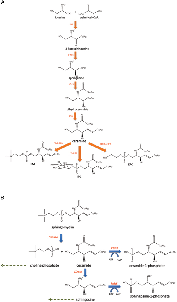
Trypanosoma brucei sphingolipid biosynthesis has long been identified as a potential drug target. Most eukaryotes are capable of synthesizing their own sphingolipids via the de novo biosynthesis pathway (Fig. 1A), which is highly conserved amongst eukaryotic organisms (Kolter and Sandhoff, Reference Kolter and Sandhoff1999). Although homologues of most of the enzymes involved in this biosynthetic pathway have been (putatively) identified in T. brucei, many still require biochemical characterization (Smith and Bütikofer, Reference Smith and Bütikofer2010). The first reaction in the pathway, condensation of serine and palmitoyl-CoA, is catalysed by the enzyme serine-palmitoyltransferase (SPT) (Tidhar and Futerman, Reference Tidhar and Futerman2013). A homologue of this enzyme has been identified in T. brucei, which was shown to be essential for cell cycle progression and parasite survival (Fridberg et al., Reference Fridberg, Olson, Nakayasu, Tyler, Almeida and Engman2008). Inhibiting the initial SPT-catalysed reaction of the T. brucei de novo biosynthesis pathway disrupted procyclic cytokinesis and kinetoplast segregation, validating the pathway as a drug target (Fridberg et al., Reference Fridberg, Olson, Nakayasu, Tyler, Almeida and Engman2008; Smith and Bütikofer, Reference Smith and Bütikofer2010). Although SPT is known to be essential in other parasites, such as Plasmodium falciparum (Gerold and Schwarz, Reference Gerold and Schwarz2001), these findings contrast with research involving the related kinetoplastid parasite Leishmania major (Denny et al., Reference Denny, Goulding, Ferguson and Smith2004; Zhang et al., Reference Zhang, Pompey, Hsu, Key, Bandhuvula, Saba, Turk and Beverley2007). Unusually, T. brucei has four tandemly linked genes, each encoding different sphingolipid synthases (TbSLSs1-4) (Sevova et al., Reference Sevova, Goren, Schwartz, Hsu, Turk, Fox and Bangs2010). This family of sphingolipid synthases is orthologous to the Saccharomyces cerevisiae AUR1-encoded inositol-phosphoceramide (IPC) synthase (Mina et al., Reference Mina, Pan, Wansadhipathi, Bruce, Shams-Eldin, Schwarz, Steel and Denny2009), and equivalent enzymes are found in both L. major (Denny et al., Reference Denny, Shams-Eldin, Price, Smith and Schwartz2006) and T. cruzi (De Lederkremer et al., Reference De Lederkremer, Agusti and Docampo2011). RNA interference (RNAi) against the TbSLS1-4 gene locus in bloodstream trypanosomes impeded growth, ultimately leading to parasite death (Sutterwala et al., Reference Sutterwala, Hsu, Sevova, Schwartz, Zhang, Key, Turk, Beverley and Bangs2008). This finding identified the TbSLSs as potential drug targets. The individual functional specificities of the four synthases were determined by employing a cell-free synthesis system (Sevova et al., Reference Sevova, Goren, Schwartz, Hsu, Turk, Fox and Bangs2010). TbSLSs 1 and 2 synthesize IPC and ethanolamine-phosphoceramide (EPC), respectively (Sevova et al., Reference Sevova, Goren, Schwartz, Hsu, Turk, Fox and Bangs2010). TbSLSs 3 and 4 are bi-functional, producing both sphingomyelin (SM) and EPC (Sevova et al., Reference Sevova, Goren, Schwartz, Hsu, Turk, Fox and Bangs2010). Research has indicated TbSLS substrate specificity is dictated by natural variations in a small number of active site residues, thought to be involved in acid–base catalysis (Goren et al., Reference Goren, Fox and Bangs2011). Establishing the functions of the TbSLSs has clarified how the parasite is capable of altering its sphingolipid complement during its life cycle (Mina et al., Reference Mina, Pan, Wansadhipathi, Bruce, Shams-Eldin, Schwarz, Steel and Denny2009; Sevova et al., Reference Sevova, Goren, Schwartz, Hsu, Turk, Fox and Bangs2010), an observation further confirmed by recent sphingolipidomic analysis (Guan and Mäser, Reference Guan and Mäser2017). Currently, trypanosomatids are the only organisms known to synthesize sphingolipid species with choline, ethanolamine and inositol headgroups (Serricchio and Bütikofer, Reference Serricchio and Bütikofer2011; Guan and Mäser, Reference Guan and Mäser2017).
Trypanosoma brucei sphingolipid metabolism. (A) The currently proposed pathway for T. brucei de novo sphingolipid biosynthesis is shown. SPT – serine-palmitoyltransferase (Q580D0); 3-KSR – 3-ketosphinganine reductase (Q38BJ6); CerS – ceramide synthase (Q57V92, Q583F9); DES – dihydroceramide desaturase (Q583N4); TbSLSs 1-4 – T. brucei sphingolipid synthases 1-4 (Q38E53; Q38E54; Q38E55; Q38E56). Biosynthesis of ceramide (36:1) has been used here as an example. (B) A simplified overview of eukaryotic sphingolipid degradation is provided (omitting glycosphingolipid catabolic pathways). A representative sphingolipid species [SM (36:1)] is degraded in a stepwise process involving SMases and ceramidases (CDases). Lipid headgroups, such as choline phosphate, and sphingosine can be recycled to participate in de novo biosynthesis via the salvage pathway (dashed arrows). Products ceramide and sphingosine can also be phosphorylated to produce signalling molecules Ceramide-1-phosphate and sphingosine-1-phosphate, respectively. ATP – adenosine triphosphate; ADP – adenosine diphosphate; CERK – ceramide kinase; SphK – sphingosine kinase.
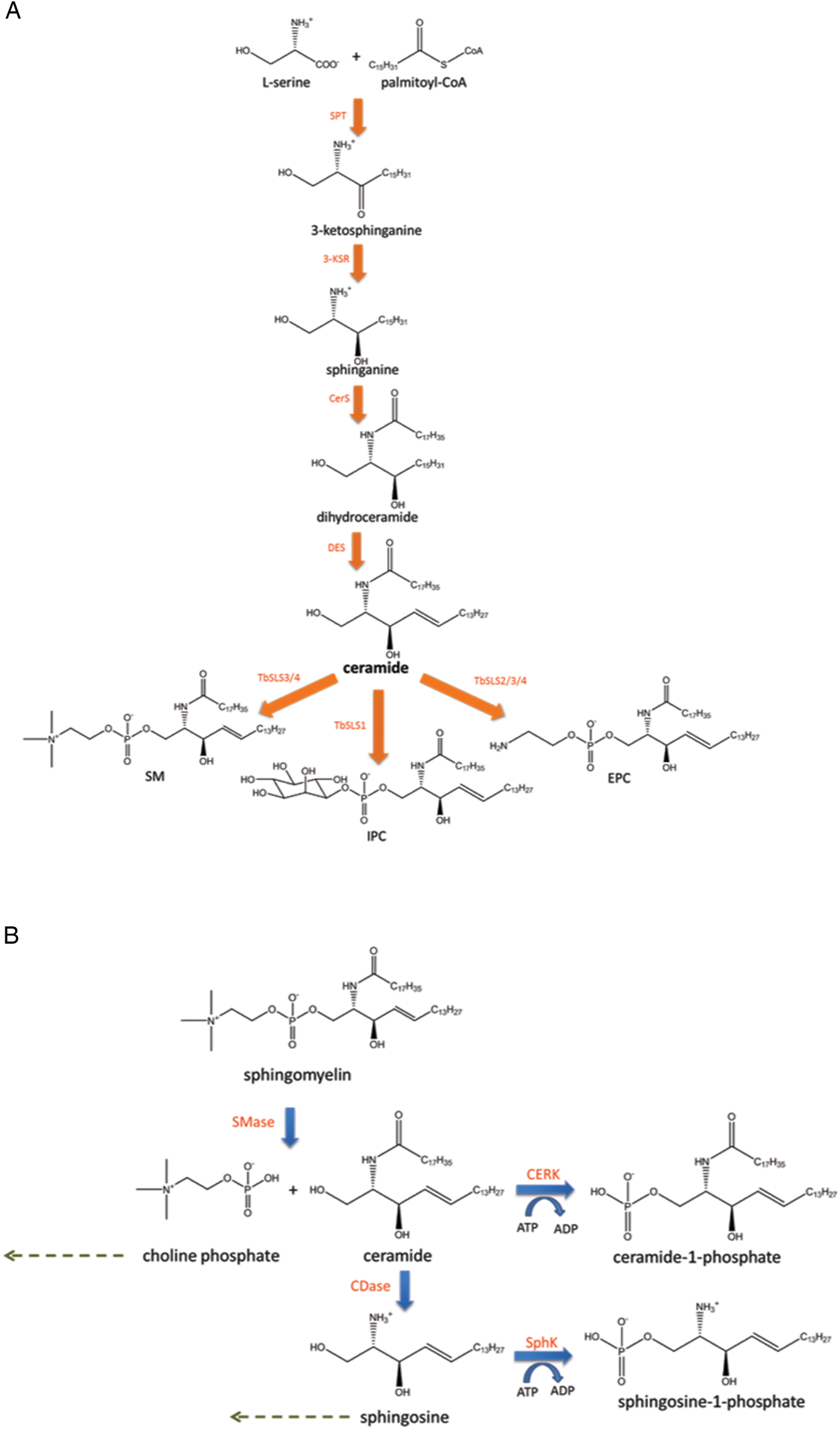
In comparison with the knowledge of T. brucei sphingolipid biosynthesis that has already been acquired, little is known of the processes involved in the parasites’ sphingolipid catabolism. The catabolism of sphingolipids in eukaryotes takes place via the degradation pathway (Fig. 1B), predominantly in lysosomes and late endosomes, but also in other cellular locations (Kolter and Sandhoff, Reference Kolter and Sandhoff1999; Jenkins et al., Reference Jenkins, Canals and Hannun2009). A number of different enzymes are involved in the degradative process (ceramidases, lyases and phospholipase D), but one major group of proteins responsible for sphingolipid turnover in mammalian cells is the sphingomyelinase (SMase) enzyme family (Kolter and Sandhoff, Reference Kolter and Sandhoff1999). As indicated by their name, the primary substrate of these enzymes in mammals is SM, which is hydrolysed to yield ceramide (Cer) and choline-phosphate (ChoP) (Jenkins et al., Reference Jenkins, Idkowiak-Baldys, Simbari, Canals, Roddy, Riner, Clarke and Hannun2011). Trypanosoma brucei has a single neutral SMase (TbnSMase) (Q57U95), a membrane protein with two identified transmembrane domains at its C-terminus (Young and Smith, Reference Young and Smith2010). TbnSMase has been localized to the ER in bloodstream form parasites, and genetically confirmed as a potential drug target (Young and Smith, Reference Young and Smith2010). The activity of this protein is thought to be essential due to its intrinsic roles in vital biochemical processes, including choline and Cer homoeostasis, and endocytosis. This is particularly relevant to the coupling of endocytic and exocytic mechanisms with post-Golgi sorting of GPI-anchored variant surface glycoprotein (VSG), which is needed to maintain VSG surface density (Young and Smith, Reference Young and Smith2010). VSG molecules form the protective coat that permits T. brucei parasites to evade the host immune system (Mugnier et al., Reference Mugnier, Stebbins and Papavasiliou2016). The importance of TbnSMase corroborates findings relating to the single leishmanial sphingolipid degradative enzyme (ISCL), another nSMase homologue (Zhang et al., Reference Zhang, Wilson, Xu, Hsu, Turk, Kuhlmann, Wang, Soong, Key, Beverley and Zhang2009; McConville and Naderer, Reference McConville and Naderer2011). ISCL IPCase activity is required for L. major promastigote stationary phase survival, most noticeably at acidic pH (Xu et al., Reference Xu, Xin, Soong and Zhang2011). The enzyme's SMase function is necessary for amastigote proliferation and virulence in mammalian hosts (Zhang et al., Reference Zhang, Wilson, Xu, Hsu, Turk, Kuhlmann, Wang, Soong, Key, Beverley and Zhang2009, Reference Zhang, Xu, Balakrishna Pillai and Zhang2012). This means that the importance of leishmanial ISCL activity is linked to cell cycle stage (Zhang et al., Reference Zhang, Xu, Balakrishna Pillai and Zhang2012). Similarly, inhibiting the nSMase/lyso-phosphatidylcholine phospholipase C of P. falciparum disrupts parasite intra-erythrocytic proliferation, suggesting the protein could serve as a drug target (Hanada et al., Reference Hanada, Palacpac, Magistrado, Kurokawa, Rai, Sakata, Hara, Horii, Nishijima and Mitamura2002). In previous research, TbnSMase was shown to catabolize SM effectively, but was inactive towards phosphatidylcholine (PC) (Young and Smith, Reference Young and Smith2010). This initial analysis has now been taken forward to provide a more comprehensive overview of TbnSMase lipid degradative activity and specificity.
Materials and methods
Unless otherwise stated, all reagents and materials were purchased from Sigma, Promega, Thermo Scientific or VWR. Ceramide-1-phosphate (C-1-P) (d18:1/16:0), EPC (d17:1/12:0), SM (brain, porcine), dipalmitoyl-PC and dimyristoyl-PC [used as a mass spectrometry (MS) standard] were purchased from Avanti Polar Lipids. SM (d18:1/6:0), lyso-phosphatidylcholine (from egg yolk), sphingosine-1-phosphate (S-1-P) (d18:1), and galactosylceramide were purchased from Sigma. EPC (from buttermilk, semi-synthetic) was purchased from Matreya LLC. Procyclic cell culture media were filter sterilized with either Millex GP 0.22 µ m syringe filters or Triple Red 0.22 µ m vacuum filtration units. Parasite cultures were maintained in Greiner Bio-One CELLSTAR® tissue culture flasks.
Recombinant expression of TbnSMase (Q57U95) in Escherichia coli
A pGEX-6P-1-TbnSMase expression construct was employed as previously described (Young and Smith, Reference Young and Smith2010). The expression construct was used to transform BL21/pLysSGold E. coli. Positive clones were selected using ampicillin-agar plates (100 µg mL−1) supplemented with chloramphenicol (34 µg mL−1). Three bacterial colonies were used to inoculate 3 × 10 mL of LB media (Miller composition, supplemented with 50 µg mL−1 ampicillin and 34 µg mL−1 chloramphenicol). After a 24 h incubation, the 10 mL overnight cultures were combined in a single tube (30 mL total volume). The combined culture was then used to inoculate 3 × 1 L auto-induction (AI) media (Formedium, supplemented with 50 µg mL−1 ampicillin and 34 µg mL−1 chloramphenicol), using 10 mL of overnight culture per litre flask of AI media. Cells were grown at 37 °C for 4 h before the temperature was decreased to 25 °C for a further 20-h incubation. Glutathione S-transferase (GST)-TbnSMase-enriched bacterial membranes were prepared from these cultures. Pellets of 500 mL spun-down culture were washed in PBS and stored at −80 °C until they could be processed.
TbnSMase-enriched bacterial membrane preparation
The protocol described is adapted from methods previously outlined (Young and Smith, Reference Young and Smith2010). Pellets of 500 mL spun-down bacterial culture were suspended and lysed in 10 mL lysis buffer [50 mm Tris-HCl (pH 8.0), 300 mm NaCl, 10% glycerol (v/v), 5 mm MgCl2, 1 mm dithiothreitol (DTT)], containing 0.2 mg mL−1 lysozyme (Sigma), Merck Millipore Benzonase® Endonuclease (250 units mL−1 lysate) and 1× protease inhibitor tablet (Roche, 11836170001). The lysis solution was incubated for 30 min at 37 °C, followed by probe sonication at 4 °C (6 min total, 30 s on/30 s off). The lysate was then centrifuged at 14 500 × g, 20 min. The product supernatant was divided amongst 3.2 mL capacity Beckman Coulter ultra-centrifuge tubes (×3) for ultra-centrifugation at 100 000 × g, 1 h. Pellets were washed with 1.5 mL PBS. Each bacterial membrane pellet was then suspended in 500 µL buffer [100 mm Tris-HCl (pH 7.4), 10 mm MgCl2, 20% glycerol] using a combination of vortexing and water bath sonication (25 °C, 4 min). The total membrane suspension (1500 µL total volume) was mixed in a single tube and was then aliquoted (50–100 µL). Aliquots were flash-frozen using liquid nitrogen and stored at −80 °C. Total protein in each membrane preparation was quantified using the bicinchoninic acid (BCA) Protein Assay Kit (Thermo Scientific). The presence of TbnSMase in bacterial membranes was confirmed by MS.
Amplex® UltraRed assay
Amplex® UltraRed assay coupling enzyme solutions were prepared from lyophilized powders using dH2O, and were stored as small aliquots at −20 °C. Alkaline phosphatase (AlkPhos) (Sigma) from bovine intestinal mucosa was prepared as a 400 units mL−1 stock solution. Choline oxidase (ChoOx) (Sigma) from Alcaligenes sp. was prepared at 20 units mL−1, horseradish peroxidase (HRP) (Sigma) at 200 units mL−1. Desiccated aliquots of Amplex® UltraRed reagent (Thermo Scientific) were suspended in 340 µL DMSO, as directed by the manufacturer, and stored at −20 °C in small aliquots. To assay the aqueous fractions of biphasically separated GST-TbnSMase SM substrate reactions, aqueous phases were dried using a Savant SPD121P SpeedVac concentrator. The fractions were suspended in 100 µL reaction buffer [100 mm HEPES Na (pH 7.4), 10 mm MgCl2] and divided between two wells of a black 96-well reaction plate (50 µL per well). Additional reaction buffer (50 µL) was then added to each well. A mastermix of Amplex® assay components (coupling enzymes and Amplex® UltraRed reagent) was prepared. The mastermix accounted for the addition of 2 µL AlkPhos, 1.5 µL ChoOx, 1 µL HRP, 0.25 µL Amplex UltraRed reagent and 95.25 µL reaction buffer per well (total reaction volume per well was 200 µL). Change in fluorescence (Ex. 560 nm, Em. 587 nm) was then monitored for 1 h using a Spectra Max Gemini XPS fluorescence plate reader at 37 °C. Results were recorded using SoftMax Pro v 5.2 software.
Lipid substrate activity assays
Lipid substrates were suspended in 2% Triton X-100 to the desired stock concentration through vortexing and water-bath sonication (10 min). Substrate mass was substrate- and analysis-dependent (20–50 nmoles), but each was added to reactions (50–100 µL total volume) to a final concentration of 0.1–0.2% Triton X-100. Substrates were incubated with TbnSMase-enriched bacterial membranes (~100 µg total protein) in 1.5 mL solvent-resistant microcentrifuge tubes. In heat-inactivated (HI) protein reactions, protein aliquots were heated to 95 °C (20 min) prior to substrate addition. Reactions were performed in 100 mm HEPES Na (pH 7.4), 10 mm MgCl2 buffer. On substrate addition, reactions were mixed briefly and sonicated for 30 s in a water bath (25 °C). Reaction tubes were then incubated at 37 °C (4 h) in a water bath. Following incubation, reactions were quenched with 800 µL CHCl3. If required, MS standards (e.g. PC 28:0) were then added (500 pmoles), prior to the addition of 250 µL dH2O. The organic phase was isolated following the Bligh and Dyer biphasic separation method (Bligh and Dyer, Reference Bligh and Dyer1959), and dried under nitrogen.
Parasite lysate N-[12-[(7-nitro-2-1,3-benzoxadiazol-4-yl)amino]dodecanoyl] (NBD)-IPC activity assays
The protocol described is adapted from methods previously outlined (Sutterwala et al., Reference Sutterwala, Hsu, Sevova, Schwartz, Zhang, Key, Turk, Beverley and Bangs2008). Cells were suspended in lysis buffer (25 mm Tris (pH 7.5), 0.1% Triton X-100, 1× protease inhibitor tablet (Roche, 11836170001)) to a density of ⩾2 × 10⁸ cells mL−1, and were incubated on ice for 5 min. Subsequently, 4 × 10⁶ parasites from each stock were transferred to each reaction. Parasites were incubated with 0.8 nmoles NBD-IPC, in 50 mm Tris (pH 7.5), 5 mm MgCl2, 5 mm DTT and 0.1% Triton X-100 reaction buffer, for 1 h at 25 °C with gentle agitation (protected from light). Following incubation, reactions were quenched with the addition of 1 mL CHCl3. Subsequently, 500 µL MeOH and 200 µL dH2O were quickly added. Samples were vortex mixed, then left to stand (protected from light). The organic phase from each sample was isolated through biphasic separation, and dried under nitrogen.
High-performance thin-layer chromatography
HPTLC analysis of EPC and NBD-IPC substrate reactions was conducted using a CHCl3:MeOH:dH2O solvent system (65:25:4). Dried lipid samples were resuspended in 2:1 CHCl3:MeOH (10–20 µL volume) and were gradually spotted onto HPTLC plates. EPC reaction results were visualized by treating HPTLC plates with ninhydrin solution (1% w/v in butan-1-ol). NBD-IPC reaction results were visualized via fluorescence imaging using a Typhoon FLA 7000 (GE), with CY2 (filter Y520, 473 nm laser) and Sypro Ruby (filter O580, 473 nm laser) settings.
Procyclic form T. brucei lipid extract preparation
Trypanosoma brucei [Lister 427 (29-13) strain] procyclic (PCF) parasites were grown in SDM-79 media at pH 7.4, as previously described (Brun and Schönenberger, Reference Brun and Schönenberger1979). Drugs G418 (15 µg mL−1) and hygromycin (50 µg mL−1) were included in the media in order to maintain the expression of a tetracycline repressor and T7 RNA polymerase (Wirtz et al., Reference Wirtz, Leal, Ochatt and Cross1999). A 10 mL culture of PCF cells (~1 × 10⁷ cells mL−1) was pelleted via centrifugation at 800 × g, 10 min. The cell pellet was resuspended in a minimal volume of media (~500 µL) and transferred to a microcentrifuge tube for further centrifugation (3800 × g, 3 min). Cells were washed in 1 mL PBS, and then re-pelleted (3800 × g, 3 min). The pellet was resuspended in 200 µL PBS and transferred to a glass vial containing 750 µL 2:1 MeOH:CHCl3 for biphasic separation, based on the method described by Bligh and Dyer (Reference Bligh and Dyer1959). The sample was dried under nitrogen and stored at 4 °C until use.
Electrospray tandem mass spectrometry
Lipid samples were analysed by electrospray ionization tandem MS (ESI-MS/MS) with an AB-Sciex Qtrap 4000 triple quadrupole mass spectrometer, incorporating an Advion TriVersa NanoMate nanoelectrospray ionization source. Single-stage MS in negative ion mode was used to obtain survey scans of phosphatidylethanolamine (PE), EPC, phosphatidylinositol (PI), IPC and Cer species (cone voltage = 1.25 kV). Positive ion mode survey scans were used to detect PC and SM species (cone voltage = 1.25 kV). In tandem MS, nitrogen was the collision gas. To examine PC/SM species, positive ion mode scans to detect precursors of m/z 184 were performed, with 50 eV collision energy (CE). In negative ion mode, precursors of m/z 196 scans allowed the identification of PE and EPC lipids, precursors of m/z 241 scans were used to observe PI and IPC species (CE = 60 eV). Spectra were acquired over or within the range of 120–1000 m/z, and each spectrum represents a minimum of 30 consecutive scans. Samples were run using a 1:1 solvent mixture of 2:1 MeOH:CHCl3 and 6:7:2 acetonitrile:isopropanol:dH2O.
Results
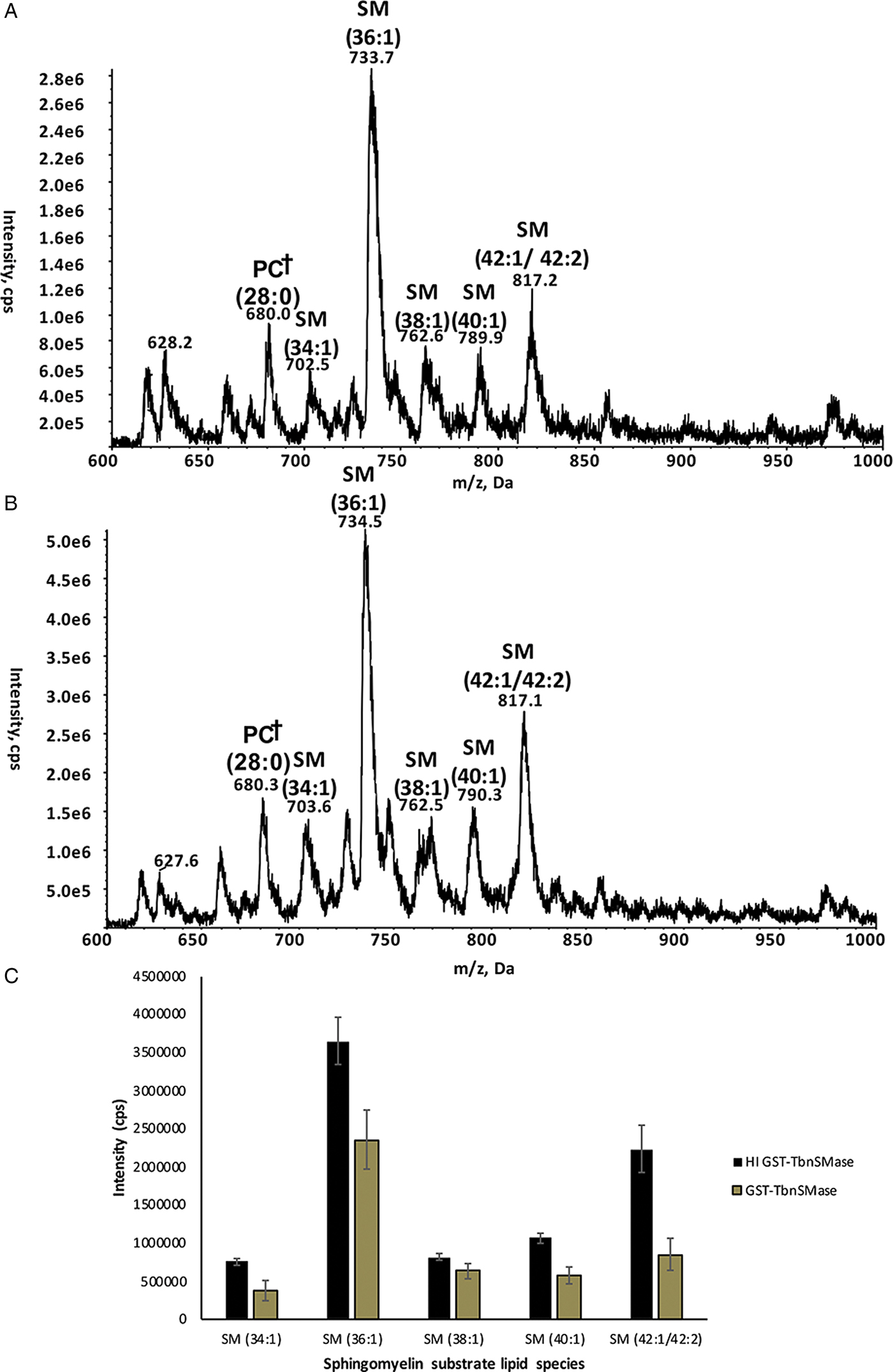
GST-TbnSMase was recombinantly expressed in E. coli to produce TbnSMase-enriched bacterial membranes, using previously described methods (Young and Smith, Reference Young and Smith2010). The presence of the GST-TbnSMase membrane protein was confirmed using MS (Fig. S1). After isolating the bacterial membranes via ultra-centrifugation, it was possible to assay GST-TbnSMase activity directly from the membranes with minimal bacteria-derived background activity, removing the need for protein purification. This had also been achieved during previous research into TbnSMase activity (Young and Smith, Reference Young and Smith2010). To confirm the GST-TbnSMase recombinant protein was active, GST-TbnSMase-enriched bacterial membranes were first incubated at 37 °C for 4 h with a SM substrate. The substrate used was Avanti Polar Lipids brain SM (product no. 860062). Although this product is enriched for SM 36:1 (d18:1/18:0), as a natural lipid preparation the product is a mixture of brain SM species. The spectra presented here are consistent with the recently released Avanti Polar Lipids fatty acid distribution analysis for this product (Fig. S2) (Avanti Polar Lipids, 2018). Following SM substrate incubation, reactions were biphasically separated using 2:1 CHCl3:MeOH to isolate the lipid-rich organic phase. ESI-MS/MS was used to detect the levels of choline-containing (SM) lipid species in lipid samples derived from both active GST-TbnSMase and HI GST-TbnSMase reactions (Fig. 2A and B, see also Fig. S3). A dimyristoyl-PC standard was added to each sample during lipid extraction, allowing SM substrate peak intensities to be normalized against the intensity of the internal standard. Relative peak intensities for each SM substrate species are shown (Fig. 2C).
ESI-MS/MS analysis of SM substrate reactions. Spectra are ESI-MS/MS precursor ion scans to detect choline-containing lipids (precursors of m/z 184) in positive ion mode. (A) GST-TbnSMase-enriched bacterial membranes plus SM substrate. (B) HI GST-TbnSMase enriched bacterial membranes plus SM substrate. †Highlights the dimyristoyl-PC (28:0) standard (500 pmoles). (C) Peak intensities (cps), normalized against the intensity of the dimyristoyl-PC (28:0) standard, for each significant SM substrate lipid species are depicted. Values are mean intensities for triplicate reactions (n = 3). Error bars represent the standard error of each mean (±).
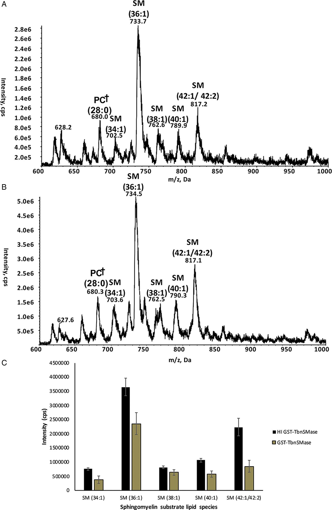
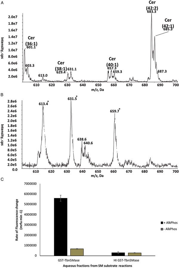
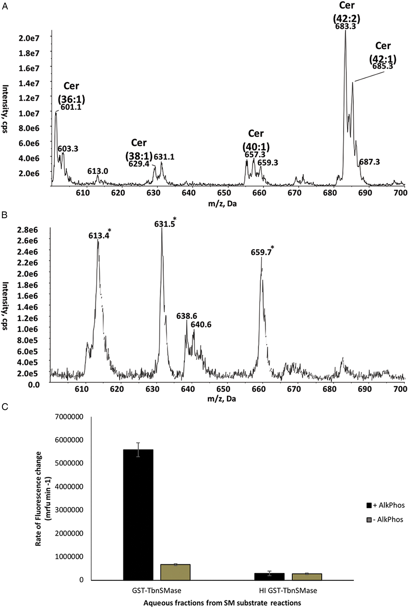
The catabolic activity of GST-TbnSMase towards SM had been demonstrated previously (Young and Smith, Reference Young and Smith2010), here providing confirmation that the GST-TbnSMase recombinant protein was active. However, the use of ESI-MS/MS allowed the relative turnover of individual SM species present in the substrate mixture to be compared (Fig. 2C). The results indicate that GST-TbnSMase is active towards SM species with a range of fatty acid chain lengths (C16–C24). There was a 2.6-fold (62%) decrease in SM 42:1 (d18:1/24:0)/SM 42:2 (d18:1/24:1), compared with a 1.5-fold (35%) decrease in SM 36:1 (d18:1/18:0), despite the latter lipid being the prevalent species in the SM substrate mixture. This may indicate GST-TbnSMase preferentially degrades sphingolipid species of specific fatty acid chain lengths. SM substrate catabolism was further confirmed by identification of product Cer chloride adducts [M + Cl]− in negative ion mode survey scans (Fig. 3). Product Cers were observed in the active GST-TbnSMase reaction (Fig. 3A), and were absent from the HI control (Fig. 3B). Additionally, aqueous fractions isolated during biphasic separation of SM substrate reactions were tested for the presence of choline phosphate. The aqueous fractions were used as substrates for the Amplex® UltraRed assay system. In this assay (Fig. S4), the coupling enzyme AlkPhos is included to dephosphorylate choline phosphate, yielding choline. The choline produced then serves as a substrate for the second coupling enzyme in the system: ChoOx. Only the aqueous fractions from the active GST-TbnSMase reactions, in the presence of AlkPhos, induced significantly increased rates of fluorescence change (Fig. 3C). These results confirm GST-TbnSMase is a phosphodiesterase C enzyme, degrading SM substrates to form Cer and choline phosphate.
GST-TbnSMase catabolism of SM yields Cer and choline phosphate. Spectra are ESI-MS/MS survey scans in negative ion mode. (A) GST-TbnSMase-enriched bacterial membranes plus SM substrate. (B) HI TbnSMase-enriched bacterial membranes plus SM substrate. Annotated Cers have formed chloride adducts [M + Cl]−. *Highlights previously identified significant contaminants of negative ion mode surveys thought to be associated with the detergent. (C) Aqueous fractions of SM substrate reactions were used as substrates for the Amplex® UltraRed assay system, plus (+) or minus (−) the coupling-enzyme AlkPhos. Change in fluorescence [millirelative-fluorescence units per minute (mrfu min−1)] was monitored spectrophotometrically. Values represent average rate of fluorescence change for aqueous fractions derived from triplicate reactions (n = 3). Error bars represent the standard error of each mean (±).
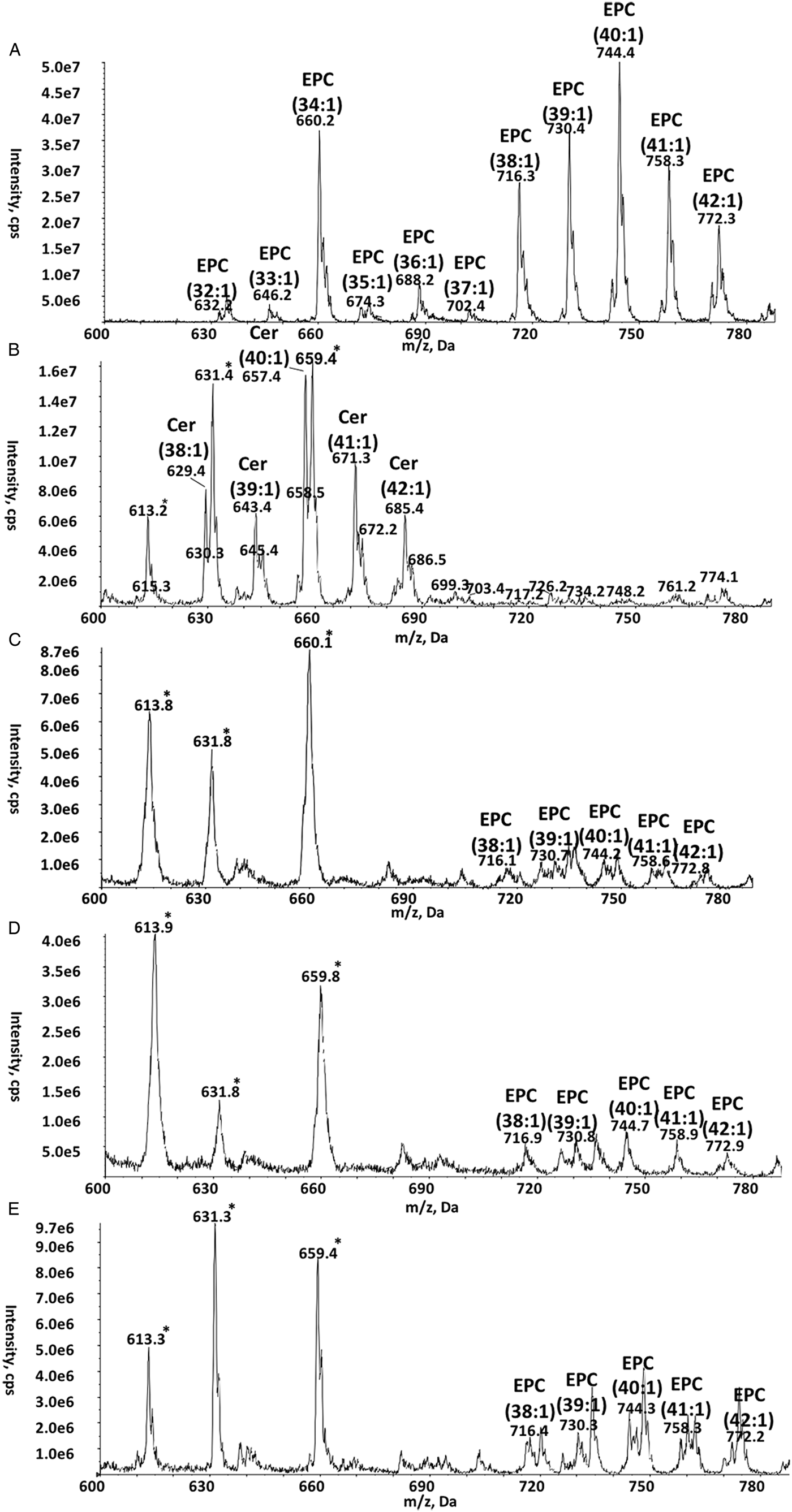
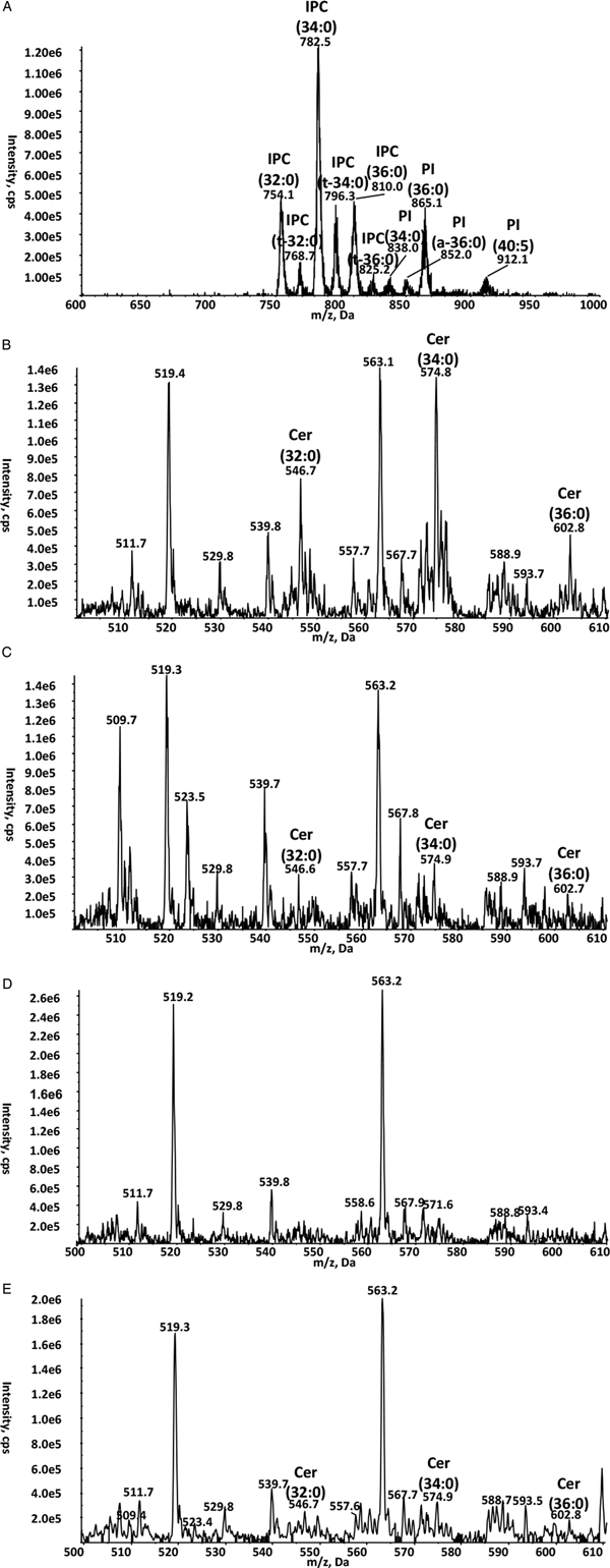
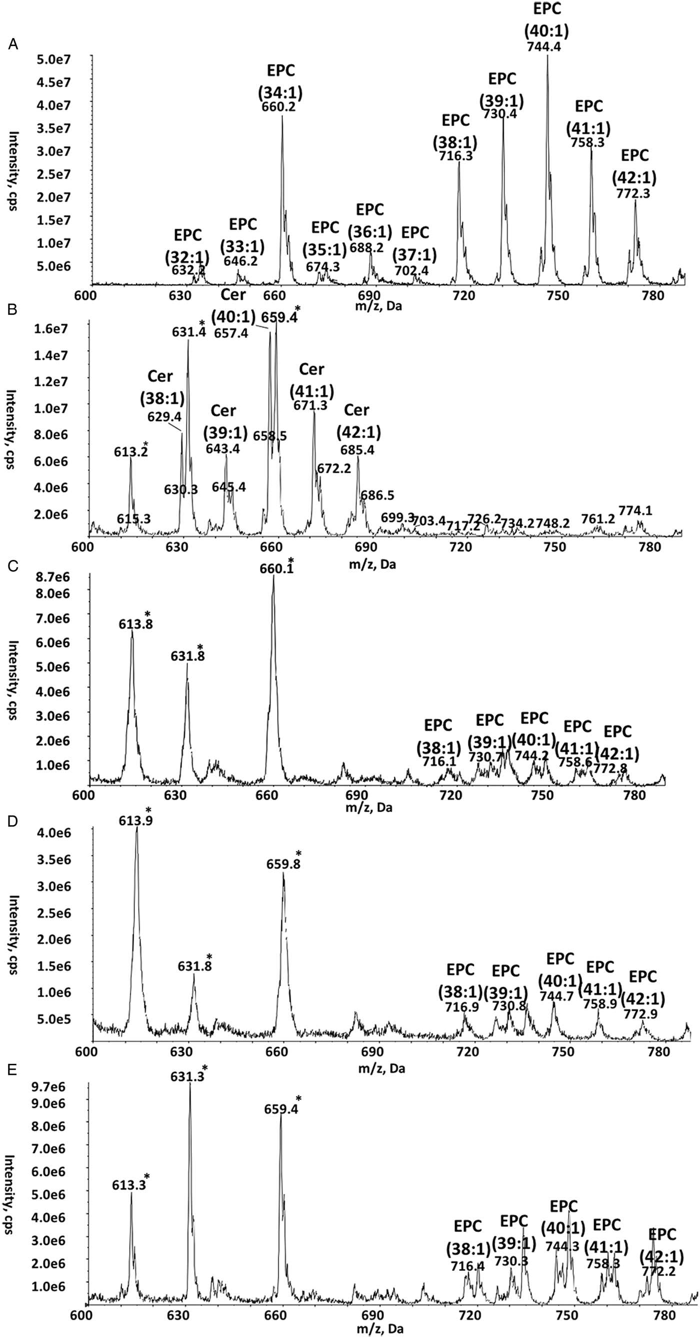
Having established the GST-TbnSMase protein was active, the enzyme's activity towards a number of different sphingolipid substrates was examined. As already highlighted, T. brucei is known to produce SM, EPC and IPC. Thus, it was important to examine the activity of TbnSMase towards EPC and IPC sphingolipids. Firstly, GST-TbnSMase was incubated with an EPC substrate. The EPC substrate used was supplied by Mattreya LLC (product no. 1327), a semi-synthetic preparation derived from bovine buttermilk. No fatty acid distribution analysis is available for this product. However, the average molecular weight reported for the product (MW = 773) appears consistent with our analysis (Fig. 4A). Lipid extracts from the EPC substrate reactions were analysed in ESI-MS/MS negative ion mode survey scans. The EPC species present in the substrate (Fig. 4A) were efficiently degraded by GST-TbnSMase, yielding Cer products (Fig. 4B). These products were absent from the HI GST-TbnSMase control, the EPC/Triton X-100 detergent mixed micelle substrate supplied to the reactions, and a non-TbnSMase-expressing control (Fig. 4C, 4D and 4E). Additionally, GST-TbnSMase degradative activity towards EPC was shown via HPTLC (Fig. S5). GST-TbnSMase was also incubated with a lipid substrate mixture containing equimolar concentrations of SM (d18:1/6:0, Avanti Polar Lipids product no. 860582) and EPC (d17:1/12:0, Avanti Polar Lipids product no. 860529). This competition assay indicated GST-TbnSMase lacks a sphingolipid substrate preference (Fig. S6). However, given the high levels of substrate turnover for both species, it is possible increasing the concentrations of both substrates may lead to a more marked preference for SM over EPC. Additionally, as EPC (d17:1/12:0) is the only pure EPC substrate commercially available, it is not possible to assess the impact of varying the sphingoid base and fatty acid composition on substrate turnover.
ESI-MS/MS analysis of EPC substrate reactions. Spectra are ESI-MS/MS survey scans in negative ion mode. (A) EPC only (minus Triton X-100 detergent). (B) GST-TbnSMase-enriched bacterial membranes plus EPC substrate. Annotated Cers have formed chloride adducts [M + Cl]−. (C) HI GST-TbnSMase-enriched bacterial membranes plus EPC substrate. (D) EPC/Triton X-100 detergent mixed micelle substrate only. (E) Non-TbnSMase-expressing bacterial membranes plus EPC substrate. *Highlights previously identified significant contaminants of negative ion mode surveys thought to be associated with the detergent.
Currently, pure IPC lipid species are only available commercially through custom synthesis. GST-TbnSMase was shown to catabolize a custom synthesized NBD-conjugated IPC substrate (kindly gifted by Dr K. Zhang) (Fig. S7). However, the synthetic nature of this IPC substrate makes it difficult to draw conclusions regarding the physiological relevance of this activity. It is well established that the prevalent sphingolipid in procyclic form T. brucei is IPC (Richmond et al., Reference Richmond, Gibellini, Young, Major, Denton, Lilley and Smith2010). Therefore, a lipid extract from procyclic parasites was used to form a mixed micelle IPC substrate for GST-TbnSMase. Negative ion mode scans to detect inositol-containing lipids (precursors of m/z 241) were used to identify the IPC species present in the procyclic T. brucei extract (Fig. 5A). Negative ion mode survey scans were then employed to search for Cer chloride adducts [M + Cl]−. Significant peaks corresponding to Cer species were observed in the active GST-TbnSMase reaction (Fig. 5B), and were only observed at low levels in the HI and non-expressing controls (Fig. 5C and 5E, see also Fig. S8). Taken together, these results suggest GST-TbnSMase is also capable of catabolizing natural IPC species to form Cer and inositol-1-phosphate.
ESI-MS/MS analysis of T. brucei procyclic-extract (IPC) substrate reactions. (A) ESI-MS/MS precursor ion scan of the procyclic lipid extract (IPC substrate) to detect inositol-containing lipids (precursors of m/z 241), in negative ion mode. The extract contains dihydroxylated Cers, as well as trihydroxylated Cers, the later denoted as ‘t-’. (B) ESI-MS/MS negative ion mode survey scans were used to detect Cers in GST-TbnSMase plus IPC substrate; (C) HI GST-TbnSMase-enriched bacterial membranes plus IPC substrate; (D) IPC/Triton X-100 detergent mixed micelle substrate only and (E) non-TbnSMase-expressing bacterial membranes plus IPC substrate reactions. Annotated Cers have formed chloride adducts [M + Cl]−.

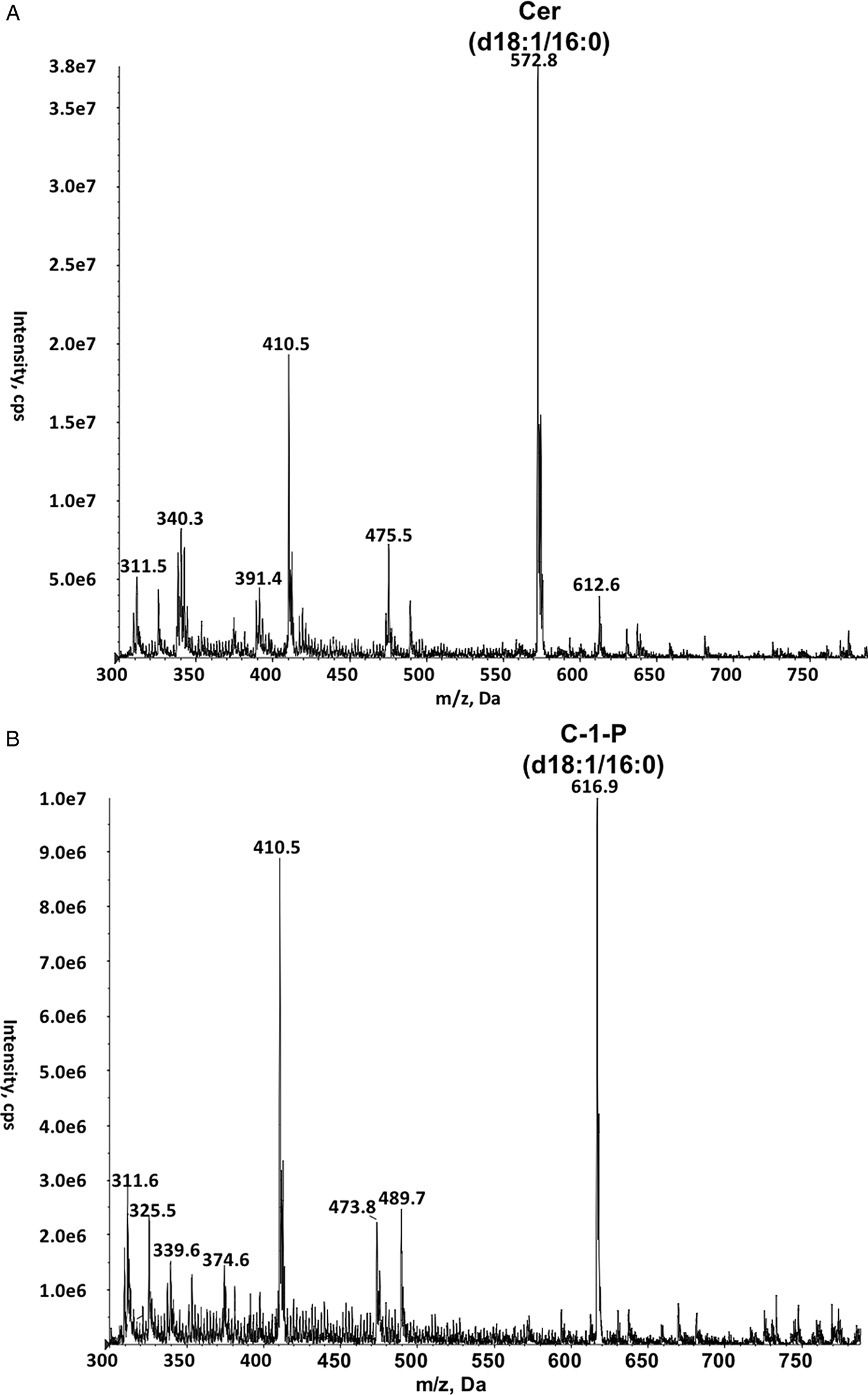
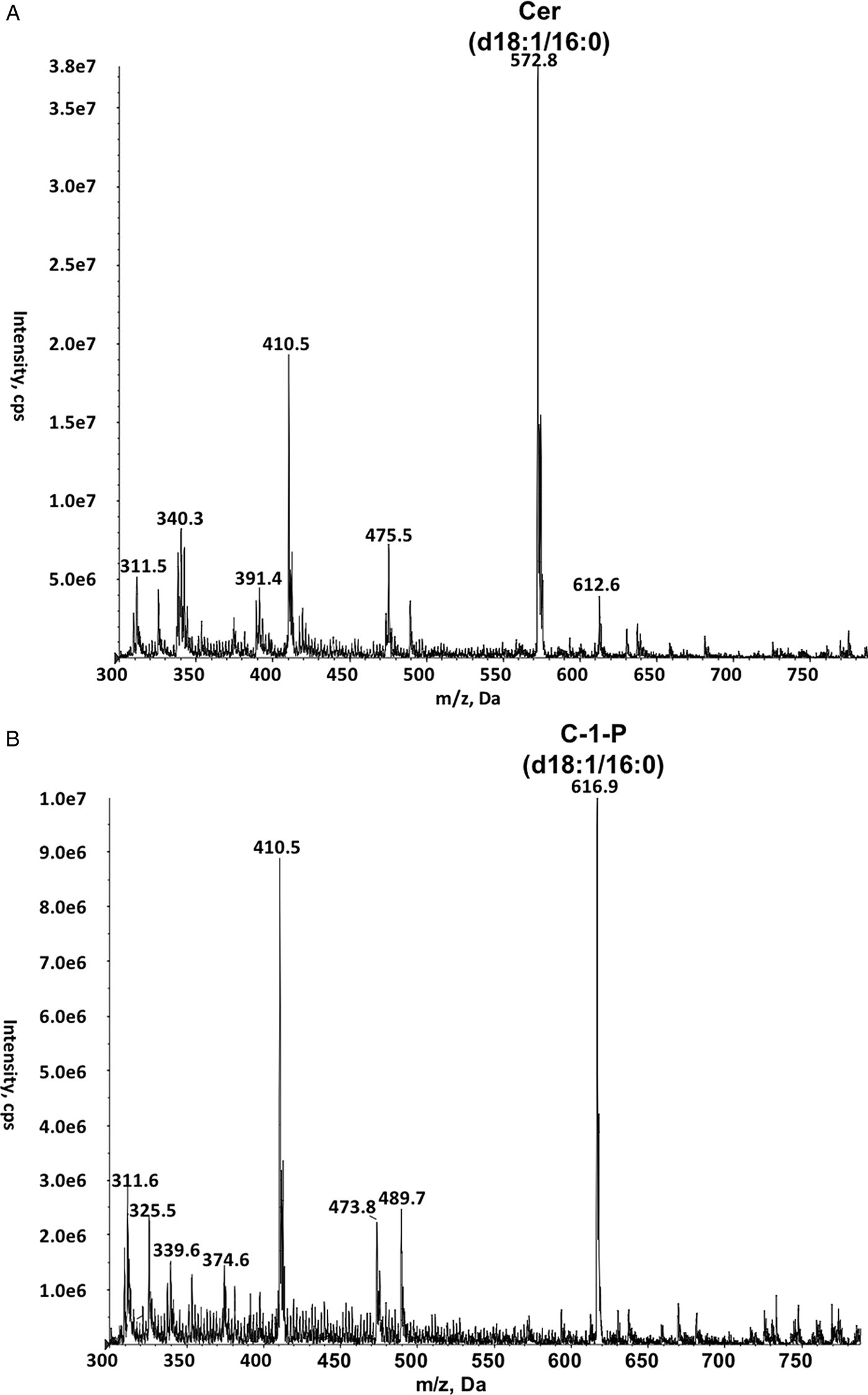
The ability of GST-TbnSMase to degrade SM, EPC and IPC indicates the enzyme does not distinguish between its sphingolipid substrates based upon headgroup identity. To further explore this finding, GST-TbnSMase was incubated with a C-1-P (d18:1/16:0) substrate (Avanti Polar Lipids product no. 860533). A Cer (d18:1/16:0) chloride adduct [M + Cl]− was apparent in the lipid extract from the active GST-TbnSMase reaction (Fig. 6A), and was absent from the control (Fig. 6B). This result suggests lipid headgroups are not involved in GST-TbnSMase substrate recognition. However, GST-TbnSMase does not appear to degrade glycosphingolipids, as no apparent catabolism was observed upon incubating GST-TbnSMase with a mixture of galactosylceramide substrate species (Sigma product no. C4905) (Fig. S9). This indicates that although GST-TbnSMase does not require a headgroup for lipid substrate turnover, features of the headgroup can impede substrate catabolism. It was also observed that the enzyme cannot degrade S-1-P (d18:1, Avanti Polar Lipids product no. 860492), as the level of the substrate species remained unchanged in lipid extracts from an active GST-TbnSMase reaction and controls (data not shown). This finding indicates Cer is a crucial component of GST-TbnSMase sphingolipid substrates.
GST-TbnSMase catabolism of C-1-P yields Cer. Spectra are ESI-MS/MS survey scans in negative ion mode. (A) GST-TbnSMase-enriched bacterial membranes plus C-1-P substrate. (B) Non-TbnSMase-expressing bacterial membranes plus C-1-P substrate. The annotated Cer product species has formed a chloride adduct [M + Cl]−.
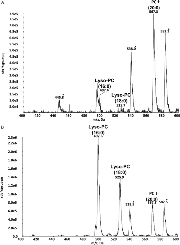
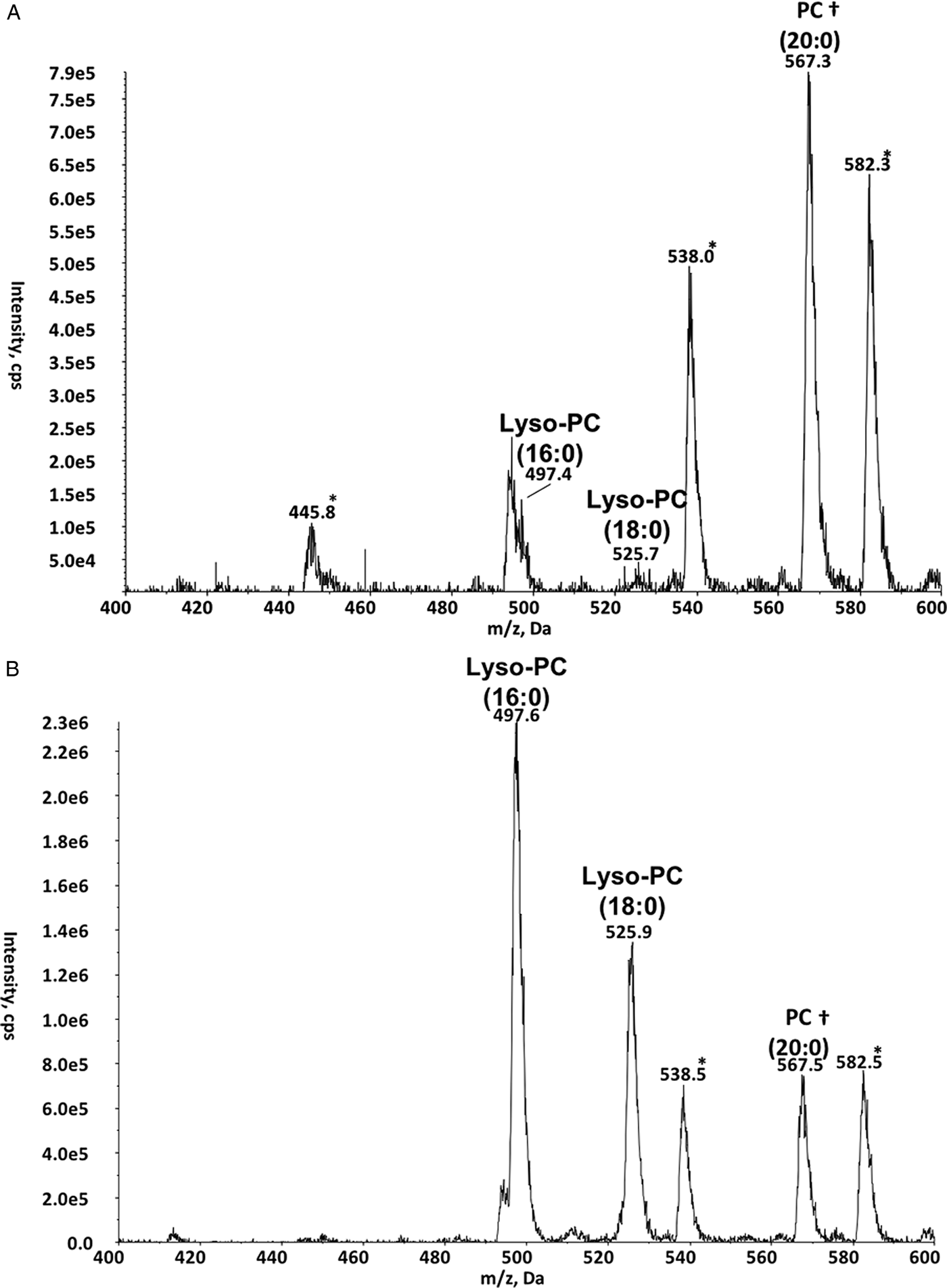
Finally, GST-TbnSMase activity towards choline-containing phospholipids was reassessed. During previous research into TbnSMase, catabolic activity towards PC and lyso-phosphatidylcholine lipid substrates could not be detected (Young and Smith, Reference Young and Smith2010). Re-assessing GST-TbnSMase activity towards PC (16:0/16:0) using ESI-MS/MS analysis failed to establish any substrate turnover (Avanti Polar Lipids product no. 850355) (data not shown). However, precursors of m/z 184 scans in positive ion mode revealed that GST-TbnSMase does turnover lyso-phosphatidylcholine species (Fig. 7). There was a marked reduction in lyso-phosphatidylcholines (16:0) and (18:0) only in the presence of active GST-TbnSMase (Fig. 7A), relative to a PC (10:0/10:0) standard included in all lyso-phosphatidylcholine reactions. This decrease did not occur in the control (Fig. 7B). Additionally, the lyso-phosphatidylcholine analogues miltefosine and edelfosine were previously shown to inhibit GST-TbnSMase (Young and Smith, Reference Young and Smith2010). This led to speculation that the previously observed inhibition of the enzyme's activity by lyso-phosphatidylcholine analogues may be due to these compounds acting as competing substrates. Testing miltefosine as a potential substrate for GST-TbnSMase (5–50 nmoles) in the Amplex® UltraRed assay system did not produce any data to suggest this lyso-phosphatidylcholine analogue is turned over by GST-TbnSMase (data not shown). This indicates lyso-phosphatidylcholine analogues are not competitive substrates for GST-TbnSMase. However, the possibility remains that these compounds may be tightly bound to the enzyme's active site, preventing the ChoP release required for assay detection.
ESI-MS/MS analysis of lyso-phosphatidylcholine substrate reactions. Spectra are ESI-MS/MS precursor ion scans to detect choline-containing lipids (precursors of m/z 184) in positive ion mode. (A) GST-TbnSMase-enriched bacterial membranes with lyso-phosphatidylcholine substrate. (B) Non-TbnSMase-expressing bacterial membranes with lyso-phosphatidylcholine substrate. †Highlights the didecanoyl-PC (20:0) standard (500 pmoles). *Highlights previously identified significant contaminants of positive ion mode scans thought to be associated with the detergent.
Discussion
Sphingolipid metabolism in kinetoplastid parasites has long been established as a potential target for anti-parasitic drug development (Smith and Bütikofer, Reference Smith and Bütikofer2010; Mina and Denny, Reference Mina and Denny2017). However, research has focused almost exclusively on the pathways involved in sphingolipid biosynthesis. Initial research into the nSMase found in T. brucei showed this enzyme has sphingolipid catabolic activity (Young and Smith, Reference Young and Smith2010). TbnSMase is the only currently identified T. brucei protein that displays this function. Unusually, the parasites appear to lack phospholipase D activity, which generally facilitates eukaryotic SM and PC catabolism. The substrate specificity of TbnSMase was examined to improve understanding of T. brucei sphingolipid catabolism and salvage.
TbnSMase is now known to turnover SM, EPC and IPC sphingolipid species. This is consistent with the established sphingolipid composition of T. brucei (Richmond et al., Reference Richmond, Gibellini, Young, Major, Denton, Lilley and Smith2010; Guan and Mäser, Reference Guan and Mäser2017): IPC predominates in procyclic parasites, whilst almost equal proportions of SM and EPC species are found in the bloodstream form (Guan and Mäser, Reference Guan and Mäser2017). In light of this knowledge, it may seem unsurprising that TbnSMase is active towards all three of these sphingolipid classes, only distinguished structurally by their headgroup (choline, ethanolamine and inositol). However, to our knowledge, this breadth of sphingolipid substrate specificity has not been documented previously for a lipid catabolic enzyme. It is possible that other sphingolipid degradative enzymes (especially those found in other kinetoplastids) share this ability but have not been tested. If this wide-ranging substrate specificity is not found in mammalian nSMase homologues, it may be possible to exploit these differences to create TbnSMase-specific substrate analogue inhibitors. The TbnSMase activity reported here precludes the need for other sphingolipid degradative enzymes in the ER and rationalizes the constitutive expression of this enzyme in both procyclic and bloodstream forms. However, no activity towards glycosphingolipids was observed, indicating phosphate groups may be an obligatory feature of TbnSMase substrates. It has been reported that T. brucei possess trace levels of glycosphingolipids, glucosylceramide species having been identified in several studies (Uemura et al., Reference Uemura, Watarai, Kushi, Kasama, Ohnishi and Kodama2006; Fridberg et al., Reference Fridberg, Olson, Nakayasu, Tyler, Almeida and Engman2008; Richmond et al., Reference Richmond, Gibellini, Young, Major, Denton, Lilley and Smith2010). These could be endocytosed host glycosphingolipids, as no glycosphingolipid biosynthetic enzymes have been formally identified in T. brucei (Uemura et al., Reference Uemura, Watarai, Kushi, Kasama, Ohnishi and Kodama2006; Richmond et al., Reference Richmond, Gibellini, Young, Major, Denton, Lilley and Smith2010; Guan and Mäser, Reference Guan and Mäser2017). Only galactosylceramide species were tested as possible substrates for TbnSMase, thus it remains possible the enzyme is active towards other glycosphingolipids. Alternatively, another (other) unidentified enzyme(s) may be responsible for T. brucei glycosphingolipid catabolism, possibly within the lysosome.
ESI-MS/MS analysis indicates that TbnSMase preferentially catabolizes SM species with specific fatty acid chain lengths. A more significant decrease in SM 42:1 (d18:1/24:0)/SM 42:2 (d18:1/24:1) occurred relative to SM 36:1 (d18:1/18:0), despite the latter species being more prevalent in the substrate mixture. This suggests that TbnSMase activity may be geared towards recycling specific long-chain fatty acids, which can then be utilized in other biosynthetic processes. Defining a crystal structure for TbnSMase may reinforce these preliminary observations, particularly if the structure provides insight into substrate binding and potential regulation. Few crystal structures for nSMase proteins are currently available, and most studies have focused on the binding of choline phosphate to the active site (Openshaw et al., Reference Openshaw, Race, Monzó, Vázquez-Boland and Banfield2005; Ago et al., Reference Ago, Oda, Takahashi, Tsuge, Ochi, Katunuma, Miyano and Sakurai2006). However, TbnSMase does not seem to distinguish between its substrates based on headgroup identity, instead appearing to preferentially degrade substrates with specific fatty acids. This finding indicates that the fatty acid composition of lipid substrates may be of greater interest when considering enzyme–substrate interactions. In support of this view, TbnSMase was shown to actively degrade C-1-P, which lacks a polar alcohol headgroup entirely. However, the enzyme was unable to catabolize S-1-P, indicating the presence of an amide bound fatty acid at the sn-2 position is vital for sphingolipid substrate recognition. Indeed, in the case of TbnSMase, the critical aspect of lipid substrate recognition appears to centre on the binding of the phosphate and fatty acid moieties to sphingosine or glycerol backbones. TbnSMase is now known to degrade lyso-phosphatidylcholine in addition to sphingolipids. This aspect of TbnSMase activity brings the enzyme in line with the nSMase found in P. falciparum parasites (PfnSMase) (Hanada et al., Reference Hanada, Palacpac, Magistrado, Kurokawa, Rai, Sakata, Hara, Horii, Nishijima and Mitamura2002). Both PfnSMase and TbnSMase are inactive towards PC substrates, despite their ability to degrade lyso-phosphatidylcholines. This points to the diacylglycerol (DAG) moiety of phospholipids impairing substrate binding to these SMase enzymes, and that removal of the fatty acid at the sn-2 position alleviates this inhibition. Thus, the requirements for glycerophospholipid recognition are the inverse of sphingophospholipid recognition.
TbnSMase is the first identified T. brucei enzyme shown to be capable of degrading lyso-phosphatidylcholines, which are believed to be a primary source of parasite choline during bloodstream infection (Bowes et al., Reference Bowes, Samad, Jiang, Weaver and Mellors1993; Smith and Bütikofer, Reference Smith and Bütikofer2010; Macêdo et al., Reference Macêdo, Schmidt, Mäser, Rentsch, Vial, Sigel and Bütikofer2013). This is due to the parasites lacking a choline de novo biosynthesis pathway (Smith and Bütikofer, Reference Smith and Bütikofer2010), and lyso-phosphatidylcholine concentration in the blood being 10-fold greater than choline (Macêdo et al., Reference Macêdo, Schmidt, Mäser, Rentsch, Vial, Sigel and Bütikofer2013). Mammalian bloodstream T. brucei requires an abundant source of choline, as over 50% of their lipid complement consists of choline-containing lipids (Smith and Bütikofer, Reference Smith and Bütikofer2010). The T. brucei phospholipase A1 was shown to degrade PC, but not lyso-phosphatidylcholine species (Richmond and Smith, Reference Richmond and Smith2007a, Reference Richmond and Smith2007b). A plasma membrane phospholipase that degrades lyso-phosphatidylcholine has been postulated (Bowes et al., Reference Bowes, Samad, Jiang, Weaver and Mellors1993) but has yet to be identified. This activity in TbnSMase indicates the enzyme may be responsible for lyso-phosphatidylcholine turnover in the ER. Research into the essentiality of TbnSMase showed that compromised enzyme function led to a concomitant decrease in the parasite's rate of endocytosis (Young and Smith, Reference Young and Smith2010). This could have wide-ranging effects on the parasites, but was thought to have particularly impacted T. brucei choline homoeostasis due to the parasites’ dependence upon endocytosed and recycled choline-containing lipids. This impact was indicated by a marked decrease in PC and increased intracellular DAG, suggesting PC de novo biosynthesis via the Kennedy pathway had been disrupted (Young and Smith, Reference Young and Smith2010). The newly discovered ability of TbnSMase to degrade lyso-phosphatidylcholine and SM species aligns with these observations, indicating decreased enzyme activity has a direct impact on choline homoeostasis. This underlines the importance of TbnSMase function in sustaining the intracellular choline metabolite levels required for parasite survival and propagation.
Given TbnSMase is able to degrade various sphingolipids and lyso-phosphatidylcholine, it seems likely that the enzyme will also be able to catabolize other lyso-phospholipids, particularly those with either an ethanolamine-phosphate or inositol-phosphate headgroup. The promiscuous activity of TbnSMase likely explains the rather unusual fact that T. brucei parasites have not retained a phospholipase D enzyme, which facilitates these degradative processes in other eukaryotes.
Further research is required to identify and characterize other enzymes that underpin lipid catabolism and salvage in these kinetoplastid parasites, which represent a valuable model system for eukaryotic lipid metabolism (Serricchio and Bütikofer, Reference Serricchio and Bütikofer2011). As in the case of TbnSMase, the activity of these enzymes may prove vital, opening new areas of T. brucei biochemistry to drug development.
Supplementary material
The supplementary material for this article can be found at https://doi.org/10.1017/S0031182018001853.
Author ORCIDs
Terry K. Smith http://orcid.org/0000-0003-1994-2009
Acknowledgements
NBD-IPC was kindly gifted by Dr K. Zhang (Texas Tech University). The Biomedical Sciences Research Complex Mass Spectrometry and Proteomics Facility, University of St Andrews, performed mass-spectrometric analysis to identify GST-TbnSMase protein.
Financial support
This work was supported primarily through the European Community's Seventh Framework Programme under grant agreements no. 602773 (Project KINDRED), with additional support from Wellcome Trust Project grant (086658); Medical Research Council (MR/Mo20118/1) and the School of Chemistry (The University of St Andrews).
Conflict of interest
None.
Ethical standards
Not applicable.