Endophenotypes were initially defined as genetically-influenced internal traits that predict behavior, but are not readily observable (John & Lewis, Reference John and Lewis1966; Ruggeri, Sarkans, Schumann, & Persico, Reference Rozga, Hutman, Young, Rogers, Ozonoff, Dapretto and Sigman2014). The endophenotype concept originated in evolutionary biology, but gained a foothold in psychiatry in schizophrenia research, where it was used to describe “internal phenotypes,” detectable biochemically or microscopically (Gottesman & Shields, Reference Gottesman and Shields1973). From there, Gottesman and Gould published their foundational paper, which described the promise of broader applications of endophenotypes to psychiatry, and presented unambiguous operational criteria (Gottesman & Gould, Reference Gottesman and Gould2003). Since then, enthusiasm surrounding the potential utility of endophenotypes has grown in basic, clinical, and translational research (Gould & Gottesman, Reference Gould and Gottesman2006). As initially defined, endophenotypes are measurable features that signal underlying genetic vulnerability to disorders (Gottesman & Gould, Reference Gottesman and Gould2003; Gould & Gottesman, Reference Gould and Gottesman2006). Such features are distinguished from risk factors, more general biomarkers, and downstream phenotypic features of disorder in several important ways.
Terminology
Risk and protection versus vulnerability and resilience
As several authors have noted, the terms “risk factor,” “vulnerability,” “biomarker,” and “endophenotype” are often conflated in the literature (Beauchaine & Constantino, Reference Beauchaine and Constantino2017; Lenzenweger, Reference Landa, Piven, Wzorek, Gayle, Chase and Folstein2013). However, each carries distinct meaning. Risk factors are exogenous variables, or features of the environment rather than the individual, that correlate with, and may or may not be causally related to, a disorder (e.g., Beauchaine, Neuhaus, Brenner, & Gatzke-Kopp, Reference Beauchaine, Neuhaus, Brenner and Gatzke-Kopp2008; Compas & Andreotti, Reference Compas, Andreotti, Beauchaine and Hinshaw2013). Common risk factors for psychiatric disorders include low socioeconomic status, neonatal teratogen exposure, and prolonged periods of stress exposure, particularly during critical developmental periods (Fryer, Crocker, & Mattson, Reference Fryer, Crocker, Mattson, Beauchaine and Hinshaw2008; Grant et al., Reference Grant, Compas, Stuhlmacher, Thurm, McMahon and Halpert2003; Compas & Andreotti, Reference Compas, Andreotti, Beauchaine and Hinshaw2013). Conversely, protective factors are exogenous influences that reduce the likelihood of disorder, such as sensitive parenting (Compas et al., Reference Compas, Champion, Forehand, Cole, Reeslund, Fear and Roberts2010) and neighborhood cohesion (Riina, Martin, & Brooks-Gunn, Reference Redcay and Courchesne2014).
In contrast to the exogenous nature of risk and protection, vulnerability and resiliency are often defined as endogenous characteristics that increase or decrease, respectively, the likelihood of psychiatric disorder (Beauchaine & Constantino, Reference Beauchaine and Constantino2017; Beauchaine et al., Reference Beauchaine, Neuhaus, Brenner and Gatzke-Kopp2008). Example vulnerabilities include genetically-influenced temperament/personality traits (e.g., impulsivity, behavioral inhibition) and cognitive strengths and weaknesses (Beauchaine & Constantino, Reference Beauchaine and Constantino2017). Common resiliencies include the use of strong cognitive coping skills such as reappraisal, strong executive function, and effective emotion regulation (e.g., Beauchaine & Cicchetti, Reference Beauchaine and Cicchetti2019; Masten, Reference Luyster, Wagner, Vogel-Farley, Tager-Flusberg and Nelson2014; Zelazo & Carlson, Reference Yucel, Belger, Bizzell, Parlier, Adolphs and Piven2012). Protective factors and resiliencies can attenuate the effects of vulnerabilities and risk factors, and consequently make penetrance of disorder less likely (Compas & Andreotti, Reference Compas, Andreotti, Beauchaine and Hinshaw2013).
Biomarkers versus endophenotypes
Biomarkers are measurable features that are often associated with a given disorder (Beauchaine & Constantino, Reference Beauchaine and Constantino2017; Lenzenweger, Reference Landa, Piven, Wzorek, Gayle, Chase and Folstein2013). Importantly, although biomarkers often indicate disorder, they are not always specific to single disorders (i.e., some are transdiagnostic), they do not necessarily indicate genetic vulnerability (e.g., a brain lesion can follow an exogenous insult), and they may or may not be causally related to disorder progression (e.g., heart disease is a tertiary not primary outcome of diabetes). An example from psychopathology research finds that elevated morning cortisol levels predict the onset of mood disorders (Ellenbogen, Hodgins, Linnen, & Ostiguy, Reference Ellenbogen, Hodgins, Linnen and Ostiguy2011). Although this association is clear, it is not clear whether elevated cortisol levels are along the causal pathway to disorder, or are merely correlated with other causal factors. Thus, biomarkers need not be heritable, specific to a single genetic etiology, or specific to a single disease state (see Beauchaine & Constantino, Reference Beauchaine and Constantino2017).
In contrast, endophenotypes are a subset of biomarkers that are associated specifically with genetic vulnerability and lie along a causal pathway from such vulnerability to observed disorder (Gottesman & Gould, Reference Gottesman and Gould2003; Gould & Gottesman, Reference Gould and Gottesman2006). Given their causal association with genetic liability, endophenotypes are state-independent and are therefore present whether an individual is in an active or remitted phase of disorder. This contrasts with many biomarkers, which are state-dependent, and therefore only observed in active phases of illness. Thus, endophenotypes are especially useful for tracking genetic vulnerability within pedigrees, and within the population. Importantly, this argues against the common conceptualization of sub-threshold symptoms of disorder as endophenotypes themselves. Rather, endophenotypes are better thought of as specific markers of function—whether neural, peripheral, or behavioral—that may not affect one's day-to-day function or be noticed either casually or clinically. In other words, endophenotypes mark genetic vulnerability regardless of the degree of penetrance. They may therefore overlap with, but are not isomorphic with, subclinical features. Several authors have suggested that the endophenotype construct is strongest when anchored tightly to specific neurobiological and behavioral substrates (e.g., neural responses to certain classes of stimuli, working memory), rather than abstract constructs or “behavioral constellations” (e.g., Beauchaine & Constantino, Reference Beauchaine and Constantino2017; Insel & Cuthbert, Reference Insel and Cuthbert2009). In particular, one study of individuals with schizophrenia and their relatives found that distinct symptom patterns (increased positive versus negative symptoms) are associated with distinct genetic markers (Wessman et al., Reference Weigelt, Koldewyn and Kanwisher2009). Although this is a worthwhile finding for subtyping efforts, elevated positive versus negative symptoms are abstract concepts. Thus, this makes functional application as an endophenotype challenging for clinical purposes in the general population (Wessman et al., Reference Weigelt, Koldewyn and Kanwisher2009).
Endophenotypes, Biomarkers, and the Research Domain Criteria (RDoC)
Both biomarkers and endophenotypes are central to the RDoC initiative, which specifies functional domains of behavior (positive valence systems, negative valence systems, cognitive systems, social processes, arousal/regulatory systems, sensorimotor systems) across levels of analysis spanning genes to self-report (National Institute of Mental Health, Reference Nadig and Shaw2019). RDoC represent migration away from defining disorders based solely on presenting symptoms in favor of specifying complex etiologies (see Cuthbert & Insel, Reference Cuthbert and Insel2013; Insel et al., Reference Insel, Cuthbert, Garvey, Heinssen, Pine, Quinn and Wang2010). Prevailing diagnostic systems, including the Diagnostic and statistical manual of mental disorders (DSM), fifth edition (American Psychiatric Association, 2013) and the International classification of diseases (ICD), tenth edition (World Health Organization, 2004), classify disorders based on enumerated phenotypic criteria, most of which were derived deductively (Morris & Cuthbert, Reference Miu, Pană and Avram2012). Diagnostic criteria have considerable overlap among disorders, which in most cases means no single symptom is necessary or sufficient for diagnosis (Kozak & Cuthbert, Reference Kimhi2016). For some disorders, criterion sets are even polythetic, yielding the same diagnosis for two people without a single overlapping symptom (see e.g., Crowell, Beauchaine, & Linehan, Reference Crowell, Beauchaine and Linehan2009). As a result, DSM and ICD diagnostic categories have limited utility for identifying biomarkers. By making etiology central to diagnoses, searches for biomarkers and endophenotypes should be more feasible. One objective of the RDoC is to define disorders based on causal mechanisms by identifying neurophysiological, neurohormonal, psychophysiological, and other biomarkers. As noted above, because endophenotypes are located along causal pathways from genes to disorders (Gottesman & Gould, Reference Gottesman and Gould2003; Gould & Gottesman, Reference Gould and Gottesman2006), they are central to the RDoC mission.
An additional, notable feature of the RDoC initiative is its explicit dimensional approach (Morris & Cuthbert, Reference Miu, Pană and Avram2012). It is now well-recognized that nearly all psychiatric syndromes expressed in childhood, adolescence, and adulthood are distributed along symptom continua, with no clear boundaries between health and dysfunction (Finucane, Challman, Martin, & Ledbetter, Reference Finucane, Challman, Martin and Ledbetter2016; Northoff & Tumati, Reference Noland, Reznick, Stone, Walden and Sheridan2019; Wright, Krueger, Hobbs, Markon, Eaton, & Slade, 2013).
Biomarkers and Endophenotypes in Autism Spectrum Disorder (ASD)
In recent decades, a large body of research has established expression of a broader autism phenotype (BAP) among relatives of those with ASD. Similarly, those who study early detection and diagnosis of ASD have identified measurable, early signs among vulnerable infants that emerge well before full-blown diagnostic criteria are met. Importantly, these lines of research partially predate popularity of the terms “endophenotype” and “biomarker” in the field of ASD research. In light of this, significant research related to the identification of endophenotypes in ASD exists, but has not yet been integrated into an inclusive framework.
Given the importance of early intervention for many children with ASD (e.g., Dawson et al., Reference Dawson, Rogers, Munson, Smith, Winter, Greenson and Varley2010), and given the utility of endophenotypes for early identification of vulnerability to disorder, specifying ASD-related endophenotypes could lead to earlier intervention and, consequently, better long-term behavioral outcomes (Beauchaine et al., Reference Beauchaine, Neuhaus, Brenner and Gatzke-Kopp2008; Iacono, Malone, & Vrieze, Reference Iacono, Malone and Vrieze2017). Additionally, valid biomarkers and endophenotypes may be crucial for identifying disorder subtypes (phenocopies) that emerge from multi-final etiological pathways (see Beauchaine, Constantino, & Hayden, Reference Beauchaine, Constantino and Hayden2018). Such pathways may have different responses to treatment and different prognoses (Beauchaine et al., Reference Beauchaine, Neuhaus, Brenner and Gatzke-Kopp2008; Faja & Dawson, Reference Faja and Dawson2015; Iacono et al., Reference Iacono, Malone and Vrieze2017). Ignoring biological and neuropsychological underpinnings forfeits the opportunity to develop targeted interventions based on valid etiological subtypes (Beauchaine & Marsh, Reference Beauchaine, Marsh, Cicchetti, Cohen, Cicchetti and Cohen2006; Beauchaine et al., Reference Beauchaine, Neuhaus, Brenner and Gatzke-Kopp2008). Some features of ASD make this disorder a particularly good candidate for identifying endophenotypes. ASD demonstrates strong heritability, with subclinical features among at least some relatives (Viding & Blakemore, Reference Thorup, Nyström, Gredebäck, Bölte and Falck-Ytter2007). Despite the recent proliferation of genetic research on ASD, diagnostic yield for genetic testing in ASD remains relatively low at around 12–15% (Kalsner, Twachtman-Bassett, Derynioski, & Chamberlain, Reference Kaiser and Pelphrey2018; Tammimies et al., Reference Tajmirriyahi, Nejati, Pouretemad and Sepehr2015); for the majority of children with autism, no clear genetic etiology is identified. A small subset of diagnosed children carries known de novo genetic differences (e.g., 22q deletion) that are associated with increased incidence of ASD, although these etiologies generally have incomplete penetrance for ASD.
The Present Review
This scoping review identifies emergent endophenotype candidates for ASD. Given the centrality of disrupted social communication, social processes, and social behaviors to ASD (e.g., Constantino & Charman, Reference Constantino and Charman2016), we focus specifically on mechanisms related to the Social Processes functional domain of behavior specified in the RDoC (National Institute of Mental Health, Reference Nadig and Shaw2019). In specifying potential endophenotypes, we rely largely on criteria proposed by Gottesman and Gould (Reference Gottesman and Gould2003). Integration and review of the available literature was undertaken per the subdomains identified under the RDoC (i.e., affiliation and attachment, social communication, perception and understanding of self, perception and understanding of others). For each subdomain, convergent evidence from a variety of paradigms, populations and units of analysis are integrated and discussed. Strengths and weaknesses in the literature for each domain are discussed and future directions are recommended. Finally, cross-cutting themes and recommendations for future research present in multiple domains are discussed in an integrated fashion. The literature reviewed in these content areas is not meant to provide a comprehensive review, but instead provide a scoping overview of active areas of investigation and a discussion of key findings as they relate to satisfying endophenotypic criteria. In this way, a broad understanding of the current state of social endophenotype research in ASD is obtained to inform promising leads and future directions.
Method
Scope
We focus on functional, task-based findings for endophenotypes related to Social Processes, as defined by the RDoC matrix (National Institute of Mental Health, Reference Nadig and Shaw2019). Task-based findings include eye-tracking, electroencephalography (EEG), patterns of neural reactivity during functional magnetic resonance imaging (fMRI), physiological measures such as heart rate, and behavioral coding and observation. It is important to note that our initial search unearthed significant literature on non-social endophenotypes. However, these articles are not covered given practical limitations of the scoping review. Promising leads not covered include sensory gating, central coherence, gamma power, and functional and structural connectivity. For the purpose of balancing a broad scope with satisfactory comprehensiveness, the literature regarding physical features, blood-based and biological endophenotypes was excluded. The literature not covered includes topics such as levels of circulating oxytocin and the presence of oxytocin receptors, macrocephaly, facial asymmetry, and digit ratio. Structural MRI findings are reviewed insofar as they augment understanding of fMRI data. In particular, future reviews should cross-examine structural and neurochemical findings with the task-based, behaviorally-observed findings reported here, as some may tap similar underlying constructs and may provide convergent evidence. As one example, oxytocin is a neuropeptide associated with social bonding that has been examined as a potential endophenotype in ASD (Oztan et al., Reference Oerlemans, van der Meer, van Steijn, de Ruiter, de Bruijn, de Sonneville and Rommelse2018; Parker et al., Reference Palermo, Pasqualetti, Barbati, Intelligente and Rossini2014) and may relate to behaviorally-measured endophenotypes.
Also of note, scale-based findings are not reviewed. Although scale measures may probe some underlying vulnerabilities, they represent a distal level of analysis with a lower signal-to-noise ratio. In fact, up to 50% of variance in scale scores is measurement error (Hoyt & Kerns, Reference Hoyt and Kerns1999). Reducing such error is a primary motivation for identifying endophenotypes (Gottesman & Gould, Reference Gottesman and Gould2003; Gould & Gottesman, Reference Gould and Gottesman2006). As noted previously, elevated scores on measures of the BAP, such as the Social Responsiveness Scale (SRS) (Constantino et al., Reference Constantino, Davis, Todd, Schindler, Gross, Brophy and Reich2003) and the Autism-Spectrum Quotient (AQ) (Woodbury-Smith, Robinson, Wheelwright, & Baron-Cohen, Reference Winder, Wozniak, Parladé and Iverson2005) are likely to correlate with endophenotypic findings. Therefore, we discuss scale-based findings of the BAP as indicators of the validity of proposed endophenotypes, but not as endophenotypes themselves.
Guiding theoretical principles
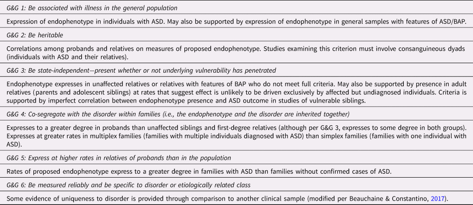
We review and discuss relevant literature with a goal of examining the extent to which specific endophenotype candidates meet some or all of the Gould and Gottesman (Reference Gould and Gottesman2006) criteria (Table 1).
Gould and Gottesman (Reference Gould and Gottesman2006) criteria (in italic) and description of relevant supporting empirical evidence

Notes: ASD = autism spectrum disorder; BAP = broader autism phenotype.
All the literature reviewed relates to known constructs and bodies of literature supporting criterion 1, association with ASD in the general population. A large body of literature is related to detailed characterization of the ASD phenotype and falls outside the scope of this review. Criterion 1 is discussed to characterize how RDoC constructs are disrupted in a broad, phenotypic sense in individuals with ASD, however; we focus below primarily on criteria 2–6. Evidence relating to specific Gould & Gottesman (Reference Gould and Gottesman2006) criteria (as listed in Table 1) are noted by, for example, (G&G 1) throughout the review.
Search strategy
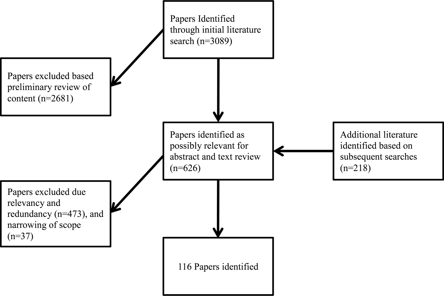
Initial searches were conducted between July and December 2017 using EBSCOhost research databases. Search databases included Medline and PsychInfo. Because several related key terms (e.g., biomarker, endophenotype, risk factor, vulnerability, intermediate phenotype) may not be used consistently in the literature, all were used as search terms. Literature regarding early detection may be informative and was included in the search. Initial searches with these terms yielded 3,089 results. Following an initial review, several exclusion terms were added to eliminate common unrelated subjects. A list of all the search terms is provided in Online Appendix A.
Next, titles and key terms were reviewed for relevancy, yielding 408 articles. We reviewed abstracts and, if necessary, text to ensure relevancy. Follow-up searches were conducted using key terms extracted from the initial searches (see Online Appendix A). Additional follow-up searches were conducted in the summer of 2018 to update the literature base, as well as probe the literature using RDoC key words (e.g., “attachment,” “affiliation”). These possibly relevant studies were reviewed for relevancy to the present review, and 218 additional potentially relevant studies were identified. All 626 identified studies from both searches were screened for relevancy to the present review, after which 473 studies were eliminated (e.g., non-empirical papers, non-ASD focus). Upon review, the majority of the initially identified reports were related to aspects of sociality (e.g., TOM, social attention, social communication, etc.), with the remaining identified literature (n = 37) distributed among other, more varied constructs (e.g., sensory processing differences, response inhibition, functional connectivity). Therefore, for the purpose of streamlining the scope and structure of the present review, we chose to review promising endophenotype candidates relating to aspects of sociality. 116 articles were included in the present review (see Figure 1 for the literature search flow chart and Table 2 for an overview of all the included literature). The included literature was organized based upon subcategories of the Social Processes functional domain of the RDoC matrix, including (a) affiliation and attachment, (b) social communication, (c) perception and understanding of self, and (d) perception and understanding of others (National Institute of Mental Health, Reference Nadig and Shaw2019).
Search strategy flow chart for reviewed literature.

Summary of reviewed literature

Note: Table detailing all referenced empirical studies organized by RDoC domains. Infants = participants under 12 months; Toddlers/Preschool age = children aged 1–4 years on average; School-age = children 5–12 years of age on average; Adolescents = participants 13–18 years of age on average; ASD = participants with a diagnosis of autism; DD = participants with developmental delay; TD = typically developing participants; BAP = broader autism phenotype; SLI = participants with specific language impairment; Parents-Down syndrome = parents of children with Down syndrome; Parents-SLI = panrents of children with specific language impairment; Relatives-ASD = relatives of people with ASD; Sibs-ASD = siblings of children with autism; Sibs-TD = siblings of typically developing children. fNIRS = functional near-infrared spectroscopy; EEG = electroencephalography; ERP = event-related potential; MRI = magnetic resonance imaging; fMRI = functional magnetic resonance imaging; FFA = fusiform face area; AQ = Autism-Spectrum Quotient = SRS Social Responsiveness Scale; RJA = responding to joint attention; IJA = initiating joint attention; FXS = Fragile X syndrome. See Table 1 for a description of the Gottesman and Gould criteria and what empirical findings constitute as in support of each criterion.
Endophenotypic Constructs
Affiliation and attachment
Affiliation. Affiliation is the propensity to engage in positive social interaction with others. At its most basic level, affiliation requires attention to social stimuli, and social motivation. Here, studies examining preference for social stimuli and responsivity to, and engagement with, social stimuli are discussed. ASD is marked by decreased social attention and motivation, including diminished attention to social stimuli, reduced reward value of social versus non-social stimuli, and aberrant patterns of engagement during real-world social interactions (G&G 1) (Jones, Carr & Klin, Reference Jones, Carr and Klin2008; Chevallier, Kohls, Troiani, Brodkin, & Schultz, Reference Chevallier, Kohls, Troiani, Brodkin and Schultz2012; Pierce, Marinero, Hazin, McKenna, Barnes & Malige, Reference Pierce, Marinero, Hazin, McKenna, Barnes and Malige2016; Lord, Luyster, Gotham, & Guthrie, Reference Lloyd-Fox, Blasi, Elwell, Charman, Murphy and Johnson2012).
Several studies indicate that individuals with a high ASD-related genetic loading (but without a confirmed diagnosis) display aberrant patterns of attention to social stimuli. This suggests that disrupted social attention may be an endophenotype of ASD. Vulnerable siblings (by virtue of having older siblings with ASD) demonstrate decreased attention to important social stimuli, including faces and speech, starting extremely early in development and persisting across time (G&G 5). Di Giorgio et al. (Reference Di Giorgio, Frasnelli, Rosa Salva, Maria Luisa, Puopolo, Tosoni and Vallortigara2016) found that vulnerable siblings demonstrated lower preference for face-like stimuli (versus inverted faces) and biological motion (versus random motion) compared with low-risk siblings (siblings of children without autism) when tested between 6 and 10 days of age, suggesting differences in inborn predispositions to attend to socially-relevant stimuli. Similarly, Rutherford (2013) examined preference for several social stimuli (faces versus non-faces; eyes versus mouth; biological versus random motion) in vulnerable and low-risk infants using eye-tracking technology. They found that low-risk siblings showed significantly larger preference for all three socially-relevant stimuli, and that the divergence between vulnerable and low-risk groups was greater at 6 months of age than at 3 months, suggesting increasing abnormality in social attention with age for the vulnerable group. Another study found that vulnerable 5-month-old infants failed to show a typical preference for direct versus averted gaze (Nele, Ellen, Petra, & Herbert, 2015). Relatedly, Noland, Reznick, Stone, Walden, and Sheridan (Reference Nichols, Ibañez, Foss-Feig and Stone2010) found better working memory for non-social compared to social targets in vulnerable siblings; however, this was driven by better performance for non-social targets in the vulnerable group, as opposed to impaired performance in memory for social targets. Nonetheless, these different patterns of attentional allocation to non-social targets could have downstream implications for social development. For studies such as these, it is unclear whether it is primarily vulnerable infants who will go on to be formally diagnosed with ASD who drive effects, or if vulnerable infants who will not go on to be diagnosed also display this difference. To establish evidence for state-independence (G&G 3), it is important that this is resolved.
Several additional studies have corroborated these findings using a variety of technologies to examine neural responses to social stimuli. In an EEG study of event-related potentials (ERPs), Key et al. (Reference Kalsner, Twachtman-Bassett, Derynioski and Chamberlain2015) found that 9-month-old vulnerable siblings showed shorter P400 latencies (potentially indicating less extensive processing) and lower Nc amplitudes (reflecting less attentional allocation) in response to subtle smiles (but not full smiles), in comparison with low-risk siblings (G&G 5). Additional ERP research found faster P400 responses to faces for low-risk siblings as compared with vulnerable siblings, and faster N290 and P400 responses to objects in vulnerable siblings as compared with low-risk siblings (McCleery, Akshoomoff, Dobkins, & Carver, Reference Masten2009), as well as diminished inferior frontal and posterior temporal responses to visual and auditory social stimuli among 4- to 6-month-old vulnerable infants, as detected by fNIRS (functional near-infrared spectroscopy) (Lloyd-Fox et al., Reference Libero, DeRamus, Deshpande and Kana2013). This fNIRS finding was recently replicated with visual stimuli in an independent sample of vulnerable 5-month-olds (Braukmann et al., Reference Braukmann, Lloyd-Fox, Blasi, Johnson, Bekkering, Buitelaar and Hunnius2018). In both cases, patterns of responding in the vulnerable groups were similar to those found in older children and adults with ASD (Lloyd-Fox et al., Reference Libero, DeRamus, Deshpande and Kana2013; McCleery et al., Reference Masten2009). Taken together, these findings indicate that vulnerable siblings show attenuated attention to visual social stimuli (faces, eyes, biological motion) beginning very early in development, establishing the presence of such behaviors in families affected by ASD (G&G 5).
Although less work has examined responses to auditory social stimuli, one study found that vulnerable infants lack normal preference for speech versus non-speech sounds (Curtin & Vouloumanos, Reference Curtin and Vouloumanos2013) as measured by look time. Another study found that when using fNIRS to measure activity in the left and right temporal regions, vulnerable female 3-month-old infants failed to habituate to repetitive sounds when compared with female low-risk infants (Edwards, Wagner, Tager-Flusberg, & Nelson, Reference Edwards, Wagner, Tager-Flusberg and Nelson2017). These findings suggest that aberrant social responding in vulnerable infants may generalize beyond specific visual processing differences and may represent a broader phenomenon of lack of attention to socially-relevant stimuli. However, ASD outcomes were not assessed, making it unclear whether effects were driven only by a subset of vulnerable siblings with eventual ASD outcomes (G&G 3). Once again, future research must demonstrate that this abnormality is also found in vulnerable infants who will not go on to develop ASD to satisfy the criteria of state-independence (G&G 3).
Addressing this, some work indicates differences in social processing among family members without ASD outcomes, although such differences are often more subtle and less consistent. For example, Elsabbagh et al. (Reference Elsabbagh, Mercure, Hudry, Chandler, Pasco, Charman and Johnson2012) found that vulnerable siblings who did not go on to develop ASD showed similar patterns of responding to eye gaze as low-risk siblings on some ERP measures. However, their ERP responses to direct gazes were slower than their responses to averted gazes—opposite the pattern seen in low-risk siblings or vulnerable siblings with an ASD outcome. This suggests some potential disruption of early social attention in vulnerable siblings, including those without ASD outcomes (G&G 3 and G&G 5). In addition, Dawson et al. (Reference Dawson, Webb, Wijsman, Schellenberg, Estes, Munson and Faja2005) found that parents of children with ASD (none of whom were themselves diagnosed with ASD) failed to show a typical stronger right-lateralized N170 response when viewing faces These findings suggest persistence of social processing differences into adulthood for family members and state-independence (G&G 3).
Key and Stone (Reference Key, Ibanez, Henderson, Warren, Messinger and Stone2012) reported that vulnerable and low-risk 9-month-olds showed similar patterns of looking at facial features, yet their patterns of brain activation differed, with low-risk infants more quickly recruiting face-specific processing mechanisms as measured by shorter N290 latencies. Neural responses were significantly correlated with parent reports of communication skills in both the vulnerable and low-risk groups, suggesting co-segregation of neural mechanisms with ASD-related symptoms (G&G 4).
Additional evidence suggests that social motivation differences are heritable, even in families without a history of ASD (G&G 2). Jones, Venema, Earl, Lowy, and Webb (Reference Jones, Venema, Earl, Lowy and Webb2017) examined social motivation behaviors (a BAP-related characteristic) in a large group of infants at 6 and 12 months of age and in their parents. Parents with features of BAP had infants who showed shorter durations of looking to faces, but not objects (G&G 2 and G&G 5). These infants also displayed reduced theta power during naturalistic social interaction and decreased difference between P400 amplitude in response to faces as compared to objects, with stronger responding to objects driving effects. Finally, Crawford et al. (Reference Crawford, Moss, Oliver, Elliott, Anderson and McCleery2016) found that eye-tracking tasks involving visual social attention differentiated between adolescents with ASD and those with other neurodevelopmental disorders.
However, some findings are mixed. Multiple studies failed to find differences in social attention between low-risk infants and vulnerable infants (including those with ASD outcomes) at around 6 months of age (Elsabbagh et al., Reference Elsabbagh, Fernandes, Webb, Dawson, Charman and Johnson2013; Ozonoff et al., Reference Oztan, Jackson, Libove, Sumiyoshi, Phillips, Garner and Parker2010; Zwaigenbaum et al., Reference Zelazo and Carlson2005). Importantly, in some of these studies, differences did emerge by 12 months (Ozonoff et al., Reference Oztan, Jackson, Libove, Sumiyoshi, Phillips, Garner and Parker2010; Zwaigenbaum et al., Reference Zelazo and Carlson2005). The studies that failed to find differences in social attention under 12 months of age primarily used eye-tracking paradigms; this may suggest that measures such as EEG and ERPs capture differences in very young infants more effectively, before more behavioral signs emerge. Several other groups report that vulnerable infants without ASD outcomes perform similarly to low-risk infants on both behavioral (Chawarska, Macari, & Shic, Reference Chawarska, Macari and Shic2013) and neural (Jones et al., Reference Jones, Venema, Earl, Lowy, Barnes, Estes and Webb2016) measures of attention to social scenes, whereas only vulnerable infants with ASD outcomes show aberrant responding. These results may indicate state-dependence (G&G 3), suggesting these characteristics could represent biomarkers rather than endophenotypes. Despite mixed findings, the preponderance of evidence suggests that social attention processes are disrupted at both neural and behavioral levels very early in development among those with a genetic liability to ASD. Disruption to social processing appears to be particularly consistent in brain-based measures targeting facial processing (N290 amplitude and latency) and temporal regions associated with speech processing.
Attachment. Although attachment is a variegated, complex construct (Ainsworth, Blehar, Waters, & Wall, Reference Ainsworth, Blehar, Waters and Wall1978), it is defined in the RDoC as “selective affiliation as a consequence of the development of a social bond” (National Institute of Mental Health, Reference Nadig and Shaw2019). In order to affiliate selectively with certain individuals over others, one must first recognize and remember familiar persons and discriminate them from strangers. Facial recognition is among the most straightforward means of making such discriminations. Individuals with ASD have difficulty with facial recognition (G&G 1), particularly discrimination of and memory for facial identity (Hedley, Brewer, & Young, Reference Hedley, Brewer and Young2011; Weigelt, Koldewyn, & Kanwisher, Reference Webb, Jones, Merkle, Namkung, Toth, Greenson and Dawson2012). Structural MRI findings indicate reduced cortical surface area in the fusiform face area (FFA) among individuals with ASD (Libero, DeRamus, Deshpande, & Kana, Reference Lenzenweger2014; Patriquin, DeRamus, Libero, Laird, & Kana, Reference Parker, Garner, Libove, Hyde, Hornbeak, Carson and Hardan2016; Sato et al., Reference Rutherford, Walsh and Lee2017), although some have not replicated this finding (Pierce, Müller, Ambrose, Allen, & Courchesne, Reference Pierce, Müller, Ambrose, Allen and Courchesne2001).
Processes of facial recognition may be similarly disrupted in family members of individuals with ASD (G&G 5). One index of facial recognition in young infants is habituation to faces; slower habituation suggests less efficient processing of facial features and may represent differences in facial scanning patterns (e.g., processing each part of the face separately, rather than more holistic processing strategies). Webb et al. (Reference Walsh, Maurer, Vida, Rhodes, Jeffery and Rutherford2010) found that slower habituation to faces is associated with greater symptom severity in toddlers with ASD. Furthermore, typically developing siblings of children with ASD show slower habituation to faces compared with siblings of children without ASD, suggesting a potentially intermediate level of impairment (G&G 4). In another study of vulnerable siblings, de Klerk, Gliga, Charman, and Johnson (Reference de Klerk, Gliga, Charman and Johnson2014) demonstrated face recognition difficulties at 3 years of age. In addition, siblings who showed longer looking to faces at age 7 months (potentially indicating face-processing challenges) had more difficulty with face processing at age 3 years. This effect was present in the entire vulnerable group (satisfying G&G 3).
Other studies have examined responses to, and processing of, familiar versus unfamiliar faces in children with ASD and their family members. Although some studies have found differences in the accuracy of facial recognition in children diagnosed with ASD and their parents (Dalton, Nacewicz, Alexander, & Davidson, 2007; Dawson et al., Reference Dawson, Webb, Wijsman, Schellenberg, Estes, Munson and Faja2005), the results are not consistent among unaffected relatives (G&G 3). For example, Dalton et al. (Reference Dalton, Nacewicz, Alexander and Davidson2007) found that adolescent siblings of children with ASD showed near perfect performance and typical latency to respond on a facial recognition task. In a study with very young siblings, Key and Stone (2012) examined ERPs to familiar versus unfamiliar faces among 9-month-old infants who were vulnerable to ASD. The vulnerable and low-risk infants showed similar patterns of face processing, although the vulnerable infants did exhibit subtle differences in the timing of P290 ERP components in response to familiar versus unfamiliar faces. In addition, Nele et al. (2015) found that, compared with low-risk infants, 5-month-old vulnerable infants showed similar preferences for their mothers’ faces over strangers’ faces. Similarly, Guy, Richards, Tonnsen, and Roberts (Reference Guy, Richards, Tonnsen and Roberts2018) reported that ERP amplitude in the N290 region when viewing familiar caregivers versus strangers differentiated infants who were vulnerable to ASD from both low-risk infants and infants with Fragile X syndrome: low-risk infants displayed increased responding to novel faces relative to the face of their parent, children with Fragile X syndrome displayed the opposite pattern, and children with ASD failed did not respond differently based on familiarity. These results suggest specificity of social attention endophenotypes to ASD (G&G 6). Others have shown that at 12 and 36 months of age, EEG responding (N290 and P400) is the same in vulnerable and low-risk infants in response to familiar and unfamiliar faces (Luyster, Powell, Tager-Flusberg, & Nelson, Reference Luyster, Gotham, Guthrie, Coffing, Petrak, Pierce and Lord2014; Luyster, Wagner, Vogel-Farley, Tager-Flusberg, & Nelson, Reference Luyster, Powell, Tager-Flusberg and Nelson2011). These results suggest that differences in the recognition of salient faces (e.g., mother's face) may not be disrupted in relatives of those with ASD. Thus, familiar face recognition may not be a viable endophenotype (per G&G 3 and G&G 5).
In contrast, other evidence suggests that more implicit patterns of encoding facial identity may be disrupted among those with ASD and their family members. The after-effects paradigm tests implicit processes involved in facial recognition. Participants view faces from three sets: (a) target faces (which are previously seen faces), (b) anti-faces (faces highly dissimilar from the target faces), and (c) neutral faces (composite faces of the other two categories). Repeated exposure to anti-faces is predictive of misidentifying neutral faces as target faces in typically developing controls. Individuals with autism display this pattern to a significantly decreased degree (Ewing, Leach, Pellicano, Jeffery, & Rhodes, Reference Ewing, Pellicano and Rhodes2013; Rhodes, Ewing, Jeffery, Avard, & Taylor, Reference Riina, Martin and Brooks-Gunn2014). Thus, individuals with ASD may use experience with faces less effectively to update facial identity norms (G&G 1). Notably, however, some studies show intact facial identity after-effects among those with ASD (Walsh et al., Reference Wallace, Sebastian, Pellicano, Parr and Bailey2015). In a general sample of adult men, those who scored high on the Autism-Spectrum Quotient (ASQ) (a measure of BAP) demonstrated lower facial identity after-effects than those who scored lower (Rhodes, Jeffery, Taylor, & Ewing, Reference Rhodes, Ewing, Jeffery, Avard and Taylor2013). More specifically, their ratings of subsequent faces were less affected by their viewing of previous faces than those who scored lower on the AQ. A similar effect was found for parents and siblings of those with ASD (Fiorentini, Gray, Rhodes, Jeffery, & Pellicano, Reference Fiorentini, Gray, Rhodes, Jeffery and Pellicano2012).
Beyond simple recognition of familiar individuals versus strangers, attachment is primarily manifested in differential behaviors toward familiar individuals (especially parents, particularly for young children) versus strangers. Historically, it has been questioned whether children with autism are able to form secure attachments (Bettelheim, Reference Bettelheim1967). The work of Marion Sigman offered important evidence demonstrating that children with autism form meaningful connections with caregivers, as demonstrated by significantly more social overtures to parents than strangers during free play and proximity-seeking after a period of separation from their caregiver (Sigman & Mundy, Reference Sheinkopf, Iverson, Rinaldi and Lester1989; Sigman & Ungerer, Reference Sigman and Mundy1984). In recent years, literature seeking to further characterize attachment in children with ASD has yielded variable findings (for a review, see Teague, Gray, Tonge, & Newman, Reference Tammimies, Marshall, Walker, Kaur, Thiruvahindrapuram, Lionel and Fernandez2017). One study found that children with ASD less frequently show secure attachment (as measured in the Strange Situation; Ainsworth et al., Reference Ainsworth, Blehar, Waters and Wall1978) as compared with typically developing children (Rutgers, Bakermans-Kranenburg, van IJzendoorn, & van Berckelaer-Onnes, Reference Ruser, Arin, Dowd, Putnam, Winklosky, Rosen-Sheidley and Folstein2004) (G&G 1). Another study found that by 11 months of age, infants who were later diagnosed with ASD exhibited less shared positive affect and sharing of toys with parents in a free-play setting, although it is likely this difference is due to atypical social responsivity broadly rather than attachment specifically (G&G 1) (Campbell, Leezenbaum, Mahoney, Moore, & Brownell, Reference Campbell, Leezenbaum, Mahoney, Moore and Brownell2016).
Complicating factors may include ASD severity, age, and degree of developmental delay (Teague et al., Reference Tammimies, Marshall, Walker, Kaur, Thiruvahindrapuram, Lionel and Fernandez2017). In regard to research examining attachment in patterns in vulnerable children, Haltigan, Ekas, Seifer, and Messinger (Reference Haltigan, Ekas, Seifer and Messinger2011) found that vulnerable siblings are just as likely to show secure attachment as low-risk siblings. However, vulnerable siblings show subtler indications of secure attachment during the Strange Situation, such as limited or atypical signs of distress during separation and somewhat less intense displays of comfort-seeking upon reunion (Esposito, del Carmen Rostagno, Venuti, Haltigan, & Messinger, Reference Esposito, del Carmen Rostagno, Venuti, Haltigan and Messinger2014; Haltigan et al., Reference Haltigan, Ekas, Seifer and Messinger2011). Overall, relatively little research has examined attachment patterns in children and adults with ASD, and even less research has examined effects in family members.
Social communication
Reception of facial communication. In addition to recognition of, and attention to, faces, individuals with ASD may also process faces and facial communication differently. Differences in facial scanning are now discussed. Paradigms with stronger emotion identification components are discussed in the section “Understanding Mental States (theory of mind).”
Typically developing individuals show a preference for looking at the eyes when scanning faces (although there are some developmental changes in typical looking patterns during the first year of life) (Rutherford, Walsh, & Lee, Reference Rutgers, Bakermans-Kranenburg, van IJzendoorn and van Berckelaer-Onnes2015). There is some evidence that this pattern is disrupted in both young children with ASD and vulnerable siblings (G&G 5). DeLaurentis (Reference DeLaurentis2008) found that 6-month-old vulnerable siblings looked more at noses and mouths in still images of emotional faces (as opposed to eyes, which were strongly preferred by low-risk infants). Gaze patterns in vulnerable siblings with no ASD outcome were similar (but not identical) to those of vulnerable siblings with an ASD outcome, and aberrant gaze patterns predicted poorer expressive language outcomes across both groups. This pattern of relatively low occurrence of aberrant gaze patterns in controls, increased presence in vulnerable siblings with no ASD outcome, and the highest presence in vulnerable siblings with an ASD outcome satisfies G&G 4 and provides evidence of co-segregation with disorder.
There is some evidence that differences in facial scanning may also extend to more dynamic and socially meaningful stimuli. Merin, Young, Ozonoff, and Rogers (Reference Mehler, Jusczyk, Lambertz, Halsted, Bertoncini and Amiel-Tison2007) examined infants’ looking patterns during the Still Face paradigm with their mothers. In the Still Face paradigm, caregivers are asked to gaze at their infants with a flat, unresponsive affect. This typically produces distress and confusion in infants. Merin et al. (Reference Mehler, Jusczyk, Lambertz, Halsted, Bertoncini and Amiel-Tison2007) identified a subgroup of 6-month-old infants with increased mouth-looking versus eye-looking during the procedure; 10 of the 11 infants in this cluster were from a group of vulnerable siblings. However, membership in this elevated mouth-looking cluster was not predictive of eventual ASD outcome (G&G 3), and actually predicted better language skills at 18 months of age (Young, Merin, Rogers, & Ozonoff, Reference Yoder, Stone, Walden and Malesa2009). Wagner et al. (Reference Wagner, Luyster, Moustapha, Tager-Flusberg and Nelson2018) also found that greater attention to the mouth (versus eyes) at 6 months was predictive of better expressive language at 18 months for vulnerable infants without an ASD outcome; however, the opposite was true for low-risk controls. Rutherford et al. (Reference Rutgers, Bakermans-Kranenburg, van IJzendoorn and van Berckelaer-Onnes2015) attempted to further elucidate these developmental patterns in gaze to eyes versus mouth by studying vulnerable and low-risk infants longitudinally at 3, 6, 9, and 12 months of age. The low-risk group showed the expected pattern of initial eye preference, which declined in favor of mouth preference until 9 months, with recovery of eye preference by 12 months. In contrast, vulnerable siblings with later symptoms of ASD showed a more consistent preference for mouth-looking across all ages (G&G 1 and G&G 5). Finally, vulnerable siblings without later symptoms of ASD showed initial preference for eye-looking that declined over the course of development, but failed to show the expected recovery by 12 months of age. These findings suggest that vulnerable infants without ASD outcomes may show a unique pattern of dysfunction in eye-looking versus mouth-looking. Given the relationship between mouth-looking and positive language outcomes, it is possible that mouth-looking could represent a compensatory strategy for vulnerable siblings (Rutherford et al., Reference Rutgers, Bakermans-Kranenburg, van IJzendoorn and van Berckelaer-Onnes2015). Alternatively, patterns of eye-looking versus mouth-looking may represent an endophenotype for communication disorders more generally rather than ASD in particular; more research on eye- and mouth-looking in non-ASD language problems, such as specific language impairment, would be needed to explore this possibility. However, at least one study has failed to find differences in eye/mouth-looking in vulnerable compared with low-risk infants (despite finding evidence of other unusual facial scanning patterns), suggesting this finding could be sample-specific (Dundas, Gastgeb, & Strauss, Reference Dundas, Gastgeb and Strauss2012).
Differences in eye-looking versus mouth-looking have also been identified in adolescent and adult relatives of individuals with ASD. Dalton et al. (Reference Dalton, Nacewicz, Alexander and Davidson2007) found that adolescent siblings of individuals with ASD showed increased gaze to the mouth rather than the eyes—a looking pattern more similar to their siblings with ASD than the low-risk sibling group (G&G 3). Similarly, Adolphs, Spezio, Parlier, and Piven (Reference Adolphs, Spezio, Parlier and Piven2008) found that parents of children with ASD who showed elements of the BAP also showed increased reliance on the mouth (as opposed to eye) region when processing emotional faces (although not to the same degree previously reported as individuals with ASD (G&G 4)). Åsberg Johnels et al. (Reference Åsberg Johnels, Hovey, Zürcher, Hippolyte, Lemonnier, Gillberg and Hadjikhani2017) examined viewing patterns of emotional faces in more detail, finding differentiation between what constituted “typical” allocation of attention depending on expression of emotion (with greater attentional allocation to the mouths of happy faces versus the eyes of angry faces in typical participants). Children and adolescents (but not adults) with ASD showed atypical patterns of this differential attention to emotional faces, and this finding extended to children/adolescents with higher-than-average autistic traits (but without diagnoses of ASD) (Åsberg Johnels et al., Reference Åsberg Johnels, Hovey, Zürcher, Hippolyte, Lemonnier, Gillberg and Hadjikhani2017). In addition, typical adults allocate more attention when eye gaze is direct (rather than averted). However, studies have indicated that adults with high scores on a measure of the BAP (Chen & Yoon, Reference Chen and Yoon2011) as well as parents/siblings of individuals with ASD (Wallace, Sebastian, Pellicano, Parr, & Bailey, Reference Wagner, Luyster, Tager-Flusberg and Nelson2010) fail to show this pattern. Moreover, attention to direct eye gaze was not associated with a measure of social anxiety (Chen & Yoon, Reference Chen and Yoon2011), suggesting specificity of this phenomenon to ASD-related characteristics (G&G 6). In addition, Constantino et al. (Reference Constantino, Kennon-McGill, Weichselbaum, Marrus, Haider, Glowinski and Gillespie2017) reported results from a series of twin studies indicating that scanning of social scenes (in particular, looking at the eye region) is under strong genetic control (G&G 2), and that the tasks showing strongest heritability were also those most clearly abnormal in toddlers with ASD (G&G 1). Taken together, these findings suggest that differences in facial scanning patterns (particularly eye-looking versus mouth-looking) may be good candidate endophenotypes for ASD.
In addition to the differences seen in behavioral tasks related to reception of facial communication, several studies have indicated that different neural regions may be used to process facial information in individuals with ASD and their family members. There is evidence that vulnerable siblings show greater autonomic arousal as measured by greater pupil size when viewing emotional faces at 9 months of age (G&G 5), which predicts poorer social-communicative functioning at 18 months (Wagner, Luyster, Tager-Flusberg, & Nelson, Reference Vouloumanos and Werker2016 (G&G 1)). Additionally, attenuated eye versus mouth gaze both in children with ASD and siblings correlated with reduced fusiform gyrus activation (Dalton et al., Reference Dalton, Nacewicz, Alexander and Davidson2007 ( G&G 3)). These findings dovetail with those of structural imaging studies suggesting abnormalities in social processing regions in individuals with ASD and their family members (Brambilla et al., Reference Brambilla, Hardan, di Nemi, Perez, Soares and Barale2003; Dalton et al., Reference Dalton, Nacewicz, Alexander and Davidson2007; Ecker, Reference Ecker2017; Stanfield et al., Reference Spencer, Holt, Chura, Suckling, Calder, Bullmore and Baron-Cohen2008). Taken together, these findings suggest a potential neural basis for the performance differences seen in individuals with ASD and their relatives during tasks involving facial scanning. Tasks related to facial emotion identification may also be an endophenotype candidate, and will be covered later in the section “Understanding mental states (theory of mind).”
Reception of non-facial communication. In addition to showing differences in reception of facial communication, individuals with ASD may also show challenges in reception of non-facial communication. One area of difference includes attention to language-relevant vocal behaviors, such as speech sounds and prosodic contours. One early and important behavior related to language learning is attention to speech-like sounds. Very early in development, typical infants show a preference for speech sounds in their own language relative to sounds specific to other languages or non-speech sounds (Mehler et al., Reference McLean1988; Vouloumanos & Werker, Reference Viding and Blakemore2009). However, this pattern may be disrupted in infants vulnerable to ASD. In one study, 5-month-old vulnerable infants did not demonstrate increased attention (as measured by eye gaze and head turning) to verbal sounds with a typical speech pattern compared with verbal sounds without a typical speech pattern, as low-risk infants did (Ference & Curtin, Reference Ference and Curtin2013) (G&G 1 and G&G 5). In addition, vulnerable infants’ ability to use language-typical lexical stress patterns for word mapping at 12 months predicted better expressive language at 24 months (Ference & Curtin, Reference Ference and Curtin2015). This finding of reduced attention to speech-like sounds was also replicated with heart rate; the heart rate of low-risk infants decelerated (indicating allocation of attentional resources) with detection of speech stimuli, while the heart rate of vulnerable infants did not (Perdue, Edwards, Tager-Flusberg, & Nelson, Reference Paul, Fuerst, Ramsay, Chawarska and Klin2017). In another study, vulnerable infants preferred infant-directed speech (animated, high affect) to adult-directed speech, but began to exhibit this preference later in development than low-risk infants (18 months as compared to 12 months of age) (Droucker, Curtin, & Vouloumanos, Reference Droucker, Curtin and Vouloumanos2013). Relatedly, Guiraud et al. (Reference Guiraud, Kushnerenko, Tomalski, Davies, Ribeiro and Johnson2011) found that vulnerable 9-month-olds showed atypical patterns of audiovisual integration of speech sounds with corresponding mouth movements, suggesting differences in speech attention and processing early in development.
Some neural findings also suggest aberrant responding to verbal social stimuli among relatives of individuals with ASD. For example, Guiraud et al. (Reference Guiraud, Kushnerenko, Tomalski, Davies, Ribeiro and Johnson2011) used an ERP paradigm and found reduced habituation to speech sounds and less responsiveness to new speech sounds among vulnerable 9-month-olds, suggesting possible over-responsivity to some stimuli, paired with under-responsivity to other (speech-relevant) stimuli (G&G 5). In a different study, among infants aged 6 to 12 months, vulnerable infants did not display typical left-lateralized ERP (P150) response to language (Seery, Vogel-Farley, Tager-Flusberg, & Nelson, Reference Seery2013). This finding was not related to later ASD diagnosis (Seery, Reference Seltzer, Abbeduto, Krauss, Greenberg and Swe2015), suggesting state-independence and higher rates in affected families (G&G 3 and G&G 5). Of note, vulnerable infants as a group displayed larger ERPs in response to speech and the magnitude of the ERP was positively correlated with subsequent language development (Seery et al., Reference Seery2013). As pointed out by Seery et al. (Reference Seery2013), this suggests that lateralization may be an endophenotype, while an increased magnitude of response may be a protective factor demonstrated by vulnerable infants who do not go on to have speech or social delays.
One fMRI study found that while sleeping toddlers listened to speech sounds, toddlers diagnosed with ASD used the right-hemisphere to a greater degree and that greater usage of the right hemisphere was counterintuitively positively related to language development (Redcay & Courchesne, Reference Quigley, McNally and Lawson2008). This suggests that right-hemisphere-lateralization in response to speech may also be best cast as a protective factor for language delays associated with ASD development (Redcay & Courchesne, Reference Quigley, McNally and Lawson2008). In line with this hypothesis, another study examining toddlers who went on to receive provisional or confirmed diagnoses of ASD displayed attenuated responding in left-lateralized speech areas and no evidence of increased response in right-lateralized areas (Eyler, Pierce, & Courchesne, Reference Eyler, Pierce and Courchesne2012). Taken together, the finding that toddlers with ASD display attenuated left-lateralized responsiveness to speech appears robust. The potentially protective effect of increased right-hemisphere response to speech against language delay in vulnerable individuals is speculative and in need of further investigation.
Production of facial communication. In addition to difficulties with interpreting facial expressions, there is some evidence that individuals with ASD also have unusual production of facial expressions, including a flattened affect and lack of social smiling (G&G 1) (for a review, see Baron-Cohen, Reference Baron-Cohen1988). Findings related to reduced social smiling may also extend to family members (G&G 5). Yirmiya et al. (Reference Wright, Krueger, Hobbs, Markon, Eaton and Slade2006) and Cassel et al. (Reference Cassel, Messinger, Ibanez, Haltigan, Acosta and Buchman2007) used the Still Face procedure to examine social response in siblings of children with ASD. Yirmiya et al. (Reference Wright, Krueger, Hobbs, Markon, Eaton and Slade2006) found that 4-month-old vulnerable siblings produced more neutral affect and were less likely to cry and fuss during the procedure compared with low-risk infants, while Cassel et al. (Reference Cassel, Messinger, Ibanez, Haltigan, Acosta and Buchman2007) found reduced smiling during the procedure in 6-month-old vulnerable infants. However, neither study examined the effects in children with an ASD outcome versus those without (G&G 3). Addressing this, Lambert-Brown et al. (Reference Kozak and Cuthbert2015) found that, compared with low-risk infants, vulnerable infant siblings without an ASD outcome showed reduced contingent smiling during a Still Face procedure with their mothers but, interestingly, vulnerable infants with an ASD outcome did not show this pattern. In contrast, Nichols, Ibañez, Foss-Feig, and Stone (Reference Nele, Ellen, Petra and Herbert2014) found reduced social smiling in vulnerable infant siblings, regardless of ASD outcomes, suggesting shared genetic vulnerability for this specific behavior. However, only vulnerable siblings with later ASD symptoms showed reduced eye contact and non-social smiling, potentially conflicting with state-independence (G&G 3). Another study found that vulnerable siblings were less likely than low-risk siblings to produce anticipatory smiles (i.e., smiling, then turning this smile to a partner to share positive affect) at 8, 10, and 12 months (Gangi, Ibañez, & Messinger, Reference Gangi, Ibañez and Messinger2014). Interestingly, it was joint attention in the absence of positive affect (and not anticipatory smiles) that correlated with later ASD symptoms in the vulnerable group. In contrast to these findings, Filliter et al. (Reference Filliter, Longard, Lawrence, Zwaigenbaum, Brian, Garon and Bryson2015) found reduced smiling only in infant siblings who went on to be diagnosed with ASD; vulnerable siblings who did not receive a later ASD diagnosis showed typical rates of smiling at 6, 12, and 18 months. Similarly, Rozga et al. (2011) found differences in frequency of smiling only in vulnerable siblings who went on to be diagnosed with ASD; siblings without a later ASD diagnosis showed no differences from low-risk controls at 6 or 12 months (failing G&G 3). Given conflicting results, although differences in affective response for young children at genetic risk for ASD may exist, this may not be the most promising lead for a robust ASD endophenotype.
Production of non-facial communication. There is substantial evidence of atypical vocal prosody in individuals with ASD (Fusaroli, Lambrechts, Bang, Bowler, & Gaigg, Reference Fusaroli, Lambrechts, Bang, Bowler and Gaigg2017), including increased pitch range (Nadig & Shaw, Reference Nadig, Ozonoff, Young, Rozga, Sigman and Rogers2012) and prosodic patterns that are perceived as “odd” by listeners (Filipe, Frota, Castro, & Vicente, Reference Filipe, Frota, Castro and Vicente2014) (G&G 1). Further, there is evidence that unusual vocal patterns begin to develop early, with toddlers with ASD (aged 18–36 months) showing a greater number of non-speech vocalizations compared with age- and language-matched peers (Schoen, Paul, & Chawarska, Reference Sato, Kochiyama, Uono, Yoshimura, Kubota, Sawada and Toichi2011). Several studies have found that some of these vocal differences may extend to first-degree relatives. Quigley, McNally, and Lawson (Reference Presmanes, Walden, Stone and Yoder2016) found differences in the trajectory of vocalization intensity from 12 to 18 months in vulnerable infants during interaction with their mothers—vulnerable infants showed increases in intensity during this period, while low-risk infants showed intensity decreases (G&G 5). Diagnostic outcomes were not reported, so it is unclear whether these findings were driven only by a subset of infants with an eventual ASD diagnosis. In another study, Sheinkopf, Iverson, Rinaldi, and Lester (Reference Seery, Vogel-Farley, Tager-Flusberg and Nelson2012) examined cry vocalizations in 6-month-old vulnerable infants versus low-risk infants. They found that pain-related cries of vulnerable infants had a higher and more variable pitch than those of low-risk infants (G&G 3); this finding was only partially driven by infants with a later ASD diagnosis, as group differences remained even after removing those infants from the analyses (G&G 3, G&G 4, and G&G 5). Another study found that 15-month-old vulnerable siblings produced cries with a higher fundamental frequency than low-risk toddlers in response to a social stressor (brief separation from a parent); again, these findings were present in (but not fully driven by) the subsample of toddlers later diagnosed with ASD (Esposito et al., Reference Esposito, del Carmen Rostagno, Venuti, Haltigan and Messinger2014). Further, Paul, Fuerst, Ramsay, Chawarska, and Klin (Reference Patriquin, DeRamus, Libero, Laird and Kana2011) found atypical vocal patterns in vulnerable siblings at both 9 and 12 months of age. While some atypical features (production of emerging consonant types) predicted later ASD diagnosis, other features (such as an abnormally high proportion of non-speech vocalizations) were related to risk-group membership, but not predictive of later ASD symptoms. Previous studies have identified communication deficits and abnormalities in adult relatives of individuals with ASD (Landa et al., Reference Lambert-Brown, McDonald, Mattson, Martin, Ibañez, Stone and Messinger1992; Ruser et al., Reference Ruggeri, Sarkans, Schumann and Persico2007). The finding that communication differences may emerge in infancy strengthens arguments they are genetically-linked markers of risk status (G&G 2).
Perception and understanding of self
Self-knowledge. Literature on self-knowledge constructs in ASD is sparse. However, there are a number of reports of alexithymia (difficulty identifying emotional experience in oneself) in individuals with ASD (Griffin, Lombardo, & Auyeung, Reference Griffin, Lombardo and Auyeung2016; Hill, Berthoz, & Frith, Reference Hill, Berthoz and Frith2004) (G&G 1). A few studies have also examined alexithymia in first-degree relatives. One study identified a cluster of parents of children with ASD who experienced both social anhedonia and alexithymia, suggesting that these traits may be more common in ASD family members (Berthoz, Lalanne, Crane, & Hill, Reference Berthoz, Lalanne, Crane and Hill2013) (G&G 5). Another research group found that parents of children with ASD scored higher on an alexithymia measure than parents of children with Prader–Willi syndrome (G&G 6) and that alexithymia in fathers was associated with ASD symptoms in their children (Szatmari et al., Reference Swanson, Serlin and Siller2008) (G&G 2). In contrast, Hill et al. (Reference Hill, Berthoz and Frith2004) did not find elevated levels of alexithymia in relatives of individuals with ASD.
Studies have also found links between self-reported alexithymia and physiological arousal (Gaigg, Cornell, & Bird, Reference Gaigg, Cornell and Bird2018) in individuals with ASD, as well as links between alexithymia and specific brain regions implicated in interoception and empathy (Bernhardt et al., Reference Bernhardt, Valk, Silani, Bird, Frith and Singer2014). However, these connections may not be ASD-specific, calling into question whether alexithymia is specific enough to ASD to be considered an endophenotype (Bird et al., Reference Bird, Silani, Brindley, White, Frith and Singer2010). Alternatively, those with ASD and heritable anhedonia/alexithymia may represent an ASD subtype. Overall, alexithymia may be an interesting endophenotypic lead to pursue; however, much more research is needed in this area to determine whether alexithymia could constitute an ASD endophenotype.
Perception and understanding of others
Animacy perception. At a basic level, the first step in understanding and perceiving others as social beings involves recognition of animacy. Typically developing individuals consistently attribute animacy to stimuli whose actions approximate biological motion. Using eye-tracking, a recent proliferation of literature reports hypo-responsivity to biological motion in children with ASD, and several studies have found a relationship between biological motion processing and brain activity in the superior temporal sulcus (STS) (for a review, see Kaiser & Pelphrey, Reference Kaiser, Hudac, Shultz, Lee, Cheung, Berken and Pelphrey2012). In particular, Kaiser et al. (Reference Jones, Carr and Klin2010) found specific neural signatures related to biological motion processing that were unique to individuals diagnosed with ASD (G&G 1) and others that were shared between individuals with ASD and their unaffected siblings (G&G 3 and G&G 5), suggesting possible endophenotype candidates. In addition, individuals with elevated scores on the SRS (a BAP measure) fail to personify objects “acting out” a social scene in the same way as typically developing individuals (Foley, Reference Foley2011).
Understanding mental states (theory of mind)
Deficits in theory of mind (TOM) include difficulty with perspective-taking, limited insight into typical social interactions, and challenges with identification of emotions in others (overlapping with reception of facial and non-facial communication), each of which are well-documented in ASD (Baron-Cohen, Reference Baron-Cohen, Baron-Cohen, Tager-Flusberg and Cohen2000; Bölte & Poustka, Reference Bölte and Poustka2003; Kimhi, Reference Key and Stone2014; Miu, Pană, & Avram, Reference Miller, Iosif, Hill, Young, Schwichtenberg and Ozonoff2012; Palermo, Pasqualetti, Barbati, Intelligente, & Rossini, Reference Ozonoff, losif, Baguio, Cook, Hill, Hutman and Young2006) (G&G 1). Many of these constructs have also been explored in detail in relatives of individuals with ASD, and have generated mixed findings.
Facial emotion recognition. One key element of TOM is the ability to infer emotional states from external indicators such as facial expressions. To test this, a common experimental paradigm is the “Reading the Mind in the Eyes” (Eyes) task, where emotional state is inferred from a cropped picture of a person's eyes. Findings of decreased accuracy on this task are robust in individuals with ASD (Baron-Cohen, Jolliffe, Mortimore, & Robertson, Reference Baron-Cohen, Jolliffe, Mortimore and Robertson1997; Baron-Cohen, Wheelwright, Hill, Raste, & Plumb, Reference Baron‐Cohen, Wheelwright, Hill, Raste and Plumb2001). However, findings have been mixed for first-degree relatives, with some studies indicating deficits in inferring emotional state from cropped pictures of eyes (Baron-Cohen & Hammer, Reference Baron-Cohen and Hammer1997) or other facial emotion recognition tasks (Wallace et al., Reference Wagner, Luyster, Tager-Flusberg and Nelson2010) (G&G 3 and G&G 5) and others showing intact performance (Baron-Cohen et al., Reference Baron-Cohen, Ring, Chitnis, Wheelwright, Gregory, Williams and Bullmore2006; Gokcen, Bora, Erermis, Kesikci, & Aydin, Reference Gokcen, Bora, Erermis, Kesikci and Aydin2009; Holt et al., Reference Holt, Chura, Lai, Suckling, von dem Hagen, Calder and Spencer2014; Tajmirriyahi, Nejati, Pouretemad, & Sepehr, Reference Szatmari, Georgiades, Duku, Zwaigenbaum, Goldberg and Bennett2013). In one study, deficits on the Eyes task were found only in the subset of relatives who showed other elements of the BAP, suggesting that performance on the Eyes task co-segregates with other ASD-related features (Losh & Piven, Reference Losh and Piven2007), providing some indication for G&G 4. Another study found intact emotion recognition performance in simplex families, but emotion recognition difficulties in family members from multiplex families (Bölte & Poustka, Reference Bölte and Poustka2003) (G&G 5).
While findings regarding behavioral performance on emotion recognition tasks in relatives are mixed, multiple studies have found more subtle differences in neural processing of facial emotion stimuli in relatives. Spencer et al. (Reference Sigman and Ungerer2011) found that siblings of individuals with ASD showed attenuated fMRI responses to happy versus neutral faces in brain areas related to face processing and empathy; siblings’ neural responses to happy faces were significantly different than those of the controls, but not distinguishable from the neural responses of their siblings with ASD (G&G 5 and G&G 3). A different study found increased activation in parents of children with ASD compared with control individuals in two core face-processing regions (amygdala and fusiform gyrus) during a face-matching task (Yucel et al., Reference Young, Merin, Rogers and Ozonoff2015 (G&G 3 and G&G 5)). Other brain areas (lateral occipital cortex and right fusiform gyrus) appeared to show different activation patterns only in parents who also showed BAP characteristics, suggesting that these areas are related to behavioral features of ASD (G&G 4), while the amygdala and fusiform gyrus might be implicated in genetic liability for ASD, regardless of behavioral features (G&G 3) (Yucel et al., Reference Young, Merin, Rogers and Ozonoff2015). In several other studies, neural responses during facial emotion identification tasks appeared to be moderated by gender. Holt et al. (Reference Holt, Chura, Lai, Suckling, von dem Hagen, Calder and Spencer2014) found brain activation differences between individuals with ASD and controls in response to the Eyes task; unaffected female siblings’ neural responses were intermediate between the typical controls and their siblings with ASD (G&G 4). However, this pattern was less pronounced for male siblings (Holt et al., Reference Holt, Chura, Lai, Suckling, von dem Hagen, Calder and Spencer2014). Another study found that typical sex differences in facial emotion processing (men evidence more activity in the left inferior frontal gyrus) are attenuated in unaffected relatives, with female relatives showing this more characteristically “male” activation pattern (Baron-Cohen et al., Reference Baron-Cohen, Ring, Chitnis, Wheelwright, Gregory, Williams and Bullmore2006).
Recognition of emotion in vocal prosody. Verbal and language-based tasks are an area of relative weakness for individuals with ASD (G&G 1) (Ankenman, Elgin, Sullivan, Vincent, & Bernier, Reference Ankenman, Elgin, Sullivan, Vincent and Bernier2014). If this weakness is heritable, verbal TOM tasks may tap into weaker skill sets for both probands and their relatives, and result in more behavioral penetrance in unaffected relatives. In one variant of the Eyes task, parents of probands inferred emotion from vocal prosody and the results indicated below-average performance relative to controls (Tajmirriyahi et al., Reference Szatmari, Georgiades, Duku, Zwaigenbaum, Goldberg and Bennett2013) (G&G 3 and G&G 5). Gokcen et al. (Reference Gokcen, Bora, Erermis, Kesikci and Aydin2009) found subtle deficits in parents of children with ASD on a task requiring verbal reasoning about complex emotional conversations, despite intact performance on more straightforward emotion-reading tasks such as the Eyes task. Another study found that unaffected siblings performed at an intermediate level (better than siblings with ASD, but worse than controls) on facial and vocal emotion processing measures (Oerlemans et al., Reference Oerlemans, Droste, Van Steijn, De Sonneville, Buitelaar and Rommelse2014) (G&G 4). Interestingly, sibling cross-correlations between social communication and executive functioning measures suggest that emotion processing abilities co-segregate with executive function abilities in this sample (Oerlemans et al., Reference Northoff and Tumati2013). The results of these three verbal emotion-reading tasks indicate TOM-related deficits for at least some relatives of individuals with ASD.
False-belief tasks. Another common way to examine TOM abilities is the use of false-belief tasks. These tasks typically involve stories or scenarios in which a character's knowledge of a situation differs from the knowledge of the listener. Individuals with delayed TOM skills are more likely to assume the character has the same knowledge as the listener due to difficulty in understanding that other individuals may have knowledge or beliefs that differ from one's own. Deficits on false-belief tasks are common in young children with ASD (Baron-Cohen, Reference Baron-Cohen1988; Happé, Reference Happé1995) (G&G 1), although this effect is moderated by development, with older children with ASD and those with higher verbal skills passing at greater rates (Baron-Cohen, Reference Baron-Cohen1991a; Happé, Reference Happé1995). With a modified paradigm suitable for adults, one study examined the performance of children with ASD (probands) and their parents on a series of false-belief tasks. In this study, only about two thirds of parents of probands performed at expected levels and only 11% of probands scored in the typical range; in contrast, 100% of parents in the control groups passed these tasks (Di Michele, Mazza, Cerbo, Roncone, & Casacchia, Reference Di Michele, Mazza, Cerbo, Roncone and Casacchia2007) (G&G 3 and G&G 5). Other studies have examined performance on false-belief tasks in younger children, where subtle deficits may be more detectable. On one such task, 3-year-old vulnerable siblings were asked to visually indicate where someone would look for an object, after the person was out of the room when the object was moved (Gliga, Senju, Pettinato, Charman, & Johnson, Reference Gliga, Senju, Pettinato, Charman and Johnson2014). Young siblings of children with ASD, regardless of their own eventual ASD outcome, performed at chance, indicating immature TOM understanding (G&G 3 and G&G 5). Similar to the findings in parents described by Di Michele et al. (Reference Di Michele, Mazza, Cerbo, Roncone and Casacchia2007), there is evidence that vulnerable siblings with a typical developmental outcome performed at an intermediate level between those with an ASD outcome and low-risk controls (who performed above chance on this task). These findings satisfy G&G 4, as co-segregation with disorder in families posits that probands should show the highest endophenotype rates, followed by first-degree relatives. These patterns of performance may also be marked by recruitment and use of atypical brain regions during TOM tasks. For example, unaffected siblings in one fMRI study did not differentiate between biological movement paired with a congruent emotional cue (an actor smiling at a cup and then reaching for it) versus an incongruent emotional cue (an actor frowning at a cup but then reaching for it anyway); in controls, greater attention to the surprising incongruent trials was evident in the posterior STS (Ahmed & Vander Wyk, Reference Ahmed and Vander Wyk2013) (G&G 3 and G&G 5).
Empathic responding. Because more complex behaviors associated with TOM do not emerge in typically developing children until preschool years, several research groups interested in very early development have identified “developmental precursors” to TOM. One such precursor is empathic responding. To probe early signs of empathy, one paradigm has young toddlers observe an examiner feigning distress or experiencing an event likely to cause distress (e.g., feigning accidental injury during play), and then behaviorally measures the child's response. Typically developing children intently visually attend to the adult (visibly anticipating a distress response) or may offer comfort. Several studies have found that toddlers later diagnosed with ASD show fewer behavioral signs of empathetic responding compared with low-risk toddlers (Campbell, Moore, Northrup, & Brownell, Reference Campbell, Moore, Northrup and Brownell2017; Hutman et al., Reference Hutman, Rozga, DeLaurentis, Barnwell, Sugar and Sigman2010) (G&G 1). With regard to vulnerable siblings without a later ASD diagnosis, Campbell, Leezenbaum, Schmidt, Day & Brownell 2015, (Reference Campbell, Leezenbaum, Schmidt, Day and Brownell2015) found that, at 22 and 28 months, empathic responses from these siblings fell in between low-risk siblings and those later diagnosed with ASD, but normalized relative to the controls by 34 months (Campbell et al., Reference Campbell, Moore, Northrup and Brownell2017) (G&G 3, G&G 4, and G&G 5). By contrast, Hutman et al. (Reference Hutman, Rozga, DeLaurentis, Barnwell, Sugar and Sigman2010) found that vulnerable toddlers without an ASD outcome consistently showed similar empathic responding as low-risk toddlers between 12 and 36 months (and actually showed more affective responses at 12 months of age compared with low-risk toddlers). This finding suggests that empathic responding may be a biomarker (specific to disorder risk) rather than an endophenotype for ASD. Similarly, McDonald, Murphy, and Messinger (Reference McCleery, Akshoomoff, Dobkins and Carver2017) found no differences between vulnerable siblings without an ASD outcome and low-risk siblings on an empathic response task administered at 4–6 years of age; furthermore, compared with parents of low-risk siblings, parents of vulnerable siblings without an ASD outcome actually reported higher levels of empathic responding in their children. Accordingly, McDonald et al. (Reference McCleery, Akshoomoff, Dobkins and Carver2017) proposed that empathic responding may actually serve as a protective factor for ASD in vulnerable siblings.
Joint attention. Joint attention skills (i.e., sharing awareness of an object or event with another person, typically demonstrated through visual or gestural referencing) have been proposed as another precursor to TOM, as an ability to share common awareness of stimuli is likely required in order to later achieve complex reasoning about another person's thoughts about an event or situation. Further, some evidence has suggested that early joint attention skills are correlated with later TOM performance in children vulnerable to ASD (Baron-Cohen, Reference Baron-Cohen and Whiten1991b; McLean, Reference McDonald, Murphy and Messinger2009; Mundy, Reference Morris and Cuthbert2018). Joint attention skills are compromised in young children with ASD (Mundy, Reference Morris and Cuthbert2018), to the degree that they are central to diagnosis in young children (Lord et al., Reference Lloyd-Fox, Blasi, Elwell, Charman, Murphy and Johnson2012; Luyster et al., Reference Luyster, Gotham, Guthrie, Coffing, Petrak, Pierce and Lord2009) (G&G 1). Therefore, these early-emerging skill sets have received considerable attention as endophenotype and/or biomarker candidates.
One of the first skills related to joint attention that emerges in young children is responding to joint attention (RJA). RJA is typically measured by examining a child's ability to respond to an adult's attentional bids, such as pointing to or gazing at a distal object or orienting when their name is called. Previous studies have found that vulnerable infants are less responsive to their names, although the results are unclear as to whether response to name is an ASD-specific feature or is found in vulnerable infants more generally (Miller et al., Reference Merin, Young, Ozonoff and Rogers2017; Nadig et al. Reference Nadig, Ozonoff, Young, Rozga, Sigman and Rogers2007). Cassel et al. (Reference Cassel, Messinger, Ibanez, Haltigan, Acosta and Buchman2007) found that vulnerable siblings scored lower on an RJA task at 18 months than low-risk siblings (G&G 5). Similarly, Presmanes, Walden, Stone, and Yoder (Reference Plomin2007) found reduced RJA scores in vulnerable siblings between 12 and 23 months of age. However, ASD outcome was not assessed in these studies, making it unclear whether these results were driven only by siblings who would go on to atypical developmental outcomes rather than also presenting to some degree in unaffected siblings (G&G 3). Swanson, Serlin, and Siller (Reference Sullivan, Finelli, Marvin, Garrett-Mayer, Bauman and Landa2013) found that allocation of attention to a gazed-at object during an eye-tracking task was associated with parent-reported ASD symptoms in children without a diagnosis of ASD (G&G 4), suggesting that gaze-following deficits are not limited to children with an ASD outcome (G&G 3). Further, Ibañez, Grantz, and Messinger (Reference Ibañez, Grantz and Messinger2013) found that vulnerable siblings with no later diagnosis displayed significantly attenuated responses to joint attention between 8 and 18 months of age, as compared with low-risk siblings (G&G 3). However, several other studies of vulnerable infant siblings suggest that gaze-following deficits may only be present in children who go on to be diagnosed with ASD or other developmental challenges (Bedford et al., Reference Bedford, Pickles, Gliga, Elsabbagh, Charman and Johnson2014; Sullivan et al., Reference Sucksmith, Allison, Baron-Cohen, Chakrabarti and Hoekstra2007; Yoder, Stone, Walden, & Malesa, Reference Yirmiya, Gamliel, Pilowsky, Feldman, Baron-Cohen and Sigman2009). For example, Bedford et al. (Reference Bedford, Pickles, Gliga, Elsabbagh, Charman and Johnson2014) found that 13-month-old vulnerable siblings with atypical developmental outcomes allocated less attention to stimuli gazed at by an adult; however, vulnerable siblings with typical developmental outcomes performed similarly to low-risk controls. This conflicting evidence regarding the specificity of early RJA deficits to only children who go on to develop ASD (versus presence in unaffected relatives) suggests that more research is needed to determine whether RJA deficits represent an endophenotype or a biomarker for ASD risk.
Another key element of joint attention in young children is initiating joint attention (IJA), in which a child attempts to draw another person's attention to an object or event of interest through the use of behavior such as alternating eye gaze, pointing, and showing. IJA is also clearly impaired in young children with ASD (e.g., Mundy, Sigman, & Kasari, Reference Mundy1994) (G&G 1). Thorup, Nyström, Gredebäck, Bölte, and Falck-Ytter (Reference Teague, Gray, Tonge and Newman2018) found that 10-month-old vulnerable siblings engaged in less gaze alternation (an early IJA behavior) during naturalistic social interaction as compared with low-risk siblings, suggesting potential disruption of IJA behaviors very early in development (G&G 3). In addition, Cassel et al. (Reference Cassel, Messinger, Ibanez, Haltigan, Acosta and Buchman2007) found that, compared with low-risk siblings, vulnerable siblings engaged in fewer IJA behaviors at 15 months of age (G&G 5). Goldberg et al. (Reference Goldberg, Jarvis, Osann, Laulhere, Straub, Thomas and Spence2005) found that young vulnerable siblings (aged 14–19 months) had IJA scores that fell between those of low-risk siblings and children with ASD (G&G 4), while Stone, McMahon, Yoder, and Walden (Reference Stanfield, McIntosh, Spencer, Philip, Gaur and Lawrie2007) found group differences in a variety of early communicative behaviors, including IJA, in vulnerable infants. While several studies noted that low scores on IJA measures were found in a large proportion of vulnerable siblings (rather than just a few outliers) (Goldberg et al., Reference Goldberg, Jarvis, Osann, Laulhere, Straub, Thomas and Spence2005; Stone et al., Reference Stanfield, McIntosh, Spencer, Philip, Gaur and Lawrie2007) (G&G 3), these studies did not assess developmental outcome, so it is unclear to what extent the behaviors of siblings with typical outcomes versus those without contributed to these findings. Supporting the possible specificity of IJA deficits to siblings with aberrant developmental outcomes, several studies have reported that delays in IJA are found primarily in vulnerable infants who go on to receive a diagnosis of ASD or language delay, and are not clearly present in vulnerable siblings with a typical developmental outcome (Cornew, Dobkins, Akshoomoff, McCleery, & Carver, Reference Cornew, Dobkins, Akshoomoff, McCleery and Carver2012; Ibañez et al., Reference Ibañez, Grantz and Messinger2013; Winder, Wozniak, Parladé, & Iverson, Reference Wessman, Paunio, Tuulio-Henriksson, Koivisto, Partonen, Suvisaari and Peltonen2013).
Discussion
This review has identified multiple promising areas of research, although all identified constructs require further research. Broadly, infant research regarding social attention and responsivity (RDoC domain of affiliation) and response to speech sounds (social communication/non-facial communication) finds consistent abnormality in vulnerable infant siblings, although frequently only one or two studies have probed whether the differences are state-independent and present in infants who will not receive a diagnosis. Regarding paradigms related to attachment, there is some research supporting behavioral and brain-based measures indicating that recognition and response to familiar faces is disrupted in infants and young children who go on to develop ASD. Findings are mixed on whether this extends to unaffected vulnerable siblings. Similarly, in the TOM/mental states domain, both response to joint attention and initiation of joint attention yield inconsistent findings for vulnerable individuals (other than those who will go on to receive a diagnosis). Other behavioral findings such as empathic responding (TOM domain) and production of facial communication (social communication domain) may be limited in their viability due to challenges with specific, operationalized definitions to measure results with the necessary sensitivity and specificity.
Some behavioral paradigms that do show promising results include false-belief tasks and perception of animacy tasks (perception and understanding of self-domain). Research in these areas has previously found abnormality in unaffected relatives. Additionally, both constructs contain paradigms that are suitable for young children, as well as paradigms that can be “scaled-up” to measure similar underlying constructs in older children and adults. By contrast, alexithymia (the perception and understanding of self) is extremely challenging to measure in individuals younger than school-age. Similarly, identification of emotions and affect in speech, while showing frequent abnormality in adult first-degree relatives, may be hard to measure with existing paradigms in infants and toddlers. While endophenotypes in these domains may be suitable for some purposes with older populations, other directions will be needed for identifying endophenotypes for the purposes of early identification of at-risk infants for early intervention.
One lead that shows promise in infants and young children is paradigms examining attention to faces (attention to eyes [social communication domain]), particularly in young children and infants, although some findings fail to replicate. Brain-based findings including fMRI tasks generally dovetail with behavioral findings and may relate to important structural abnormalities in probands (i.e., amygdalar volume), as well as differences in family members, suggesting heritability and co-segregation with disorder. Overall, facial scanning paradigms are developing robust evidence and present a promising lead for endophenotype identification.
Future Directions
Important considerations for future endophenotype research include consideration of age and sex, careful design and selection of measures, thoughtful participant selection, and the utility of data-sharing efforts.
Consideration of endophenotypes by sex
Sex alters the validity of endophenotypes (Chawarska, Macari, Powell, DiNicola, & Shic, Reference Chawarska, Macari, Powell, DiNicola and Shic2016; Edwards et al., Reference Edwards, Wagner, Tager-Flusberg and Nelson2017; Holt et al., Reference Holt, Chura, Lai, Suckling, von dem Hagen, Calder and Spencer2014; Palermo et al., Reference Ozonoff, losif, Baguio, Cook, Hill, Hutman and Young2006; Rhodes et al., Reference Rhodes, Jeffery, Taylor and Ewing2013). Emotion identification, infant attention to social stimuli, and adaptive facial coding are endophenotype constructs for which trends for males do not hold for females. Of the remaining endophenotype candidates, it is unclear whether they vary by sex because adequate data on females has not been collected to probe sex differences. Consistent with this, one prospective infant study found that common early indicators of ASD for male infants were not related to outcomes for female infants (Bedford et al., Reference Bedford, Jones, Johnson, Pickles, Charman and Gliga2016). Because more males than females are diagnosed with ASD, ASD samples are often largely male whereas control groups (such as unaffected relatives) and typically developing controls are usually divided evenly between males and females. This discrepancy may impact findings to a larger degree than intended, as vulnerabilities and etiological pathways likely vary by sex. Going forward, researchers should plan to actively investigate differential effects of sex. Recent ongoing data collection efforts, such as the Autism Sisters Project, which aims to examine the “female protective effect” by studying unaffected sisters alongside their brothers with ASD, are likely to be fruitful in gaining a better understanding of these important sex differences and identifying both endophenotypes and protective factors.
Age dependencies
In addition to sex, the effects of age will be an important variable to consider moving forward. It may be that endophenotypes function like windows into genotypes, which open and close across the lifespan. Therefore, the predictive value of each endophenotype measure is developmentally contingent. Many endophenotypes may be most evident in unaffected populations early in development, when compensatory behavior is at its least practiced and developed. This is evident in the current literature, as young vulnerable siblings show behavioral differences independent of later diagnosis (Ahmed & Vander Wyk, Reference Ahmed and Vander Wyk2013; de Klerk et al., Reference de Klerk, Gliga, Charman and Johnson2014; Esposito et al., Reference Esposito, del Carmen Rostagno, Venuti, Haltigan and Messinger2014; Gliga et al., Reference Gliga, Senju, Pettinato, Charman and Johnson2014; Lambert-Brown et al., Reference Kozak and Cuthbert2015; Nichols et al., Reference Nele, Ellen, Petra and Herbert2014; Seery Reference Seery2015; Sheinkopf et al., Reference Seery, Vogel-Farley, Tager-Flusberg and Nelson2012; Webb et al., Reference Walsh, Maurer, Vida, Rhodes, Jeffery and Rutherford2010). Many of these behavioral weaknesses may not be as apparent in adulthood, as evidenced by average performance of relatives on many facial and emotional recognition tasks (Dalton et al., Reference Dalton, Nacewicz, Alexander and Davidson2007; Gokcen et al., Reference Gokcen, Bora, Erermis, Kesikci and Aydin2009; Holt et al., Reference Holt, Chura, Lai, Suckling, von dem Hagen, Calder and Spencer2014), and by normalization of empathic responding by 36 months (Campbell et al., Reference Campbell, Moore, Northrup and Brownell2017). These developmental effects mean that it is important for researchers (a) to consider carefully the age groups selected for their studies, especially in young infants and toddlers, when developmental change is rapid and (b) to identify paradigms that are appropriate or can be adapted for very young children. In cases where examination of endophenotypes early in development is not possible, the use of subtler, more sensitive paradigms (e.g., those that tap into speed or methods of responding, or neural signatures) may be most fruitful when examining endophenotypes.
These challenges related to age dependency may be ameliorated by careful selection of methodology. As an example, vulnerable infants demonstrate decreased preference for faces (Rutherford et al., Reference Rutherford2013) and may struggle to anticipate the emotions and actions of others (Campbell, Leezenbaum, Mahoney, Moore, & Brownell, Reference Campbell, Leezenbaum, Mahoney, Moore and Brownell2016; Campbell et al., Reference Campbell, Moore, Northrup and Brownell2017). Conversely, adult first-degree relatives usually demonstrate intact emotion and facial recognition (Miu et al., Reference Miller, Iosif, Hill, Young, Schwichtenberg and Ozonoff2012; Sucksmith, Allison, Baron-Cohen, Chakrabarti, & Hoekstra, Reference Stone, McMahon, Yoder and Walden2013; Tajmirriyahi et al., Reference Szatmari, Georgiades, Duku, Zwaigenbaum, Goldberg and Bennett2013; Yucel et al., Reference Young, Merin, Rogers and Ozonoff2015). This may be an example of behavioral weakness early in life becoming undetectable later in development. Developmental science paradigms that use creative methodologies and response types (e.g., eye-tracking, heart rate, or neurophysiological markers) to understand how infants and young children process information before they can provide explicit responses may be useful in identifying early endophenotypes. In addition, more precise and idiosyncratic tasks may continue to expose weaknesses later in development. Specifically, the facial after-effects paradigm, which taps implicit processing of faces, reveals an aberrant lack of responsivity in adult relatives (Fiorentini et al., Reference Fiorentini, Gray, Rhodes, Jeffery and Pellicano2012; Rhodes et al., Reference Riina, Martin and Brooks-Gunn2014). Similarly, neurophysiological measures such as EEG and fMRI reveal atypical patterns of processing when viewing faces and processing emotionality, likely suggesting compensatory mechanisms that prevent weaknesses from penetrating to a behavioral level (Ahmed & Vander Wyk, Reference Ahmed and Vander Wyk2013; Baron-Cohen et al., Reference Baron-Cohen, Ring, Chitnis, Wheelwright, Gregory, Williams and Bullmore2006; Holt et al., Reference Holt, Chura, Lai, Suckling, von dem Hagen, Calder and Spencer2014; Spencer et al., Reference Sigman and Ungerer2011; Yucel et al., Reference Yucel, Belger, Bizzell, Parlier, Adolphs and Piven2015).
In sum, endophenotypes may be most perceptible in older children and adults when behavioral findings are enhanced by looking “under the hood,” using techniques such as EEG and fMRI (Billeci et al., Reference Billeci, Calderoni, Conti, Gesi, Carmassi, Dell'Osso and Guzzetta2016). Alternatively, some researchers are able to enhance the sensitivity of traditional behavioral measures by circumventing potential compensatory strategies, using paradigms involving implicit processing, restricting time, and incorporating additional specificity (e.g., whether emotion recognition is facilitated by direct eye-gaze). These alterations to classic paradigms may constrict the ability of compensatory processes to mask behavioral differences and result in penetrance of idiosyncratic behavioral differences. Finally, examination of genetically-vulnerable groups very early in development may reveal differences that are obscured by compensatory mechanisms later in life.
Selection of participants
Related to the importance of paradigm specificity, it is important to be thoughtful about the role of questionnaires measuring the BAP in general samples. Many studies seek to validate candidate endophenotypes by examining correlations between their endophenotype candidate and elevations on the AQ or SRS in general samples. While this may be a reasonable step in early stages, critics accurately point out that individuals from general samples may present as elevated on AQ and SRS due to other psychopathology, and seeming subclinical traits of ASD could arise from various etiologies (Gregory & Plaisted-Grant, Reference Gregory and Plaisted-Grant2016).
In addition to the limitations of using general samples elevated on the AQ or SRS as analogous to individuals with latent genetic vulnerability for ASD, researchers also need to be thoughtful about selecting the most informative control groups. Most often, control groups consist of typically developing individuals or infants with no family history of ASD. In addition, several studies have used parents of children with Down syndrome as controls for parents of children with ASD (Di Michele et al., Reference Di Michele, Mazza, Cerbo, Roncone and Casacchia2007; Losh & Piven, Reference Losh and Piven2007; Tajmirriyahi et al., Reference Szatmari, Georgiades, Duku, Zwaigenbaum, Goldberg and Bennett2013). The use of individuals with Down syndrome as controls has been noted and discussedby experts in the field (Dykens, Hodapp, & Finucane, Reference Dykens, Hodapp and Finucane2000; Seltzer, Abbeduto, Krauss, Greenberg, & Swe, Reference Seltzer, Abbeduto, Krauss, Greenberg and Swe2004). Down syndrome is a de novo genetic change that virtually always is not heritable. Therefore, for the purpose of studying potential genetic links vulnerabilities in families, this choice may not be the most informative.
While control groups such as typically developing infants or children with Down syndrome are useful initially, additional focus may be needed on discriminability from other diagnostic classes with heritable vulnerability (Seltzer et al., Reference Seltzer, Abbeduto, Krauss, Greenberg and Swe2004). In keeping with the RDoC perspective initially laid out in this review, complete discretion from related classes of disorder should not be expected (Beauchaine & Constantino, Reference Beauchaine and Constantino2017). Further, many disorders are comorbid at rates higher than chance, and there may be some overlap for even seemingly unrelated classes of disorders (Faja & Dawson, Reference Faja and Dawson2015). Even so, identifying phenotypes that best discriminate among distal classes of disorders is likely to be most useful. One possibly utile control group may be families with a history of anxiety, as genetic links exist and many phenotypic features of anxiety overlap with ASD. Therefore, finding endophenotype markers that discriminate between the two is likely to produce findings with the greatest validity. In a recent study, anxiety was found to potentially better explain the relationship between amygdalar volume and ASD (Herrington et al., Reference Herrington, Maddox, Kerns, Rump, Worley, Bush and Miller2017). This suggests that structural findings regarding amygdalar volume could be associated to a greater degree with comorbid symptoms of anxiety, rather than core ASD symptoms. The potential implications of findings such as these underscore need for validation of endophenotype candidates against other clinical samples.
Subtyping and endophenotypes
As noted at the outset of this review, multiple etiological pathways probably exist for ASD. This heterogeneity in samples has likely led, and will continue to lead, to mixed findings and potential occlusion of true relationships (Beauchaine & Constantino, Reference Beauchaine and Constantino2017). Researchers may hope to eliminate some of this variability by restricting samples to individuals with average cognitive or language functioning. For some reviewed studies, cognitive ability is an explicit exclusion criterion. For others, features of the methodologies likely constrict the range of individuals able to participate (e.g., tolerance of more invasive measurement techniques, reliance on verbal instruction). Thus, verbal and cognitive skills become implicit inclusion criteria. This has resulted in systematic exclusion of individuals with ASD and comorbid intellectual disability from the research base, likely to the detriment of the field (Jack & Pelphrey, Reference Jack and Pelphrey2017).
In the case of explicit exclusion for the purpose of sample homogeneity, this may be less successful than anticipated. This is because phenotypic characteristics hold relatively little predictive power for genotype (Plomin, Reference Pierce, Müller, Ambrose, Allen and Courchesne2005; Viding & Blakemore, Reference Thorup, Nyström, Gredebäck, Bölte and Falck-Ytter2007). Therefore, restriction based on most phenotypic symptoms is a weakly powered way to improve homogeneity when interested in characteristics of genotype. Researchers are encouraged to think deeply about underlying constructs in more cognitively demanding tasks, and to identify whether analogous paradigms exist that are accessible to broader populations or that at least correlate reasonably with original methodologies.
Some potentially more powerful ways to improve etiological homogeneity may be examining simplex versus multiplex families, or families with similar patterns of comorbidities under genetic control (e.g., epilepsy, ADHD). In addition, a subset of diagnosed children carries known de novo (e.g., 22q deletion) and inherited (e.g., Fragile X syndrome) genetic conditions that result in an increased presence of ASD symptoms. It is likely that children with identified genetic conditions have unique components to their etiological pathways to ASD. Given potential etiological differences, it is possible that the endophenotypes they display are different from other diagnosed children. Conversely, it is also possible that de novo mutations such as 22q deletion do not represent a unique etiological pathway, but facilitate expression of autism in families that carry other latent vulnerabilities (e.g., features of BAP) (Finucane et al., Reference Finucane, Challman, Martin and Ledbetter2016). In this case, endophenotypic convergence among single-gene mutation cases of ASD and idiopathic ASD may be found. Future research will benefit from exploring consistency or divergence of endophenotypic expression in children with known genetic vulnerabilities. In addition, researchers investigating endophenotypes should be sure to screen for known genetic vulnerabilities in their samples to potentially explain or limit sources of known etiological heterogeneity.
Researchers studying endophenotypes in ASD should anticipate that only a subset of their ASD group likely share common etiological underpinnings, and should therefore plan for the possibility that their endophenotype candidate may only express in one particular subset. To combat this, more highly powered designs (i.e., with more participants) should be used. Relatedly, small effect sizes in studies and inconsistencies among studies should be tolerated before endophenotypic leads are discarded (Beauchaine & Constantino, Reference Beauchaine and Constantino2017; Viding & Blakemore, Reference Thorup, Nyström, Gredebäck, Bölte and Falck-Ytter2007). Post hoc, researchers may benefit from identifying those family members and individuals with ASD who flag on measures of potential endophenotypes, and attempt to identify other ways in which they are systematically distinct from families with positive ASD history who are negative for the tested endophenotype. In this way, the goals of genetic, endophenotypic, and subtyping efforts are convergent.
Considerations for quantitative analyses
A potential problem with many of the endophenotypic candidates discussed in the present review is that they are described conceptually as continuous and appear to express continuously in reported data. Thus, no natural “cut-point” for endophenotype-positive and endophenotype-negative individuals exists. This was one of the main reasons for eliminating subtypes based on cognitive and adaptive functioning in the move from DSM-IV to DSM-5, and it would be disappointing if endophenotypes did not offer any greater precision. The lack of natural cut-points makes when to intervene, and how to vary treatment by endophenotype expression, more confusing. This challenge as it relates to endophenotypes has been noted, and several statistical recommendations for identifying valid cut-points have been made (Beauchaine & Marsh, Reference Beauchaine, Marsh, Cicchetti, Cohen, Cicchetti and Cohen2006). One strength of some studies in this array is their attempt to identify cut-points in data that meaningfully separate a portion of first-degree relatives from controls. This is not to say that a cut-point should be drawn that separates the most elevated first-degree relatives from all controls with 100% specificity because a certain number of controls in any given sample ought to express endophenotypes, even if no history of ASD has reached penetrance in their family. Similarly, we should not expect 100% sensitivity, as many first-degree relatives likely carry no genetic vulnerability. Instead, researchers may wish to identify cut-points that separate a subset of higher scoring relatives from most but not all controls, and then attempt to validate predictive power of these cut-points on a new sample.
Another quantitative challenge for endophenotype researchers is state-independence, or endophenotypic expression in unaffected or subclinical first-degree relatives. When examining state-independence of a potential endophenotype in ASD, three groups are typically examined: (a) low-risk controls, (b) first-degree relatives (e.g., siblings of children with autism), and (c) individuals with confirmed ASD. The Gould and Gottesman (Reference Gould and Gottesman2006) criteria prescribe that endophenotype presence should be most frequent in confirmed cases, present but to a potentially attenuated degree in first-degree relatives, and at low base rates in low-risk populations. Omnibus tests of group differences such as the t test and analysis of variance (ANOVA) are not equipped to test these hypotheses in a sensitive manner, particularly with the small sample sizes that are common in research with disability populations. Alternatively, Dunnet's test, which compares two groups to a single control (Dunnett, Reference Dunnett1955), may help test relationships in a more power-conserving manner by testing if both probands and first-degree relatives are significantly different from controls on endophenotype measures. Similarly, this test could be used to test whether infant siblings, both who do and do not go on to be diagnosed, are statistically different than controls. This would address state-independence (G&G 3) and elevated expression in affected families (G&G 5), but would not address co-segregation with disorder within affected families, or if probands with confirmed cases express the endophenotype more frequently than first-degree relatives (G&G 4). To accomplish this, increased power through larger sample sizes for more sophisticated model testing may be required.
Data sharing and secondary data analysis
Given the challenges described above and the difficulties inherent in recruiting large numbers of participants, data sharing will likely be a key consideration in moving endophenotype research forward (Iacono et al., Reference Iacono, Malone and Vrieze2017). Data sharing on large projects is now standard (and often required by funders), and large pools of ASD-related data are available to researchers through platforms and studies such as the Simons Simplex Collection (Fischbach & Lord, Reference Fischbach and Lord2010), Autism Genetic Resource Exchange (Geschwind et al., Reference Geschwind, Sowinski, Lord, Iversen, Shestack, Jones and Spence2001), and the National Database for Autism Research (Hall, Huerta, McAuliffe, & Farber, Reference Hall, Huerta, McAuliffe and Farber2012). In recognition of both the promise and challenge of working with existing data, major funders such as the National Institute of Mental Health have encouraged secondary data analysis projects examining RDoC criteria (e.g., PAR-17-158). Continuation and expansion of these types of data sharing efforts and funding opportunities will be crucial in advancing endophenotype research.
One area in which secondary data analysis may be particularly fruitful is among infant sibling research groups. The Baby Sibling Research Consortium (BSRC) was created in 2003 with the explicit mission to advance infant sibling research through collaboration, and is likely to help advance collaborative research in this area. Over the years, one substantial focus of infant sibling research has been the pursuit of biomarkers that separate vulnerable infants who go one to be diagnosed from those that do not. This emphasis has produced cutting-edge research, the dissemination of which will undoubtedly improve early detection, treatment, and subsequent longer-term outcomes for children with ASD. Past research studying vulnerable infants sometimes notes surprise when results support endophenotype findings or differences in vulnerable infants as a group, rather than in vulnerable infants with an ASD outcome specifically (Gangi et al., Reference Gangi, Ibañez and Messinger2014; Lambert-Brown et al., Reference Kozak and Cuthbert2015; Young et al., Reference Yoder, Stone, Walden and Malesa2009). In recent years, literature from the BSRC has begun to study differing phenotypes seen in vulnerable infants without and ASD outcome (Messinger et al., Reference Messinger, Young, Ozonoff, Dobkins, Carter, Zwaigenbaum and Landa2013)), and it is likely that this group will be at the forefront of future endophenotypic research into ASD. Prospective follow-up is needed to determine if the effects seen in vulnerable infant populations are present in both infants with ASD outcomes and non-ASD outcomes. As noted throughout this review, it may be the case that differences between vulnerable and low-risk sibling groups are driven in some cases by just the group of infants that go on to develop ASD. The BSRC has facilitated this process in the past, and will likely continue to further elucidate viable biomarkers and endophenotypes.
Limitations
This scoping review attempted to broadly identify areas of strength, weakness, and future direction in an available array of studies. Search terms were carefully selected to cast a wide net, with targeted follow-up searches to identify additional potentially relevant literature. However, it is likely that the search terms used did not identify all possible literature studies. In addition, hand searches of references and quality review were not conducted, as is common in systematic reviews. The field may benefit in the future from more precise, comprehensive, and systematic review or individual systematic reviews regarding the most promising endophenotype leads. The present review attempted to identify converging evidence for RDoC constructs across variable methodologies. Although this organizational choice was judged to be worthwhile, other organizational choices may yield additional insight. In particular, it may be beneficial for future researchers to attempt to identify convergent evidence for specific neural networks, as this may provide additional insight into mechanisms driving variability in outcomes.
Conclusions
The features of ASD make this disorder fruitful ground for endophenotype research. Strong heritability and a clear existence of subclinical features in relatives indicate the presence of endophenotypes. Phenotypic efforts focused on broad constructs such as cognitive ability and symptom severity have largely failed to explain etiological origins; thus, identifying latent endophenotypes may aid subtyping efforts and subsequent differential intervention responses in the future. Promising endophenotype leads exist in the literature. In some cases, these leads may have grown incidentally, out of biomarker and early detection efforts. However, none of the endophenotypes reviewed here as of yet have comprehensively and robustly satisfied Gottesman and Gould's criteria. Future areas of research should include establishing heritability via correlations among affected individuals and their unaffected relatives, greater use of individuals impacted by other disorders (e.g., anxiety, ADHD) as control groups, and investigation into the differential effects of age and sex. Given the complexity and heterogeneity of ASD, efforts to combine existing datasets for secondary analysis are likely to be necessary.
Supplementary material
The supplementary material for this article can be found at https://doi.org/10.1017/S0954579420000577.
Acknowledgements
We would like to thank Dr Theodore Beauchaine for his advisement, consultation, and feedback. We would also like to thank Dr Luc Lecavalier and Dr Andrea Witwer for their edits and feedback on earlier drafts of this manuscript.
Conflicts of interest
None.
Financial support
This research received no specific grant from any funding agency, commercial or not-for-profit sectors.