Implications
The relatively recent availability of genome sequence information for many agriculturally important species has permitted physiological investigations related to the process of digestion in ruminants at the gene transcript level. In this review, recent investigations of mRNA and protein expression of candidate genes important in normal function of the ruminant gastrointestinal tract are discussed, as well as genes affected by dietary manipulation, growth and development, parasitic infection and disease states. In addition, some areas of future investigation are identified.
Introduction
The relatively recent availability of genome sequence information has permitted physiological investigations related to the process of digestion for many agriculturally important species at the gene transcript level. Knowledge of genes involved in the gastrointestinal tract (GIT) development and differentiation, as well as conditions that alter the digestion process, can provide insight into mechanisms to enhance nutrient uptake and production efficiency of ruminants. Similarly, an understanding of genes involved in the response of the ruminant GIT to disease and parasitic infection can increase our ability to treat or prevent these conditions and promote animal health.
Many of the gene expression studies of ruminant species have focused on characterization of expression patterns of candidate genes involved in normal, physiological processes of importance to ruminants, such as the absorption and transport of nutrients (e.g. glucose, volatile fatty acids and minerals) across the epithelial cell barrier in the different segments of the alimentary canal, maintenance of rumen pH, regulation of GIT motility and regulation of cell proliferation within the GIT. For instance, Ontsouka et al. (Reference Ontsouka, Blum, Steiner and Meylan2004a) applied quantitative real-time reverse transcription-polymerase chain reaction (qRT-PCR) to estimate the abundance of lactase mRNA throughout the GIT of calves due to its importance in lactose digestion in milk-fed animals. Their work showed that lactase mRNA is expressed only in the duodenum, jejunum and ileum, as has been reported in monogastric species, and is restricted to the epithelial layer of the intestinal mucosa. Furthermore, their work was among the first to identify stably expressed housekeeping genes across tissues of the GIT of cattle that could be used for normalization in qRT-PCR assays. These types of investigations have identified many of the critical proteins participating in the digestive process and characterized their distribution within the various segments and cell types of the GIT, which has provided insight into their physiological functions. In the following sections, recent studies evaluating mRNA and protein expression of candidate genes important for normal function of the ruminant GIT will be discussed, as well as genes affected by dietary manipulation, growth and development, parasitic infection and disease states.
Gene expression in normal physiology
Absorption of short-chain fatty acids (SCFAs)
Acetate, propionate and butyrate are the principal SCFA produced by rumen microbes during carbohydrate fermentation and subsequently serve as the principal energy source for ruminants (Bergman, Reference Bergman1990). However, their uptake by epithelial cells lining the alimentary canal is partially dependent upon carrier-mediated transporters (Müller et al., Reference Müller, Huber, Pfannkuche, Aschenbach, Breves and Gäbel2002; Graham et al., Reference Graham, Gatherar, Haslam, Glanville and Simmons2007) in addition to passive diffusion across the apical membrane of epithelial cells (Sehested et al., Reference Sehested, Diernaes, Møller and Skadhauge1999). The existence of multiple monocarboxylate transporter (MCT) isoforms has been demonstrated in various mammalian cell types (Halestrap and Price, Reference Halestrap and Price1999), and mRNA expression of seven MCT isoforms (MCT1 through MCT7) was evaluated by Graham et al. (Reference Graham, Gatherar, Haslam, Glanville and Simmons2007) in bovine rumen epithelium by RT-PCR. In bovine rumen, only MCT1 and MCT2 mRNA transcripts were detected. Expression of MCT1 mRNA in ovine rumen epithelium was demonstrated also by Müller et al. (Reference Müller, Huber, Pfannkuche, Aschenbach, Breves and Gäbel2002). At the protein level, immunohistochemistry (IHC) studies indicated localized expression of the MCT1 protein almost exclusively in the stratum basale of the stratified epithelial layers of the rumen (Müller et al., Reference Müller, Huber, Pfannkuche, Aschenbach, Breves and Gäbel2002; Graham et al., Reference Graham, Gatherar, Haslam, Glanville and Simmons2007) and diffuse, weak staining for MCT2 in the three epithelial layers beneath the stratum corneum (Graham et al., Reference Graham, Gatherar, Haslam, Glanville and Simmons2007). Using primary cultures of ovine ruminal epithelium treated with β-hydroxybutyrate, acetoacetate and lactate with and without competitive blockers of MCT1, Müller et al. (Reference Müller, Huber, Pfannkuche, Aschenbach, Breves and Gäbel2002) demonstrated MCT1-mediated uptake of ketone bodies and lactate. Their uptake was associated with a drop in intracellular pH. Furthermore, intracellular pH recovery of cells preloaded with ketones and lactate occurred only in the absence of MCT1 inhibitors. Combined, these findings indicted a dual role of MCT1 in the movement of these energy sources from the epithelial cells into the bloodstream and maintenance of intracellular pH.
Similarly, through use of inhibitory molecules and a series of immunohistochemical and RT-PCR studies, Kirat et al. (Reference Kirat, Masuoka, Hayashi, Iwano, Yokota, Taniyama and Kato2006b) demonstrated that absorption of SCFA, particularly acetate and propionate by the GIT epithelium is regulated by MCT1. Expression of MCT1 mRNA was detected in all tissues sampled from the GIT of goats (Kirat et al., 2006Reference Kirat, Masuoka, Hayashi, Iwano, Yokota, Taniyama and Katob), pre-ruminating calves (Kirat et al., Reference Kirat, Inoue, Iwano, Hirayama, Yokota, Taniyama and Kato2005) and sheep (Kirat et al., Reference Kirat, Inoue, Iwano, Hirayama, Yokota, Taniyama and Kato2006a) by RT-PCR, with highest expression of the protein in the rumen and reticulum, moderate expression in the omasum, cecum, colon and abomasum, and very low expression in the small intestine, suggesting a positive association between SCFA concentration and MCT1 expression. In addition, localization of the transport protein to the stratum basale and stratum spinosum of the caprine rumen epithelium, the two cell layers closest to the blood-rumen interface, further indicated a role of MCT1 in the transport of SCFA across the rumen epithelium into the blood (Kirat et al., Reference Kirat, Masuoka, Hayashi, Iwano, Yokota, Taniyama and Kato2006b).
In a similar study, expression of MCT1 and its function in SCFA transport was evaluated specifically in large intestine of cattle (Kirat and Kato, Reference Kirat and Kato2006) and found to exist primarily in the basolateral membranes of the large intestinal epithelium, where protein expression decreased from the cecum to the distal colon. This is quite different from the expression in monogastric species where MCT1 was localized to the lumenal membrane of colonic epithelium (Ritzhaupt et al., Reference Ritzhaupt, Wood, Ellis, Hosie and Shirazi-Beechey1998). Using MCT1 inhibitors and Ussing chambers to simulate mucosal v. serosal propionate exposure of cecal epithelium, Kirat and Kato (Reference Kirat and Kato2006) measured the degree of acetate absorption and concluded that the cecum, which harbors a large microbial population capable of fermentation and SCFA production similar to the rumen, may be important in providing SCFA substrates for hepatic gluconeogenesis and require transport mechanisms for their absorption.
In addition to MCT1, Koho et al. (Reference Koho, Maijala, Norberg, Nieminen and Pösö2005) evaluated protein expression of two other MCT isoforms; MCT2 and MCT4 in rumen and small intestine of captive and free-ranging reindeer. The MCT1 and MCT4 proteins were expressed in both the rumen and small intestine, but expression was greater in rumen where concentrations of SCFA are higher. The expression of MCT2, which is known to be involved in SCFA transport at low concentrations (Sepponen et al., Reference Sepponen, Koho, Puolanne, Ruusunen and Pösö2003), was limited to the small intestine. In addition, free-ranging reindeer feeding on a diet consisting of lichens, which provide a rich source of digestible carbohydrate, were found to exhibit higher expression of MCT1 in rumen than captive, concentrate-fed reindeer. These finding support the role of MCT1 in SCFA absorption and its apparent coordinated expression with SCFA concentration. Interestingly, Koho et al. (Reference Koho, Maijala, Norberg, Nieminen and Pösö2005) concluded that the presence of multiple MCT isoforms (MCT1, MCT2 and MCT4) in small intestine, which have varying affinities for SCFA, may permit the absorption of seasonally fluctuating concentrations of SCFA supplied by varying carbohydrate sources in the free-ranging ruminant diet.
A role of MCT4 in SCFA transport in the ruminant GIT was further characterized by Kirat et al. (Reference Kirat, Matsuda, Yamashiki, Hayashi and Kato2007) who found protein expression in all segments of the GIT, with greatest expression in the rumen and cecum, followed by abomasum and ileum. Again, these findings are consistent with the concentrations of SCFA in these segments of the GIT in ruminants. Interestingly, distribution of MCT4 within the cell layers of the various segments of the GIT indicated a distinct role from that of MCT1. Namely, expression of MCT4 was primarily in the epithelial layers associated with the luminal side of the epithelial barrier, whereas MCT1 has been associated with the layers of the blood side, suggesting that MCT4 is responsible for SCFA transport from the lumen into the epithelial cells lining the GIT. A modified summary of MCT isoform expression and localization in the ruminant GIT epithelium from Kirat et al. (Reference Kirat, Matsuda, Yamashiki, Hayashi and Kato2007) is depicted in Figure 1, based on the current literature in various ruminant species.
Proposed model of monocarboxylate transporter (MCT) isoform expression and localization in the ruminant gastrointestinal epithelium involved in the uptake and extrusion of short-chain fatty acids (SCFA−) based on real-time reverse-transcription PCR and immunohistochemical studies in cattle, sheep, goats and reindeer (modified from Kirat et al., Reference Kirat, Matsuda, Yamashiki, Hayashi and Kato2007). Dashed line depicting MCT2 indicates location of transporter (basolateral or apical) is unknown. Font size of SCFA− is related to SCFA− concentration and transporter size is related to relative abundance in that tissue. MCT1 = monocarboxylate transporter 1; MCT2 = moncarboxylate transporter 2; MCT4 = moncarboxylate transporter 4; H+ = proton.

Nutrient transport
In addition to SCFA, transport of nutrients such as urea is of particular importance to ruminant nutrition and physiology as the availability of nitrogen and amino acid supply for ruminants is greatly influenced by microbial metabolism of urea in the rumen (Reynolds and Kristensen, Reference Reynolds and Kristensen2008). Therefore, knowledge of mechanisms that regulate its supply to the rumen could be used to improve nitrogen balance in production animals. Based on the role of urea transporters in monogastric species, Stewart et al. (Reference Stewart, Graham, Cattell, Smith, Simmons and Smith2005) characterized the mRNA expression of the urea transporters, UT-A and UT-B in bovine rumen using RT-PCR, Northern blotting and IHC. The authors found that two UT-B mRNA isoforms, B1 and B2, are expressed, as has been described in humans, but found no expression of UT-A mRNA or protein using murine-specific probes and antibodies. In addition, the UT-B protein was localized within the cell membrane of the stratum basale, spinosum and granulosum of the rumen epithelium and the vasculature by IHC, suggesting a role in transport of urea from the blood into the rumen. Finally, using Ussing chambers to simulate the flow of urea across the epithelial cells between the blood and the ruminal lumen, Stewart et al. (Reference Stewart, Graham, Cattell, Smith, Simmons and Smith2005) found that bi-directional trans-epithelial urea transport was substantially reduced by the facilitative transporter inhibitor, phloretin. These results indicate the presence of a functional urea transporter in bovine rumen that the authors suggested might play a role in urea nitrogen scavenging and regulation of microbial growth and metabolism by facilitating direct passage of liver-derived urea from the blood to the lumen of the rumen.
Similarly, the facilitated transport of Ca2+ from the gastrointestinal lumen to the blood is critical to meet the increase in calcium demands of early lactation in ruminants, particularly dairy cattle, which exhibit a high incidence of hypocalcemia or ‘milk fever’. It has been estimated that preintestinal Ca2+ absorption may contribute to more than half of the Ca2+ uptake in ruminants and that absorption in the rumen increases with increased calcium intake, and is stimulated by SCFA in the diet (Leonhard-Marek et al., Reference Leonhard-Marek, Becker, Breves and Schröder2007). In addition, the calcium-binding protein, calbindin-D9k (CaBP-9k) is one of the multiple calcium-binding proteins that facilitates calcium absorption in the intestine of mammals (Choi and Jeung, Reference Choi and Jeung2008) and its expression in GIT of dairy cattle of various ages, parities and degrees of clinical disease was evaluated by Yamagishi et al. (Reference Yamagishi, Yukawa, Ishiguro, Soeta, Lee, Boshi and Yamada2002) by Northern blotting. Their results indicated expression of CaBP-9k mRNA in only duodenum with highest levels in the proximal duodenum. This distribution is more limited than that reported for rats (Yamagishi et al., Reference Yamagishi, Yukawa, Ishiguro, Soeta, Lee, Boshi and Yamada2002). However, due to small sample size and inconsistencies in the sources of tissues for the study, their results were inconclusive regarding the relationship between CaBP-9k mRNA and serum levels of hormonal vitamin D3, one of its regulators, which had previously been detected in other species. This study provides interesting data on potential species differences in Ca2+ absorption in the gut, although more sensitive methods of detection, such as RT-PCR are warranted before a definitive conclusion can be made. If, however, Ca2+ transport by this calcium-binding protein is limited to such a small region in the GIT of cattle, then a means to enhance local expression of the CaBP-9k protein or improve Ca2+ availability specifically to the proximal duodenum may be beneficial for improving Ca2+ uptake in parturient dairy cows.
Finally, glucose is absolutely required for milk synthesis in ruminants where it has been estimated that up to 85% of total body glucose is used for the synthesis of lactose (Annison and Linzell, Reference Annison and Linzell1964). However, the majority of glucose in ruminants is derived from propionate via hepatic gluconeogenesis rather than the diet, as nearly all dietary glucose is converted to SCFA by rumen microbes (Young, Reference Young1977). Despite this fact and the current practice of feeding high-starch rations to productive ruminants, it appears that some glucose does escape fermentation by rumen microbes on the basis of the expression of specific glucose transporters in the bovine GIT. Two transporters for six-carbon sugars including SGLT1, required for glucose uptake from the gut, and GLUT5, a fructose transporter, were evaluated in the adult dairy cow by Northern or Western blotting by Zhao et al. (Reference Zhao, Okine, Cheeseman, Shirazi-Beechey and Kennelly1998) and found to be expressed in all three segments of the small intestine. In addition, the expression of SGLT1 and GLUT5 mRNAs in the rumen, omasum and cecum suggested that these tissues might also contribute to uptake of six-carbon sugars from the GIT, which had not previously been considered. Through a series of functional in vivo experiments, Aschenbach et al. (Reference Aschenbach, Bhatia, Pfannkuche and Gäbel2000a) demonstrated direct absorption of glucose from the ovine rumen. The uptake was shown to be mediated by a Na+-dependent transporter in which glucose absorption could be competitively inhibited by the presence of galactose. Based on their findings, it was estimated that SGLT1-mediated absorption of glucose from the rumen could contribute to a significant proportion of dietary energy and reduce the requirement for gluconeogenesis in ruminants. However, because the experiments were conducted in isolated epithelial cells, extrapolation of results to conventional systems and diets must be done with caution. In addition, measurement of glucose uptake by isolated brush border membrane vesicles from the ruminant small intestine showed Na+-dependent, carrier-mediated transport with affinities for D-glucose similar to those reported in monogastric species (Zhao et al., Reference Zhao, Okine, Cheeseman, Shirazi-Beechey and Kennelly1998). Glucose absorption in isolated ovine ruminal epithelium was also found to be similar to that in the small intestine, that is, mediated by SGLT1 for apical uptake from the ruminal lumen and likely mediated by GLUT2 for basolateral transfer into the bloodstream (Aschenbach et al., Reference Aschenbach, Wehning, Kurze, Schaberg, Nieper, Burckhardt and Gäbel2000b). Thus, results from these works provide evidence for novel roles of various segments of the ruminant GIT related to glucose metabolism, although one explanation for the presence of these functional glucose transporters is to provide glucose directly to the cells lining the digestive tract for their own use and metabolism.
Maintenance of rumen pH
Maintaining an optimal rumen pH is critical for volatile fatty acid absorption and growth and metabolism of rumen microbes, which are essential for cellulolytic activity in the rumen (Van Soest, Reference Van Soest1994). The production of SCFA and lactate during fermentation of carbohydrates in the rumen lowers rumen pH, which is neutralized through SCFA uptake, production of saliva with a high-buffering capacity (Van Soest, Reference Van Soest1994), and by the secretion of HCO3− from rumen epithelial cells (Huhn et al., Reference Huhn, Müller, Honscha, Pfannkuche and Gäbel2003). The exact mechanisms of HCO3− transport have not been fully elucidated, although Bilk et al. (Reference Bilk, Huhn, Honscha, Pfannkuche and Gäbel2005) have demonstrated the mRNA expression of three proteins known to be involved in HCO3− export in mammalian tissues, namely anion exchanger 2 (AE2), downregulated in adenoma (DRA), and putative anion transporter (PAT1), in ovine rumen epithelium by RT-PCR. On the basis of the cellular localization of these proteins in other species, the authors suggest that DRA and PAT1, which participate in transport of HCO3− from the apical plasma membrane, are likely expressed in the stratum granulosum and are responsible for movement of HCO3− from the rumen epithelium to the ruminal lumen. On the contrary, AE2 is expressed in the basolateral plasma membrane in other species, suggesting that this transporter may be involved in bicarbonate secretion between the rumen epithelium and the blood. Certainly, additional study is needed to determine the cellular localization of these proteins in the layers of the ruminal epithelium and their specific functions in maintaining rumen pH.
Maintenance of rumen intracellular pH is accomplished partly through exchange proteins, such as Na+/H+ exchange (NHE) proteins and MCT1 that regulate cellular H+ efflux (Gäbel and Aschenbach, Reference Gäbel and Aschenbach2006) and transport of SCFA metabolites into the blood (Müller et al., Reference Müller, Huber, Pfannkuche, Aschenbach, Breves and Gäbel2002), respectively. The NHE proteins, in concert with Na+/K+-ATPases (Albrecht et al., Reference Albrecht, Kolisek, Viergutz, Zitnan and Schweigel2008), counteract the increase in H+ concentration in the cytosol during transport of SCFA into the rumen epithelium (Müller et al., Reference Müller, Aschenback and Gabel2000). Graham et al. (Reference Graham, Gatherar, Haslam, Glanville and Simmons2007) characterized the mRNA expression of the NHE1, NHE2, NHE3, NHE4, and NHE8 isoforms by RT-PCR in bovine rumen and found all but NHE4 to be expressed. The NHE1 protein was primarily associated with the apical plasma membrane of the stratum granulosum of the rumen epithelium with decreasing expression in layers towards the blood side, whereas NHE2 protein was intracellularly located in the stratum basale, spinosum and granulosum (Graham et al., Reference Graham, Gatherar, Haslam, Glanville and Simmons2007). The authors suggested that NHE1 functions in reducing the pH of the extracellular space of the stratum granulosum, which promotes formation of non-ionized SCFA and their uptake by the rumen epithelium via diffusion. Then, NHE2 and other NHE maintain intracellular pH through H+ export as the cytosol becomes acidified by SCFA uptake. In support of this hypothesis, Etschmann et al. (Reference Etschmann, Heipertz, von der Schulenburg and Schweigel2006) demonstrated that NHE1 and NHE3 are the predominant isoforms expressed in cultured primary rumen epithelial cells, with expression of NHE2 being considerably lower, as determined by realtime qRT-PCR. The authors demonstrated through the use of specific inhibitors that NHE1 and NHE3 are responsible for 50% and 20% of the export of H+ from epithelial cells, respectively, in the absence of HCO3− and work in concert with other transporters described below in the maintenance of intracellular pH.
In addition to the exchange proteins, the Na+/HCO3− cotransporter 1 (NBC1) mediates cellular HCO3− import in rumen epithelial cells to counteract the intracellular decline in pH with uptake of SCFA and their metabolism to ketones and lactate (Gäbel and Aschenbach, Reference Gäbel and Aschenbach2006). Huhn et al. (Reference Huhn, Müller, Honscha, Pfannkuche and Gäbel2003) demonstrated the presence of HCO3− exchange proteins in cultured ovine rumen epithelial cells and confirmed NBC1 mRNA expression by RT-PCR. They also proposed that NBC1 protein is located in the stratum basale of the rumen epithelium and functions in the transport of HCO3− from the blood to the epithelial cell, based on its basolateral expression patterns in epithelial cells of other species. To date, however, this hypothesis has not been tested by immunohistochemical studies of rumen tissues.
Albrecht et al. (Reference Albrecht, Kolisek, Viergutz, Zitnan and Schweigel2008) recently evaluated mRNA and protein expression of Na+/K+-ATPases and their function in bovine rumen epithelial cells and demonstrated, through the use of a specific inhibitor (ouabain), their requirement for full activity of NHE in the regulation of intracellular pH. The Na+/K+-ATPase proteins were localized by IHC to the plasma membrane of all epithelial layers of the rumen excluding the stratum corneum, with highest density in the stratum basale. The expression of the Na+/K+-ATPase, as determined by realtime qRT-PCR and Western blotting was approximately 10-fold greater than levels of an alternate ATPase, vacuolar-type ATPase (vH+-ATPase) expressed in the rumen.
Recent in vitro studies of primary ovine rumen epithelial cells showed that the vH+-ATPase contributes to approximately 30% of H+ extrusion (Etschmann et al., Reference Etschmann, Heipertz, von der Schulenburg and Schweigel2006) for maintenance of intracellular pH in the absence of HCO3−. Expression of vH +-ATPase mRNA was detected by RT-PCR in both isolated ovine and bovine rumen epithelial cells, as well as whole rumen epithelium collected at slaughter (Etschmann et al., Reference Etschmann, Heipertz, von der Schulenburg and Schweigel2006; Albrecht et al., Reference Albrecht, Kolisek, Viergutz, Zitnan and Schweigel2008). Studies of vH+-ATPase protein distribution in bovine rumen epithelium indicated expression in small groups primarily within the cell membranes, but also within the cytosol in the epithelial layers of the stratum spinosum and granulosum, and in a few epithelia of the stratum basale (Albrecht et al., Reference Albrecht, Kolisek, Viergutz, Zitnan and Schweigel2008). On the basis of the results of this and earlier functional studies by Kramer et al. (Reference Kramer, Michelberger, Gürtler and Gäbel1996), it was concluded that the vH+-ATPase also contributes to the transport of SCFA through the rumen epithelium by supplying lumenal H+ for protonation of SCFA, facilitating their uptake. Figure 2 summarizes the proteins involved in maintenance of rumen intracellular pH and their putative roles.
Proteins participating in maintenance of rumen intracellular pH and their putative functions. AE2 = anion exchanger 2; DRA = downregulated in adenoma; H+ = proton; HCO3− = bicarbonate ion; MCT1 = monocarboxylate transporter 1; NBC1 = Na+/HCO3− cotransporter 1; NHE1 = Na+/H+ exchange protein 1; NHE2 = Na+/H+ exchange protein 2; NHE3 = Na+/H+ exchange protein 3; PAT1 = putative anion transporter; vH+ATPase = vacuolar-type ATPase.

Intestinal motility
It is well known that serotonin affects peristalsis in mammals (Hansen, Reference Hansen2003) and GIT motility is influenced by adrenergic pathways (reviewed by De Ponti et al., Reference De Ponti, Giaroni, Cosentino, Lecchini and Frigo1996). In ruminants, a functional role for serotonin has been suggested based on expression of its various receptor subtypes in multiple segments of the GIT (Reist et al., Reference Reist, Pfaffl, Morel, Meylan, Hirsbrunner, Blum and Steiner2003; Meylan et al., Reference Meylan, Georgieva, Reist, Blum, Martig, Georgiev and Steiner2004a; Ontsouka et al., Reference Ontsouka, Korczak, Hammon and Blum2006a), as well as the effects of agonists and antagonists of serotonin and its receptors on GIT motility (briefly summarized by Reist et al., Reference Reist, Pfaffl, Morel, Meylan, Hirsbrunner, Blum and Steiner2003). Because of their gastrointestinal effects, serotonin (5-HT) and its receptors (5-HTR) and the various adrenergic receptors (ADR) may mediate disorders such as displaced abomasum and cecal dilation-dislocation in dairy cattle, which are believed to be related to disruption of normal GIT motility (Steiner, Reference Steiner2003). Thus, a limited number of studies have characterized 5-HTR and ADR subtype expression in the bovine GIT.
Meylan et al. (Reference Meylan, Georgieva, Reist, Blum, Martig, Georgiev and Steiner2004a and Reference Meylan, Georgieva, Reist, Blum, Martig, Georgiev and Steiner2004b) evaluated the mRNA expression of eight of the 14 known 5-HTR subtypes (1A, 1B, 1D, 1F, 2A, 2B, 2C and 4) and nine ADR subtypes (α1A, α1B, α1D, α2AD, α2B, α2C, β1, β2 and β3) relative to the housekeeping gene GAPDH in whole tissues from 10 locations of the bovine GIT including the abomasum, small intestine and colon by realtime qRT-PCR. Transcripts for 5-HTR1A were not detectable in any of these tissues despite its expression by GIT tissues of other species, including reticulum and rumen of sheep (Brikas et al., Reference Brikas, Fioramonti and Bueno1994; Meylan et al., Reference Meylan, Georgieva, Reist, Blum, Martig, Georgiev and Steiner2004a). Further, expression of 5-HTR1B and ADRα2AD was greatest in abomasum, and the 5-HTR1B ,5-HTR2B and 5-HTR4 mRNA 5-HTR subtypes and the ADRα2AD and ADRβ2 mRNA subtypes of ADR were most abundant among the intestinal tissues. Overall, distribution of the transcripts for both 5-HTR and ADR varied by subtype and by location within the GIT, but were very low relative to GAPDH mRNA. In addition, distribution of 5-HTR subtypes was clearly different from previous reports in humans and sheep, suggesting functional differences across species. Subsequent studies evaluating the mRNA and protein expression of 5-HTR4 (Ontsouka et al., Reference Ontsouka, Korczak, Hammon and Blum2006a) and three of the ADRα2 subtypes (Ontsouka et al., Reference Ontsouka, Philipona, Hammon and Blum2006b) in isolated smooth muscle from bovine abomasum, ileum, cecum and colon by realtime qRT-PCR and binding studies indicated differential distribution of two 5-HTR4 forms with high v. low binding affinity throughout the segments of GIT. In addition, expression of functional ADRα2 proteins in bovine intestine was showed, although correlation of mRNA and protein levels was only significant for the ADRα2AD subtype, which comprises > 90% of ADR α2 mRNA. Findings from these studies suggested variability in function of the 5-HTR and ADR isoforms among different segments of the bovine digestive system.
Recently, the relative mRNA expression of the 5-HTR1A, 5-HTR1B, 5-HTR1D, 5-HTR1F, 5-HTR2A, 5-HTR2B and 5-HTR4 isoforms (Engel et al., Reference Engel, Kobel, Ontsouka, Graber, Blum, Steiner and Meylan2006) and nine ADR isoforms (Kobel et al., Reference Kobel, Engel, Ontsouka, Graber, Blum, Steiner and Meylan2006) was evaluated in the small intestines and colon of healthy cows v. cows with cecal dilation-dislocation by realtime qRT-PCR. Lower abundance of 5-HTR1B, 5-HTR2B and 5-HTR4 isoforms was observed in the cows with cecal dilation-dislocation, particularly with regard to the 5-HTR2B and 5-HTR4 isoforms in the colon. In addition, cows with cecal dilation-dislocation exhibited lower expression of ADRα1B, ADRα2AD, ADRα2B, ADRβ1 and ADRβ2 mRNA in the ileum and colon. Thus, it appears that expression of these receptors is involved in normal GIT motility of ruminants and that changes in their expression may contribute to this disease condition. However, more conclusive evidence is needed in order for a definitive conclusion to be made.
Finally, contraction of the GIT is also modulated by acetylcholine from the enteric nervous system, which binds to muscarinic acetylcholine receptors in intestinal smooth muscles (Steiner, Reference Steiner2003). To quantify the expression of these receptors, Ontsouka et al. (Reference Ontsouka, Sauter, Blum and Hammon2007) evaluated the transcript levels of the five known subtypes of the muscarinic cholinergic receptors (Chrm) by realtime qRT-PCR and measured receptor binding in smooth muscles from the GIT of dairy cattle. Their results indicated a lack of subtype four (Chrm4) mRNA expression, although Chrm4 has been detected in rabbit ileum (Ontsouka et al., Reference Ontsouka, Sauter, Blum and Hammon2007). Very low expression of Chrm1 and Chrm5 mRNA was detected, whereas Chrm2 and Chrm3 mRNA comprised over 97% of the total Chrm transcripts (Ontsouka et al., Reference Ontsouka, Sauter, Blum and Hammon2007). Expression of Chrm2 and Chrm3 mRNA also varied by location within the GIT with lowest expression in ileum compared with abomasum, cecum and colon. Binding studies indicated both high- and low-affinity receptors presumably corresponding to Chrm3 and Chrm2 proteins, respectively, where Chrm2 abundance was approximately five-fold greater than Chrm3. Similar to 5-HTR and ADR, additional study is needed to clarify differences among species in Chrm function and the roles of these receptors in GIT motility-related disease.
Cell proliferation and growth
A few studies have evaluated the mRNA expression of candidate genes in the GIT of ruminants contributing to proliferation and growth of the intestinal mucosa, including those of the growth hormone (GH) axis and the insulin receptor (Pfaffl et al., Reference Pfaffl, Georgieva, Georgiev, Ontsouka, Hageleit and Blum2002; Georgieva et al., Reference Georgieva, Georgiev, Ontsouka, Hammon, Pfaffl and Blum2003; Ontsouka et al., Reference Ontsouka, Blum, Steiner and Meylan2004b, Reference Ontsouka, Bruckmaier, Steiner, Blum and Meylan2004c and Reference Ontsouka, Hammon and Blum2004d), and receptors for steroid hormones estrogen, progesterone and androgens (Pfaffl et al., Reference Pfaffl, Lange and Meyer2003). In addition, preliminary results have been reported on the potential role of glucagon-like peptide 2 (GLP2) in growth of the ruminant GIT on the basis of the expression of its precursor and receptor in various intestinal tissues (Taylor-Edwards et al., Reference Taylor-Edwards, McLeod, Matthews, Holst and Harmon2007 and Reference Taylor-Edwards, Edwards, Doig, Vanzant, McLeod, Boling, Matthews and Harmon2008; Connor, unpublished results) and its effects on intestinal cell proliferation and apoptosis in monogastric species. The GLP2 is produced by intestinal L-cells of the distal GIT in response to feeding and improves nutrient uptake by intestinal cells (Burrin et al., Reference Burrin, Stoll and Guan2003); thus understanding its expression and function in ruminant species could have significant implications for management to enhance production efficiency.
First, because the hormones of the GH axis function in the growth and development of the GIT of young animals (Blum and Baumrucker, Reference Blum and Baumrucker2008), Pfaffl et al. (Reference Pfaffl, Georgieva, Georgiev, Ontsouka, Hageleit and Blum2002) evaluated mRNA expression of insulin-like growth factor I (IGF-I), its receptor (IGF-IR), IGF-2 and its receptor (IGF-2R), the IGF binding proteins 1, 2 and 3 (IGFBP-1, IGFBP-2, and IGFBP-3), and the receptors for GH (GH-R) and insulin (INS-R) by absolute realtime qRT-PCR in ileum of newborn calves. The abundance of IGF-2R and IGFBP-2 mRNA was very low at copy numbers less than 1000 transcripts, whereas expression of IGF-2 was greatest at approximately 570 000 copies. Transcript abundance of the remaining genes ranged from approximately 1300 to 77 000 copies. Assays developed in this study were then used to compare expression of these genes in ileum, duodenum, jejunum and colon of newborn calves at three stages of development (day 277 of gestation, day eight of life after 277 days of gestation, or at birth after a normal 290-day gestation; Georgieva et al., Reference Georgieva, Georgiev, Ontsouka, Hammon, Pfaffl and Blum2003). The work illustrated expression of all genes in each tissue and all developmental stages examined with expression of genes increasing from the proximal to distal gut, indicating differences in the functions of these hormones in the various parts of the GIT. This finding was also supported by differential expression of INS-R, IGFPB-2 and IGFPB-3 between villous tips and crypts in jejunum and expression of IGF-2, IGFBP-2 and IGFBP-3 between villous tips and crypts in ileum of newborn calves (Ontsouka et al., Reference Ontsouka, Blum, Steiner and Meylan2004b), as well as differential expression of IGF-1, IGF-2, IGFBP-2, IGFBP-3, GH-R, INS-R, IGF-1R and IGF-2R with postnatal age and among different segments of the GIT in neonatal calves (Ontsouka et al., Reference Ontsouka, Bruckmaier, Steiner, Blum and Meylan2004c). Furthermore, Georgieva et al. (Reference Georgieva, Georgiev, Ontsouka, Hammon, Pfaffl and Blum2003) found that of these genes, only GH-R and IGF-I mRNA did not differ by developmental stage. Most notably, higher levels of IGF-2 and INS-R in pre-term calves suggested a role of IGF-2 and its potential interaction with the INS-R in pre-term intestinal growth. Of interest, many of the expression patterns were consistent with previous reports on expression of these genes in GIT of rodents and humans (Georgieva et al., Reference Georgieva, Georgiev, Ontsouka, Hammon, Pfaffl and Blum2003), which would suggest similar roles of the somatotropic axis in intestinal development of ruminants and monogastrics.
Second, because previous reports in rats showed a potential role of estrogen and its receptor in calcium absorption in the gut and on the basis of the known functions of steroid hormones in cellular growth and differentiation, Pfaffl et al. (Reference Pfaffl, Lange and Meyer2003) quantified the mRNA expression of the estrogen receptors alpha and beta (ERα and ERβ), progesterone receptor (PR) and androgen receptor (AR) in 10 segments of the bovine GIT by absolute realtime qRT-PCR in cows treated with an estrogenic compound and untreated controls. Notably, expression of AR mRNA was most highly expressed in the rumen, reticulum and omasum, which may indicate an important role for androgens in ruminants, although the specific function currently is unknown. The mRNA for PR was essentially below the level of reliable detection of the assay and neither AR nor PR mRNA was responsive to estrogen treatment. However, expression of ERα and ERβ mRNA was moderate in colon, rectum and ileum and abundance of the estrogen receptors was responsive to estrogen treatment in abomasum, rectum and jejunum. This report provided the first evidence for a role of estrogen in ruminant GI function and cell proliferation. Certainly, additional research is needed to determine the specific roles of the estrogen receptors in ruminant GIT and their potential to mediate intestinal calcium absorption, particularly in periparturient dairy cattle.
Finally, the potential role of GLP2 in bovine GIT function and epithelial cell proliferation has been an area of recent investigation due to the importance of this hormone in GI growth and adaptation in monogastric species (Yazbeck et al., Reference Yazbeck, Howarth and Abbott2009). The peptide is derived from cleavage of the prohormone glucagon (GCG) and its actions are mediated by a G-protein-coupled receptor (Burrin et al., Reference Burrin, Stoll and Guan2003). Taylor-Edwards et al. (Reference Taylor-Edwards, McLeod, Matthews, Holst and Harmon2007 and Reference Taylor-Edwards, Edwards, Doig, Vanzant, McLeod, Boling, Matthews and Harmon2008) were first to report the mRNA expression of the GLP2 precursor, GCG, in ruminant intestine by relative realtime qRT-PCR and showed expression of GCG mRNA in biopsied bovine duodenal and ileal epithelium, and non-detectable levels in ruminal epithelium. Similarly, a survey of GCG expression in bovine GIT by relative qPCR in our laboratory indicated transcript expression in small intestine, cecum and colon, but only at reliably quantifiable amounts in ileum and rectum. Expression levels in rectum were two-fold greater than in ileum, nearly nine-fold greater than in duodenum and approximately 20-fold greater than in abomasum (Connor, unpublished results). Taylor-Edwards et al. (Reference Taylor-Edwards, Edwards, Doig, Vanzant, McLeod, Boling, Matthews and Harmon2008) reported 5000-fold greater expression of GCG mRNA in intestinal epithelium relative to forestomachs.
In addition to expression of the GLP2 precursor, mRNA expression of the GLP2 receptor (GLP2-R) has been detected in bovine GIT with mRNA levels nearly 50-fold greater in intestine relative to forestomachs (Taylor-Edwards et al., Reference Taylor-Edwards, Edwards, Doig, Vanzant, McLeod, Boling, Matthews and Harmon2008). Similarly, we found GLP2-R mRNA was essentially non-detectable in forestomachs of cattle but highly expressed in duodenum, jejunum, ileum, cecum and rectum (Connor, unpublished results). Of the intestinal epithelium, highest expression of GLP2-R mRNA was in jejunum and lowest expression was in rectum. Our preliminary results also indicated positive relationships between ileal GCG mRNA expression and ileal expression of genetic markers of cell proliferation (CCND1 and SOX9), intestinal blood flow (NOS3) and peptide transport (PEPT1), as well as negative associations with ileal CASP6 mRNA expression, an indicator of cellular apoptosis. Because GLP2 treatment has been shown to affect these physiological processes in monogastric species, our results suggest that the GLP2 pathway may also be functional in ruminant GIT. Future investigations of the effects of GLP2 treatment on ruminant intestinal cell proliferation; apoptosis and morphology should provide insight into the importance of this growth factor to intestinal function and digestion of ruminant species.
Dietary effects on gene expression
In addition to surveys of expression of genes of interest in ruminant GIT, the effects of dietary factors on the expression of a few gene families of physiological importance to growth and development has also been evaluated. For instance, the effects of short-term feeding (4 days) of vitamin A and colostrum on relative mRNA expression of nuclear receptors including retinoic acid receptors, retinoid × receptors, pregnane × receptor, constitutive androstane receptor and peroxisome proliferator-activated receptor-α and their downstream gene targets cytochrome p450, sulfotransferases, UDP glucuronosyl transferases, cytochrome p450 reductase and lecithin:retinol acy-transferase in jejunum and colon of newborn calves was examined by realtime qRT-PCR (Krüger et al., Reference Krüger, Blum and Greger2005). Despite the broad range of biological effects of vitamin A, treatment with vitamin A only increased expression of cytochrome P450 2B6 mRNA in colon of neonatal calves and colostrum feeding increased expression of retinoic acid receptor α mRNA in colon relative to formula-fed calves. In fact, the majority of genes evaluated were not detectable in jejunum or colon. The authors suggested the general lack of an effect in intestine might have been due to the early developmental stage examined and the absence of fully established pathways, which could differ in more mature animals. However, this hypothesis has not been tested to date.
In similar studies, the effects of short-term (4 days) colostrum feeding (Ontsouka et al., Reference Ontsouka, Hammon and Blum2004d) and long-term (60 days) dietary protein, fat and energy level (Velayudhan et al., Reference Velayudhan, Daniels, Horrell, Hill, McGilliard, Corl, Hiang and Akers2008) on the mRNA expression of members of the GH axis and INS-R in GIT of newborn calves were examined by realtime qRT-PCR. Colostrum contains biologically active IGF-I, IGF-II and IGF binding proteins, which may affect the expression of genes in the GH axis within the GIT (Blum and Baumrucker, Reference Blum and Baumrucker2008). In ruminants, substantial changes occur in the growth and function of the GIT in early life and during the establishment of a fully functional rumen. Thus, Ontsouka et al. (Reference Ontsouka, Hammon and Blum2004d) quantified expression of IGF-I, IGF-II, IGFBP-2 and IGFBP-3, IGF-1R, IGF-2R, GH-R and INS-R mRNA in whole tissues of esophagus, rumen, abomasum, duodenum, jejunum, ileum and colon of calves fed colostrum or formula during the first 4 days of life. In general, colostrum feeding resulted in lower mRNA expression of IGF-I, IGFBP-2, IGFBP-3 and all four receptors in most tissues evaluated. The authors suggested that changes might have resulted from negative feedback inhibition of gene expression due to the presence of GH, IGF and IGFBP proteins in colostrum. Because energy intake was similar between treatment groups, reductions in gene expression likely were not due to differences in energy intake. Of interest, Hammon and Blum (Reference Hammon and Blum2002) found that protein expression of receptors for IGF-I, IGF-II and insulin in the intestinal mucosa of calves was increased with colostrum intake. Therefore, these results suggest that diet affects the intestinal GH axis of developing calves and may ultimately influence GIT growth and development. On the contrary, Velayudhan et al. (Reference Velayudhan, Daniels, Horrell, Hill, McGilliard, Corl, Hiang and Akers2008) found that expression of IGF-I, IGF-1R, GH-R and IGFBP-1 to IGFBP-4 and IGFBP-6 mRNA in segments of the small intestinal mucosa did not vary in response to changes in dietary protein, fat or energy level of milk replacer (MR) in young dairy heifers. Although, expression of IGFBP-5 mRNA was reduced by a diet of high protein, fat and energy relative to controls. Like previous studies of GH axis gene expression in the GIT, Velayudhan et al. (Reference Velayudhan, Daniels, Horrell, Hill, McGilliard, Corl, Hiang and Akers2008) confirmed the majority of differences in mRNA expression were among different intestinal locations rather than due to dietary effects.
More definitively, measurement of SGLT1 mRNA by Northern blot and protein expression by Western blot in jejunum of lambs and calves during the transition from a pre-ruminant to ruminant state revealed significant declines in both SGLT1 mRNA and protein after establishment of a functional rumen (Wood et al., Reference Wood, Dyer, Hofmann and Shirazi-Beechey2000), presumably due to limited quantities of six-carbon sugars reaching the intestine after fermentation (Shirazi-Beechey et al., Reference Shirazi-Beechey, Hirayama, Wang, Scott, Smith and Wright1991). Furthermore, analysis of SGLT1 protein expression in jejunum of six ruminant species varying in their carbohydrate intake showed a strong positive correlation between dietary carbohydrate content and SGLT1 protein expression (Wood et al., Reference Wood, Dyer, Hofmann and Shirazi-Beechey2000). Similarly, maintaining lambs on milk prevented the normal decline in SGLT1 protein expression that occurs with maturation of the rumen (Lescale-Matys et al., Reference Lescale-Matys, Dyer, Scott, Freeman, Wright and Shirazi-Beechey1993). Of interest, these changes appeared to be regulated primarily at the translational or post-translational level as increases in SGLT1 mRNA expression in response to d-glucose infusion did not reflect the magnitude of changes observed in SGLT1 protein expression (Lescale-Matys et al., Reference Lescale-Matys, Dyer, Scott, Freeman, Wright and Shirazi-Beechey1993).
Mechanisms regulating translation of the SGLT1 glucose transporter remain unclear, although more recent studies suggest that the glucose-dependent transcriptional responses of the ovine SGLT1 gene are mediated in part by the transcription factor hepatic nuclear factor-1 (Vayro et al., Reference Vayro, Wood, Dyer and Shirazi-Beechey2001) and activation of murine SGLT1 gene may be via histone acetylation (Honma et al., Reference Honma, Mochizuki and Goda2009). Thus, dietary glucose indeed positively influences the expression of its apical membrane transporter in ruminant GIT, although changes at the transcript level appear to play a minor role relative to translational or post-translational responses. Similar results were observed in goats fed varying levels of dietary energy where no effect was observed on IGF-R mRNA expression, but significant effects were seen on IGF-R number as measured by receptor binding (Zanming et al., Reference Zanming, Seyfert, Löhrke, Schneider, Zitnan, Chudy, Kuhla, Hammon, Blum, Martens, Hagemeister and Voigt2004). These studies reinforce the known limitations of interpreting gene expression results at only the mRNA level, without corroborative protein analysis.
Moreover, due to the importance of nucleosides as substrates for intestinal epithelial cell proliferation and growth, Liao et al. (Reference Liao, Alman, Vanzant, Miles, Harmon, McLeod, Boling and Matthews2008a) characterized epithelial mRNA expression of five nucleoside transporters from bovine small intestine by realtime qRT-PCR in response to ruminal and abomasal infusion of starch. The transporters included concentrative nucleoside transporters CNT1, CNT2 and CNT3 and equilibrative nucleoside transporters ENT1 and ENT2, which function in the uptake of nucleosides from the intestinal lumen and their transport across the basolateral membrane of intestinal epithelium. In ruminants, the supply of nucleosides is high due to the digestion of RNA and DNA from rumen microbes and as a result, the supply may be increased by ruminal starch infusion. Alternatively, abomasal infusion of starch may increase nucleoside uptake by serving as an energy source for active transport by intestinal epithelial cells. Expression of mRNA for all five nucleoside transporters was detected in bovine duodenum, jejunum and ileum, which was consistent with previous findings in rodents; although CNT3 activity could not be detected in isolated jejunal membrane vesicles of dairy cattle in two studies by previous investigators (Liao et al., Reference Liao, Alman, Vanzant, Miles, Harmon, McLeod, Boling and Matthews2008a). Thus, there appear to be differences in expression at the mRNA and protein levels. In addition, increased expression of CNT3 mRNA in duodenum and ileum, ENT1 mRNA in duodenum, and ENT2 mRNA in duodenum and ileum in response to ruminal starch infusion were observed; however, abomasal starch infusion only tended to increase expression of ENT2 mRNA in ileum (Liao et al., Reference Liao, Alman, Vanzant, Miles, Harmon, McLeod, Boling and Matthews2008a). These findings indicate that mRNA expression of nucleoside transporters is affected by substrate supply and may be influenced by cellular energy supply, but the responses are tissue- and transporter-specific. As the number of studies examining these transporters and dietary effects on their expression in ruminant GIT is limited, this may be an important area of future investigations to understand mechanisms regulating ruminant GIT cellular proliferation and growth.
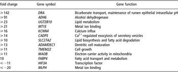
Finally, we recently evaluated changes in gene expression in rumen epithelium of young calves during transition from a MR diet to a diet consisting of MR supplemented with hay (H) or grain (G) using a whole bovine genome microarray (Connor, unpublished results). In this study, rumen epithelium was collected at slaughter from three calves fed MR only for 42 days, three calves fed MR for 42 days followed by MR + H for 14 days, and three calves fed MR for 42 days followed by MR + G for 14 days. Of note were substantial increases in expression of DRA mRNA involved in maintenance of rumen pH and absorption of SCFA, alcohol dehydrogenase 6, and three fatty acid metabolism genes (UGT2B10, FABP4 and SLC27A2) in the G and H groups relative to MR (Table 1). These transcript changes are consistent with expected changes in the rumen in response to forage and concentrate feeding. For example, as SCFA are produced and rumen pH is reduced, upregulation of DRA expression would be expected in order to increase HCO3− export from epithelial cells into the luminal rumen to neutralize rumen pH. Similarly, production of alcohols and fatty acids by rumen fermentation may induce the expression of factors that promote their metabolism. Of interest, unique to the G group were changes in expression of genes controlling apoptosis (BNIP3 and FAIM), organ morphogenesis (HEY1 and LBH) and cellular proliferation (SPC25), which we believe may be associated with the substantial differentiation of rumen epithelium that occurs during papillary development in response to a grain-based diet. Conversely, unique to the H group were gene expression changes related to regulation of growth and proliferation (ARMETL1 and SPOCK1), and cellular differentiation (HES4 and DMBT1). Using data generated from this study, future work in our laboratory will focus on developing specific molecular markers of rumen growth and epithelial cell differentiation associated with maturation of the rumen. Overall, future investigations evaluating the effects of various feed ingredients on gene expression in the ruminant GIT such as the rumen should increase our ability to manipulate GIT development to improve function and animal production and health.
Gene expression changes as determined by quantitative real-time reverse-transcription PCR in rumen epithelium of newborn calves fed milk replacer for 42 days followed by grain or hay for 14 days relative to calves fed milk replacer for 42 days

Effects of development on gene expression
Although the ruminant GIT represents less than 10% of empty BW (McLeod and Baldwin, Reference McLeod and Baldwin2000), its metabolic activity accounts for 25% to 35% of whole body protein synthesis (Lobley et al., Reference Lobley, Connell, Milne, Newman and Ewing1994) and 30% of whole body oxygen consumption (Burrin et al., Reference Burrin, Ferrell, Eisemann, Britton and Nienaber1989). The high-metabolic energy demands of the GIT appear to be met largely by amino acid oxidation in lieu of carbohydrate oxidation, resulting in nitrogen excretion and amino acid re-synthesis; although the extent of their use may be affected by the availability of SCFA and glucose substrates. Despite the strong influence of the GIT on efficiency of nutrient use in ruminants, very few studies have examined changes in gene expression of the ruminant GIT during development. One difficulty in conducting such an investigation is separating dietary effects from those due to ontogeny. For instance, in most studies discussed below, gene expression was evaluated in ruminant GIT at multiple developmental stages, but variations in diet also occurred during the same evaluation period that could be causing the differences in observed gene expression. An appropriately designed study to evaluate ontogeny of gene expression requires collection of tissues from multiple animals of equal age maintained on a constant diet over time. Lane et al. (Reference Lane, Baldwin and Jesse2002) accomplished this by performing a serial slaughter of lambs from birth to 84 days of age discussed in detail below.
As the rumen matures, the ruminal epithelium becomes capable of performing ketogenesis from butyrate produced by rumen microbes. Two enzymes believed to be critical for ketogenesis in ruminants are acetoacetyl-CoA acetyltransferase 1 (ACAT-1), which catalyzes the first step of ketogenesis, and HMG-CoA synthase (HMGCS1), which is the rate-limiting step for hepatic ketogenesis in monogastric species (Lane et al., Reference Lane, Baldwin and Jesse2002). To gain insight into developmental effects on the expression of the genes encoding these enzymes, groups of lambs were evaluated for their mRNA expression of ACAT-1 and HMGCS1 in rumen epithelium during development including pre-ruminating milk-fed lambs and conventionally weaned lambs (grass hay- and barley-fed at day 49 of age) by Northern blotting. Three lambs from each group were slaughtered at each of days 0, 4, 7, 14, 28, 42, 49, 56 and 84 of age for collection of rumen epithelium and transcript analysis. In addition, at 49 days of age, three milk-fed lambs were infused with SCFA and three milk-fed lambs were fed pelleted lamb starter until slaughter at day 84. From this study, it was evident that expression of both genes increases with age, particularly after 42 days, independent of dietary treatment, and that abundance of HMGCS1 mRNA parallels production of the ketone β-hydroxybutyrate. The latter finding suggests that HMGCS1 may be the rate-limiting enzyme in rumen epithelial ketogenesis. Most interesting, because there was no effect of SCFA feeding or SCFA production on expression of ACAT-1 and HMGCS1 mRNA by day 84, expression of these ketogenic enzymes in the rumen was shown to be independent of the actions of SCFA. However, these results did not provide insight into probable alternative mechanisms regulating the ontogeny of their expression, which identifies an interesting area for future investigation.
In a more descriptive study, Roh et al. (Reference Roh, Kuno, Hishikawa, Hong, Katoh, Obara, Hidari and Sasaki2007) used differential display to identify mRNA transcripts differentially expressed in rumen, reticulum, omasum and abomasum of adult cattle and then characterized their expression in calves of 3 to 13 weeks of age and adult cattle. The goal of the work was to identify genes involved in development of the mature ruminant GIT. However, only five transcripts were identified including RPS19, RPS23, BHLHB2, NDUFV2 and EXOSC9, which generally varied in their expression across tissues and age groups of cattle, but the effect of age was confounded by diet. Overall, the work identified transcripts differentially expressed among the four chambers of the bovine stomach, but provided no insight into genetic mechanisms contributing to rumen development and function. This highlights again the need for dietary control for interpretation of results or difficulties to be faced in such studies.
Liao et al. (Reference Liao, Vanzant, Boling and Matthews2008b) evaluated the mRNA expression of cationic amino acid transporter 1 (CAT-1), the primary intestinal transporter of cationic amino acids such as l-Lys, in bovine small intestine at different production periods. Epithelial scrapings were collected from duodenum, jejunum and ileum of steers of four ages at slaughter including day 32 (suckling), day 184 (weanling), day 248 (growing) and day 423 (finishing) for relative quantification of CAT-1 mRNA by realtime RT-PCR. To minimize the differential effects of cell proliferation and growth factors on CAT-1 mRNA expression, each developmental stage was fed to produce equal growth rates; however, the composition of the diet was different for each stage, again confounding effects of developmental stage with diet. The work showed differential CAT-1 mRNA expression only in jejunum of growing steers, which exhibited 1.6 to 2.3 times the expression level in jejunum of the other developmental stages, as well as the duodenum and ileum within the growing group. Transcript expression was not affected by production stage in duodenum or ileum. Interestingly, the expression of CAT-1 mRNA across the intestinal segments mirrored the predicted availability of luminal amino acid supply based on earlier studies in sheep, where it was shown that highest substrate concentrations occur in the jejunum (Tagari and Bergman, Reference Tagari and Bergman1978). Although the study only evaluated mRNA expression of CAT-1, previous research had showed a high correlation between CAT-1 mRNA expression and CAT-1 transporter activity in vitro (Hyatt et al., Reference Hyatt, Aulak, Malandro, Kilberg and Hatzoglou1997). Ultimately, results of this work suggested similar intestinal CAT-1 function in ruminants compared to monogastric species based on the distribution and relative abundance within the GIT, and should support optimized feeding of essential amino acids to ruminants to target maximal capacity for their uptake. Although, further study is necessary to determine whether increased expression of CAT-1 mRNA in jejunum of growing animals (248 days of age) is independent of dietary effects.
Finally, the abundance of IGF-1R, IGF-2R and INS-R mRNA and protein expression of the receptors in small intestine and colon of 2-week pre-term calves v. normal, full-term calves was evaluated by absolute real-time qRT-PCR and receptor radioligand binding to determine developmental effects on their expression (Georgiev et al., Reference Georgiev, Georgieva, Pfaffl, Hammon and Blum2003). At the transcript level, differences were detected solely in IGF-2R mRNA expression in colon where concentrations in full-term calves were 2.1-fold greater than in pre-term calves, whereas at the protein level, decreases were detected in binding of IGF-1R in colon and increases were found in IGF-2R in duodenum and ileum, and INS-R in total intestine in full-term v. pre-term calves. Additionally, correlations between mRNA and protein levels for the receptors were both positive and negative, indicating that differences in receptor activity or number are not regulated at the transcript level. On the basis of these findings, it appears that there are some tissue-specific changes in IGF-1R, IGF-2R and INS-R protein expression during intestinal development of ruminants, which likely play a role in gestational GIT development.
Overall, the number of definitive studies evaluating developmental changes in gene expression in the ruminant GIT and their relationships with protein expression are extremely limited to date. Future studies should incorporate adequate controls and experimental designs that permit a more clear interpretation of results. Use of ruminal, duodenal or ileal cannulations should permit repeated sampling of tissues by biopsy from the same individuals over time and reduce the need for extensive slaughter studies to conduct such investigations.
Gene expression during parasitic infection
Because GI parasitism, particularly by nematodes, is a leading cause of economic loss in ruminant livestock production, and the incidence of drug resistance in parasites is increasing, identification of genes involved in host resistance or immune response to infection has become an important area of study for development of alternative strategies to battle nematode infection (recently reviewed by Li and Gasbarre, in press). Similarly, research related to GIT diseases including coccidiosis (Alcala-Canto and Ibarra-Velarde, Reference Alcala-Canto and Ibarra-Velarde2008), and the zoonotic pathogen Escherichia coli O157:H7 (Li and Hovde, Reference Li and Gasbarre2007) has recently exploited molecular approaches to identify means to prevent their occurrence in cattle. Some common experimental approaches include the application of microarrays to evaluate gene pathways activated in the GIT during infection, evaluation of candidate genes involved in the immune response to infection, and the use of selected animals for parasitic resistance to identify genetic mechanisms contributing to resistance or susceptibility to a parasitic challenge.
For instance, Diez-Tascón et al. (Reference Diez-Tascón, Keane, Wilson, Zadissa, Hyndmann, Baird, McEwan and Crawford2005) used microarray analysis to identify differentially expressed genes in duodenum of sheep from genetic lines selected for high (susceptible) or low (resistant) fecal egg count to identify putative gene pathways involved in resistance to GI nematodes. Tissues were collected at slaughter after two consecutive exposures to pastures infected with Ostertagia, Trichostrongylus, Cooperia and Nematodirus spp. that resulted in worm counts at slaughter 48 times greater in susceptible lambs than in resistant lambs. From 126 differentially expressed genes detected, two major functional pathways were represented. The first was enhanced acquired immunity as evidenced by increased major histocompatibility complex (MHC) class II gene expression, and the second was increased expression of enteric smooth muscle proteins, which may result in an enhanced ability to rapidly expel parasites from the GIT. Thus genes involved in these pathways may contribute to parasite resistance, or may be differentially expressed as a result of different parasitic loads between the two groups. To further evaluate mechanisms related to resistance, the same laboratory examined gene expression in duodenum of the genetic lines in the absence of a parasitic challenge by microarray (Keane et al., Reference Keane, Zadissa, Wilson, Hyndman, Greer, Baird, McCulloch, Crawford and McEwan2006). In this study, susceptible lambs had greater basal expression of genes involved in the stress response, which the authors interpreted to mean that susceptible lambs might be predisposed to nematode infection due to the presence of other pathogens in the GIT. However, very few differences were detected between the two lines indicating that resistance is likely related to genes induced or suppressed by parasitic infection. Of interest, no genes were consistently differentially expressed between the two studies; although a few genes related to apoptosis and maintenance of the immune system were upregulated in resistant lambs in the second study. In a follow-up analysis, Keane et al. (Reference Keane, Dodds, Crawford and McEwan2008) determined that three MHC Class II genes, Ovar-DQA1, Ovar-DQB1 and Ovar-DRA that function in antigen presentation to T helper cells were more highly expressed in duodenum of resistant compared to susceptible lambs in response to a parasitic challenge. As previous research has indicated associations between the MHC region of the genome and parasite resistance, these genes are candidates for parasite resistance worthy of additional study.
Our laboratory has also evaluated gene expression in two recent studies in resistant v. susceptible beef cattle for GI nematode infection based on selection for low or high fecal egg count in response to Ostertagia and Cooperia challenge (Gasbarre et al., Reference Gasbarre, Leighton and Sonstegard2001). First, Araujo et al. (Reference Araujo, Padilha, Zarlenga, Sonstegard, Connor, Van Tassell, Lima, Nascimento and Gasbarre2009) used an immune pathway-focused cDNA array to compare gene expression in mesenteric lymph node and small intestinal mucosa of the selected groups after an exposure to Ostertagia, Cooperia and Nematodirus spp. Results indicated differential expression in both tissues in several functional classes of genes between the resistant and susceptible animals including transcripts encoding immunoglobulin (Ig) chains, integrins such as ITGA4, antigen presentation molecules such as CD45 and CD3E, tissue factors including DMBT1, PGA@, FGFR2 and TUBA2 and proinflammatory cytokine modulators of tumor necrosis factor and interferon. Of interest, mRNA for both IgE receptor and IFI35 were found to be differentially expressed between resistant and susceptible cattle, which had also been detected in divergently selected lines of parasite resistant sheep (Keane et al., Reference Keane, Zadissa, Wilson, Hyndman, Greer, Baird, McCulloch, Crawford and McEwan2006; Ingham et al., Reference Ingham, Reverter, Windon, Hunt and Menzies2008). In general, the patterns of differential expression indicated a complex response to GI nematodes and no apparent single pathway related to resistance. In addition, immune responses were of both the TH1 and TH2 types, which differs from studies in rodents that indicate resistance to GI nematodes is related to a TH2 response (Urban et al., Reference Urban, Madden and Svetic1992). In a second study of this selected cattle population, Li et al. (Reference Li, Sonstegard, Van Tassell and Gasbarre2007) evaluated expression of 17 cytokines and cytokine receptors by qRT-PCR in five GI tissues of resistant and susceptible animals in response to a combined Ostertagia ostertagi and Cooperia oncophora challenge. It was found that heifers from the resistant group exhibited higher mRNA levels primarily in abomasum for multiple proinflammatory cytokines, as well as some anti-inflammatory cytokines, and the polymeric Ig receptor, which functions in the transport of polymeric Ig into glandular and mucosal secretions. Overall, the resistant animals appeared to have more pronounced inflammatory responses at the site of parasitic infection relative to susceptible animals, which may be a mechanism contributing to their GI nematode resistance.
Using a similar approach, Ingham et al. (Reference Ingham, Reverter, Windon, Hunt and Menzies2008) used realtime qRT-PCR to evaluate expression of 80 candidate genes in GI mucosa of two separate lines of sheep selected for resistance to GI nematodes. The two lines consisted of sheep selected for fecal egg counts after Haemonchus contortus challenge or immune response to vaccination with killed Trichostrongylus columbriformis larvae. Each line received a single challenge of each GI nematode used in selection to evaluate genes related to innate immunity, as well as a tertiary challenge to evaluate genes involved in acquired immunity. Although differences in gene expression were variable between selection lines and primary v. tertiary response to challenge with each parasitic species, the results indicated that resistant animals in general exhibit a more rapid inflammatory response, greater toll-like receptor expression, which is involved in recognition of pathogens, and increases in cellular defenses against pathogens, such as production of free radicals.
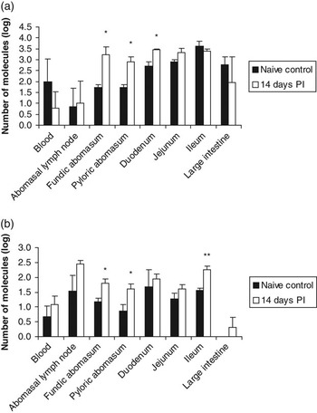
Consistent with these findings, Li and Gasbarre (Reference Li and Gasbarre2009) used microarray hybridization to evaluate gene pathways altered in the jejunum of 3-month-old calves in response to C. oncophora infection at days 7, 14, 28 and 42 post-infection (PI) and observed considerable differences in pathways affected at each time point including a decrease in expression of acute phase response and complement system genes at day 7 PI. This finding supports earlier findings that resistance may be related to the host’s ability to elicit a localized and rapid inflammatory response (Li et al., Reference Li, Sonstegard, Van Tassell and Gasbarre2007; Ingham et al., Reference Ingham, Reverter, Windon, Hunt and Menzies2008). Alternatively, by day 28 PI, the major pathways affected were related to upregulation of lipid metabolism and eicosanoid signaling genes, which the authors suggested may be related to a potential role of polyunsaturated fatty acids in the modulation of the immune system of cattle. Expression of a total of 310 genes was affected during infection with the majority of changes occurring at 14 days PI. Some specific genes of interest affected included GCNT3, an enzyme involved in mucin biosynthesis, the cell adhesion molecule cadherin-like 26 (CDH26), and the immunoregulatory cytokine IL-13. Specifically, expression of GCNT3 was upregulated more than five fold at all time points PI and is consistent with the role of mucins in providing a protective barrier to intestinal pathogens. Expression of CDH26 was induced by day 14 PI and was among the most highly upregulated genes, peaking at day 28 at 157-fold higher than control calves, and IL-13 mRNA was consistently upregulated by day 14 PI by more than seven fold. Interestingly, a strong positive correlation was detected between abundance of intestinal CDH26 mRNA and the number of jejunal eosinophils and adult C. oncophora at day 14 PI, indicating a potential role of this cell adhesion molecule in host recognition of nematodes. Further analysis of CDH26 mRNA expression in bovine blood and segments of the GIT during infection with O. ostertagi by realtime qRT-PCR also indicated induced expression at infection sites of abomasum and duodenum of calves 14 days PI with L3 larvae compared to naive controls (Figure 3A). Similarly, expression of IL-13 mRNA was significantly upregulated in abomasum and ileum of O. ostertagi infected calves relative to controls (Figure 3B). Of note, a recent study of Nelore cattle naturally infected with Cooperia punctata showed that IL-13 mRNA, along with IL-4 mRNA was more highly expressed in resistant animals relative to susceptible ones (Bricarello et al., Reference Bricarello, Zaros, Coutinho, Rocha, Silva, Kooyman, De Vries, Yatsuda and Amarante2008) and resistant sheep challenged with T. colubriformis expressed higher levels of intestinal IL-13 mRNA relative to susceptible sheep (Pernthaner et al., Reference Pernthaner, Cole, Morrison and Hein2005). Thus these findings support a role of this TH2 cytokine in nematode resistance in both sheep and cattle.
The mRNA expression of CDH26 (a) and IL-13 (b) in gastrointestinal tract of 3-month-old calves (n = 3) 14 days post-infection (PI) with 100 000 L3 Ostertagia ostertagi larvae and age-matched naive control calves as determined by absolute quantitative real-time reverse-transcription PCR. Expression values are the number of molecules per 100 ng of total RNA. Mean log10 molecule numbers are presented with s.e. as error bars. An unpaired t-test was used to detect differences between infected (14 days PI) and naive controls, *P < 0.05; **P < 0.01.

In summary, a number of recent investigations, particularly evaluating gene expression in the GIT of resistant v. susceptible populations of ruminants to GI nematodes, have identified candidate genes and mechanisms involved in host immunity contributing to parasitic resistance. A better understanding of these mechanisms or identification of structural variation in the genes should lead to alternative strategies to select for the trait, and manage parasitic infection in susceptible animals.
Conclusions
Analysis of gene expression in the ruminant GIT has provided a basic foundation for examining numerous physiological pathways impacting agriculturally important ruminant species. However, a thorough understanding of these pathways will require expansion upon characterizations of gene expression during various physiological states to well-designed, more complex functional investigations and hypothesis-driven experiments. Furthermore, use of techniques such as laser-capture microdissection and in situ hybridization will be needed to further characterize expression profiles of individual cell types within the GIT, and to remove expression biases that may occur in studies evaluating whole tissue samples, which may consist of varying proportions of different cell types contributing to the gene expression profile. Overall, outcomes of these studies should provide novel means to manage ruminant production species to optimize animal health and production efficiency, as well as select for genetically superior animals.