Introduction
The lifestyle of modern society is liable for the reduction in sleeping time of the population in general.Reference Spiegel, Leproult and L’Hermite-Baleriaux1 However, sleep restriction (SR) is related to important hormonal changes.Reference Spiegel, Leproult and L’Hermite-Baleriaux1, Reference Spiegel, Knutson, Leproult, Tasali and Van Cauter2 Studies have shown that sleep deprivation altered essential physiological mechanisms such as the regulation of cortisol secretion and sympathovagal balance.Reference Gangwisch, Heymsfield and Boden-Albala3, Reference Schussler, Yassouridis and Uhr4 Additionally, sleep deprivation seems to be able to suppress insulin production by the pancreatic cells and to act as one of the factor determinants for the development of insulin resistance and diabetes mellitus Type II.Reference Spiegel, Leproult and L’Hermite-Baleriaux1, Reference Spiegel, Knutson, Leproult, Tasali and Van Cauter2, Reference Spiegel, Leproult and Van Cauter5 During critical periods of development, hormonal changes may result in permanent malprogramming of systems critical for normal hormonal regulation.Reference Plagemann6 Thus, during pregnancy, SR can alter the maternal environment and interfere with maternal and foetal health.Reference Pires, Andersen, Giovenardi and Tufik7, Reference Pardo, Goularte and Hoefel8
Experimental data have shown that male offspring from SR mothers presented neurological,Reference Pardo, Goularte and Hoefel8 cardiovascular and renal alterations.Reference Thomal, Palma and Ponzio9–Reference Raimundo, Bergamaschi, Campos, Palma, Tufik and Gomes11 However, few studies evaluated the effects of SR during pregnancy on female offspring.
Considering the lack of studies with female offspring from SR mothers, the present study aims to evaluate the effects of SR in the last week of pregnancy on lipids and glucose homeostasis of the female offspring at adulthood. Additionally, we evaluated the effect of maternal SR on the female offspring in the absence of female hormones (after oophorectomy to abolish the influence of female hormones at the studied parameters).
Methods
The Ethical Research Committee of the Universidade Federal de São Paulo – UNIFESP analysed and approved the protocol of this study (CEUA: 7647020614). The experimental procedures were performed following the international guidelines for the care of research animals.
The rats (female Wistar rats weighing between 200 and 250 g and male Wistar rats weighing between 300 and 350 g) were obtained from CEDEME-UNIFESP (Centro de Desenvolvimento de Modelos Animais). During the experimental protocol, all the animals had free access to food and water and were maintained in a room with fixed temperature (22 °C) and light cycle (light and dark cycle alternating every 12 h, starting the light period at 7 am). The food offered was NUVILAB – special for rats (Quimtia S.A. – Colombo – Pr, Brazil).
Pregnant females were distributed into two experimental groups: control and sleep‐restricted mothers. The SR was performed in a room containing a tiled tank (123 × 44 × 44 cm) with 10 narrow circular platforms (6.5 cm in diameter) inside. The tank was filled with water to within 1 cm below the upper border of the platforms. This technique of SR is based on the muscle atony that accompanies paradoxical sleep.Reference Thomal, Palma and Ponzio9–Reference Raimundo, Bergamaschi, Campos, Palma, Tufik and Gomes11 The SR was carried out between the 14th and 20th days of pregnancy, because this is a critical period of developmentReference Pardo, Goularte and Hoefel8, Reference Thomal, Palma and Ponzio9, Reference Raimundo, Bergamaschi, Campos, Palma, Tufik and Gomes11 and susceptible to changes in the maternal environment.
After birth, the control offspring (C) and offspring from sleep‐restricted mothers (SR) were weighed, and the litters were reduced to six offspring, preferably in the ratio of four males to two females, that stayed with the mother for 28 days. After the lactation period, the female offspring were placed in collection cages containing four animals per cage. The males were not used in the present study. The total numbers of mothers were as follows: 17 for the control group and 34 for the SR group.
Half of the C and SR offspring (one female from each litter) were submitted to ovariectomy (OVX); the others underwent sham surgery. For the surgeries, the rats were anaesthetized with a mixture of xylazine (10 mg/kg) and ketamine (75 mg/kg) given intraperitoneally (i.p.). The OVX was performed at 2 months. Four experimental groups were formed: control sham (Csham), control ovx (Covx), SR sham (SRsham) and SRovx (SRovx). The rats were evaluated at the age of 8 months. Body weight data were published in reference.Reference Argeri, Nishi, Volpini, Palma, Tufik and Gomes12
Total cholesterol, LDL cholesterol, HDL cholesterol, and triglycerides were evaluated at 8 months of age. Blood samples were collected by a caudal puncture. The concentration of lipids in serum was obtained using the Cobas Lipid Panel CHOL–TRIGL–HDL–LDL SYSTEM from Roche Diagnostics GmbH (Mannheim – Germany).
Glucose tolerance test (GTT) – After an 8-hour fast, a drop of blood from the tail was collected to measure blood glucose by using a glucosimeter (Johnson & Johnson glucose monitor). This procedure was performed at 0 time and 20, 40, 60, 80, 100 and 120 min after glucose administration (2.5 g/kg i.p.).
Insulin tolerance test (ITT) – After an 8-hour fast, a drop of blood from the tail was collected to measure blood glucose by using a glucosimeter (Johnson & Johnson glucose monitor). This procedure was performed at 0 time and 30, 60, 90 and 120 min after insulin administration (1.0 U/kg i.p.).
The curves of the variation of the glucose concentrations were constructed as a function of time, and the AUCs were calculated using GraphPad Prism-6.01.
The results are presented as the means ± standard errors. Statistical testing was performed using analysis of variance (ANOVA) complemented by the Bonferroni test (GraphPad Prism software 6.01®). The level of significance was 5% (P≤0.05).
The renal function of this set of animals was evaluated and already published.Reference Argeri, Nishi, Volpini, Palma, Tufik and Gomes12
Results
Effects of SR on lipid profile
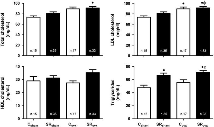
The results of serum concentration of lipids (total cholesterol, LDL cholesterol, HDL cholesterol and triglycerides) are presented in Fig. 1 and described as follows: Total cholesterol (mg/dL): Csham: 74 ± 2.3 (n = 15); SRsham: 81 ± 3.0 (n = 35); Covx: 89 ± 3.6 (n = 17); SRovx: 91 ± 3.2 (n = 33); LDL (mg/dL): Csham: 14 ± 0.77 (n = 15); SRsham: 15 ± 0.86 (n = 35); Covx: 19 ± 1.4 (n = 17); SRovx: 20 ± 1.1 (n = 33); HDL (mg/dL): Csham: 29 ± 3.3 (n = 15); SRsham: 31 ± 1.8 (n = 35); Covx: 27 ± 1.6 (n = 17); SRovx: 35 ± 2.2 (n = 33); Triglycerides (mg/dL): Csham: 47 ± 4 (n = 15); SRsham: 66 ± 3.6 (n = 35); Covx: 55 ± 4.2 (n = 17); SRovx: 71 ± 3.2 (n = 33). The SRsham group presented increased values of triglycerides in comparison to Csham; SRovx presented increased values of triglycerides in comparison to Csham and Covx. In addition, SRsham values of triglycerides were no different from SRovx, showing that maternal SR altered in the triglyceride levels of offspring but OVX did not. The LDL cholesterol concentration was increased in groups Covx and SRovx in comparison to Csham. Additionally, only SRovx had higher LDL than SRsham, suggesting that there was an interaction between maternal SR and offspring OVX on LDL concentrations. The total cholesterol was significantly increased in SRovx compared to Csham. There was no significant difference between the groups in HDL cholesterol.
Lipid profile in the offspring of control (C) and sleep-restricted (SR) mothers, submitted to ovariectomy surgery (OVX) or not (Sham). ∙P < 0.05 v. Csham; ɸP < 0.05 v. SRsham; εP < 0.05 v. Covx (Bonferroni test). The number of measurements analysed is shown inside the bars.

Glucose metabolism
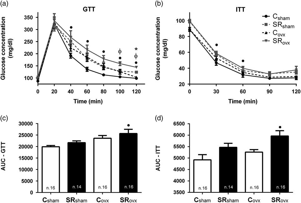
The results for glucose metabolism are shown in Fig. 2a. Basal values of glucose concentration (time 0 − GTT) were increased in sleep-restricted rats SRsham and SRovx compared to Csham and Covx (Glucose concentration, mg/dl: Csham: 87 ± 2; Covx: 92 ± 1; SRsham: 99 ± 12; SRovx: 104 ± 2). The SRovx presented increased values of glucose concentration in comparison to Csham at times 40, 60, 80, 100 and 120 min after glucose overload. The glycaemia of SRsham group at time point 120 min after glucose overload was increased compared to Csham. The glycaemia of SRovx was higher than that observed in SRsham at times 100 and 120 min. The AUC was significantly increased in SRovx compared to Csham (Fig. 2b).
Glucose tolerance test – GTT (a) and respective area under curve – AUC (b). Insulin tolerance test – ITT (c) respective area under curve in the offspring of control (d) control (C) and sleep-restricted (SR) mothers, submitted to ovariectomy surgery (OVX) or not (Sham). ∙P < 0.05 SRovx v. Csham; ɸP < 0.05 RSovx v. SRsham; *P < 0.05 RSsham v. Csham (Bonferroni test). The number of measurements analysed is shown inside the bars.

The results of the ITT are shown in Fig. 2b. The SRovx presented increased values of glucose concentration in comparison to Csham at times 30 and 60 min after insulin overload. The AUC was significantly increased in SRovx compared to Csham (Fig. 2d).
Discussion
The present study evaluated the consequences of SR at the end of pregnancy on the metabolic profiles of the female offspring submitted to OVX. The following observations were made: (1) the offspring of the rats submitted to the SR (SRsham and SRovx) showed increased values of triglyceride serum concentration; (2) the rats submitted to OVX had increased LDL plasma concentration; (3) the SRovx group showed a significant increase in total cholesterol, LDL and triglycerides compared to Csham. Regarding glucose metabolism, we observed the following: (1) the offspring of rats submitted to SR had baseline glucose values higher than the control group and presented alterations at several points on the glucose tolerance curve and (2) the SRovx group presented increased AUC for both GTT and ITT in comparison to Csham.
SR is associated with important neuroendocrine changesReference Spiegel, Leproult and L’Hermite-Baleriaux1–Reference Spiegel, Leproult and Van Cauter5, Reference Koban, Le and Hoffman13 and even during pregnancy, after SR, there is an increase in corticosterone plasma concentrationReference Calegare, Fernandes, Tufik and D’Almeida14 and hypertrophy of the adrenal glands.Reference Thomal, Palma and Ponzio9 The exposure to corticosteroids during foetal development is associated with foetal programming of neuroendocrine alterationsReference Kapoor, Dunn, Kostaki, Andrews and Matthews15 and metabolic disorders.Reference Seckl and Meaney16 The changes at the HPA axis due to stress/glucocorticoids may vary depending on the gender evaluated. In the female offspring, increased basal levels of adrenocorticotropic hormoneReference McCormick, Smythe, Sharma and Meaney17 and corticosteroneReference Bhatnagar, Lee and Vining18 and an increased response to stress (increased sympathetic responsiveness)Reference Szuran, Pliska, Pokorny and Welzl19 were observed. Chronically, these changes may lead to insulin resistance and type II diabetes. Unlike female offspring, the male offspring did not show these changes,Reference McCormick, Smythe, Sharma and Meaney17–Reference Szuran, Pliska, Pokorny and Welzl19 suggesting that female offspring subjected to glucocorticoids are more susceptible to changes in the HPA axis.Reference Kapoor, Dunn, Kostaki, Andrews and Matthews15 In addition, the neural regulation of blood pressure was shown to be altered in the male offspring of SR mothers. Raimundo et al.Reference Raimundo, Bergamaschi, Campos, Palma, Tufik and Gomes11 and Lima et al.Reference Lima, Rodrigues and Bergamaschi10 showed that SR, whether at the end of pregnancy or throughout the duration, caused an increase in sympathetic tonus to the heart and arterial hypertension in male offspring.
The rats subjected to OVX (Covx and SRovx) presented significant increase in LDL in comparison to Csham. We showed before that these rats (Covx and SRovx) present significant increase in fat deposition and in body weight compared to Csham and SRsham.Reference Argeri, Nishi, Volpini, Palma, Tufik and Gomes12 The lack of oestrogen (E) predisposes the rats to obesity by decreasing the activity of enzymes involved in the production and action of thyroid hormones (HT).Reference Ignacio, Fortunato and Neto20 In addition, it seems that E is able to regulate food intake by increasing central sensitivity to cholecystokinin (satiety hormone) and leptinReference Brown and Clegg21; thus, the absence of this hormone may predispose rats to an increase in adiposity, a known factor associated with metabolic disorders. The lack of oestrogen may contribute to weight gain, however in the present study the food intake was not measured which is a limitation of our study. New experiments with hormone replacement therapy may be able to better investigate the role of sex hormones on the results obtained.
The sleep-restricted groups (SRsham, SRovx) presented increased values of triglycerides in comparison to Csham and Covx showing that maternal SR altered the triglyceride levels of offspring. Additionally, the SRovx group also had significantly increased values of LDL compared to SRsham. It seems that SR and OVX affected lipid metabolism differently.
The sympathetic nervous system, through endings localized in liver and adipose tissue, is an important regulator of lipid mobilization, and its activation increases lipolysis in white adipose tissue and increases the hepatic production of triglycerides.Reference Geerling, Boon and Kooijman22 Considering that female offspring of dams submitted to stress have increased response to stressReference Kapoor, Dunn, Kostaki, Andrews and Matthews15 and that SR may be considered a stressful stimulus, the increase in sympathetic activity through its effects on fat mobilization could contribute to modifications in the lipid profile observed in the female offspring of sleep-restricted mothers. Nevertheless, other experiments are necessary to confirm this hypothesis.
It is possible that changes in maternal environment originating from SR have caused adaptations in the female offspring, predisposing them to metabolic alterations. The increase in plasma corticosterone concentration is probably an important causative factor. However, levels of this hormone were not measured in the present study.
Thus, we conclude that restriction of sleep during pregnancy can be considered a risk factor for the development of diseases in adult offspring. In addition, the changes in lipid deposition and profile observed after OVX suggest that this condition may be considered an extra threat. Further studies are needed to understand the mechanisms responsible for the changes observed in this study.
Acknowledgements
We thank Dr Sergio Tufik and Dr Monica Levy Andersen for allowing the use of the sleep restriction room of the Department of Psychobiology for this study and Dr Gerhardus H.M. Schoorlemmer for text revision.
Financial Support
FAPESP (2013/23622-7).