INTRODUCTION
Breast cancer is the leading cause of cancer incidence and mortality among women worldwide.Reference Ferlay, Soerjomataram and Ervik 1 Radiation therapy has been the standard treatment after breast-conserving surgery for patients with early-stage disease. Prone positioning has been studied in various groups as a way to decrease the secondary effects induced by radiation, including pulmonary, cardiac and skin toxicity.Reference Osa, DeWyngaert and Roses 2 – Reference Stegman, Beal, Hunt, Fornier and McCormick 5
Several studies have described that patients with large breasts tend to present worse cosmetic outcomes after radiation therapy, have greater dose inhomogeneity and larger doses administered to organs at risk.Reference Buijsen, Jager and Bovendeerd 6 – Reference Bergom, Kelly and Morrow 8 Patients with larger breasts have been shown to have poorer acute toxicity and cosmetic results, including breast retraction and fibrosis due to inhomogeneity in the dose distribution and the bolus effect created in the inframammary fold.Reference Grann, McCormick and Chabner 9 , Reference Johansen, Overgaard and Rose 10
At our institution, we have found that positioning the patient in prone reduces the mean dose administered to the heart and lung, especially in left-breast patients. We also found that patients with breasts larger than 750 cc benefited more from prone positioning compared with supine when receiving radiation therapy as adjuvant treatment after breast-conserving surgery. In these patients, we found a significant reduction of heart mean dose when treated prone compared with supine. In patients with breasts smaller than 750 cc we found a non-significant higher mean dose to the heart when prone. During this study, we also found large daily set-up errors when in prone, leading either to unfeasible large planning treatment volume (PTV) expansion margins or the need for daily cone-beam computed tomography (CBCT) imaging.
At our institution, prone positioning will be reserved for patients with breasts larger than 750 cc, in whom the benefit of the position outweighs the daily positioning difficulties and extra CBCT imaging. To be able to accurately predict if a patient will benefit from prone positioning without having to perform a previous CT scan to measure breast volume we conducted a correlation study. In this study, we aimed to determine if the patient’s cup size accurately correlates with the breast volume measured in the CT scan, and to determine which sizes correspond to a volume larger than 750 cc.
METHODS
Before radiation therapy, we measured the breast size of consecutive patients on the day of the simulation CT scan. Patients who had received breast-conserving surgery and did not require radiation to the supraclavicular or axillary nodes were included. Two radiation technicians measured the thoracic circumference below the breast (band), and the breast circumference (bust) through the most prominent part of the breast. These measurements were recorded in inches, as this is the unit conventionally used to determine bra size in most countries. Images were acquired using 2 mm slices on a Siemens Somatom Definition AS scanner from the lower limit of the mandibular bone to the mid abdomen.
One radiation oncologist contoured all breast volumes on the CT scans defined as all breast tissue visible in the CT image; the volume was limited anteriorly within 5 mm from the skin and posteriorly to the anterior surface of the pectoralis, serratus anterior muscle, boney thorax and lung, in concordance with international guidelines for breast clinical target volume (CTV) definition. 11 To reduce any possible bias due to the radiation oncologist knowing the patient’s breast size in advance, this procedure was done blinded to the results of the previously measured sizes. Breast volume was calculated using the Eclipse® version 11 (Varian Medical Systems, Palo Alto, CA, USA) planning software.
Sample size was calculated with 0·80 power and 0·05 significance level in order to detect a 0·4 correlation coefficient. Although we could have used commercial bra size, considering that commercial bra size estimation includes certain numeric treatment for uneven numbers of the band, for the correlation analysis we used the measured difference in inches between band and bust measure, and not the calculated commercial bra size. Pearson’s coefficient was used to determine the correlation coefficient between breast size and volume. A receiver operating characteristic (ROC) analysis was performed to determine the best cut-off point in breast size to predict a 750 cc volume. The 750 cc limit was determined in a previous study at our institution, in which this value represented the mean value for breast volume for a group of patients receiving prone breast radiation therapy; in addition, this volume was chosen to allow for comparison with other published studies that address breast volume in radiation therapy.
This study was approved by our Institutional Review Board and all patients gave their verbal consent after a brief explanation of the purpose and methods of the study.
RESULTS
Breast size data were collected on 59 patients. Two patients were excluded from the analysis because there was evidence of pectus excavatum on the CT scan, thus altering the band and cup measurements; one patient had prosthetic implants.
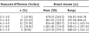
Breast volume ranged from 304·8 to 1,514·1 cc and had a mean of 851·78 cc (SD=284·83). The band circumference ranged from 29 to 40 inches with a mean of 34·77 inches (SD=2·77) and the bust circumference ranged from 32 to 47 with a mean of 39·47 inches (SD=3·19). The difference between the bust and band measurements had a mean value of 4·7 inches (SD=1·17; range 2–7).
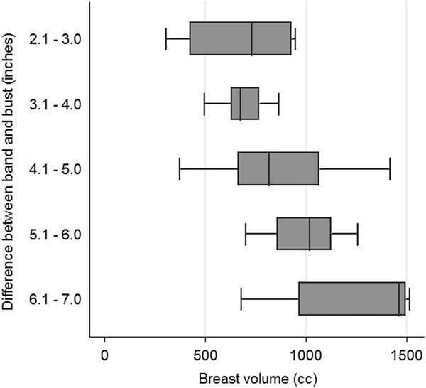
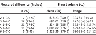
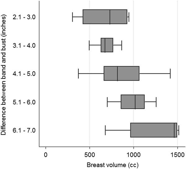
As shown in Table 1, most patients in this study had a difference between the band and bust measurements ranging from 4·1 to 5 inches, which corresponds to a D or DD bra size. The mean breast volume for each bra size increased according to increasing cup sizes. However, not all patients with a small cup size had a small breast volume and not all with a large cup size had a large volume, as shown by the volume distributions in Figure 1.
Boxplot for breast volume according to measured size difference.
Difference between band and bust and corresponding volume
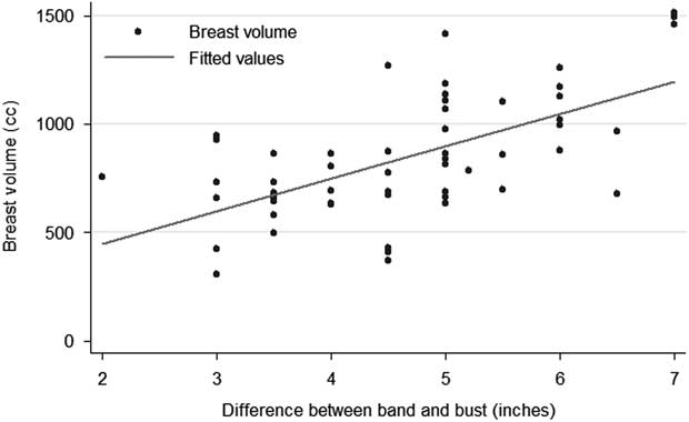
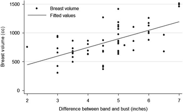
Both the differences between bust and band and breast volumes were normally distributed. Pearson’s coefficient was used to evaluate the correlation between both variables. The correlation coefficient r=0·61 was statistically significant (p<0·001). Figure 2 presents the correlation between both variables.
Correlation between breast volume and measured difference.
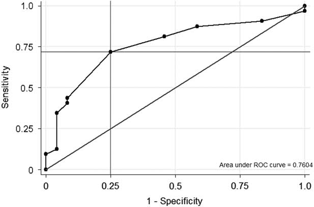
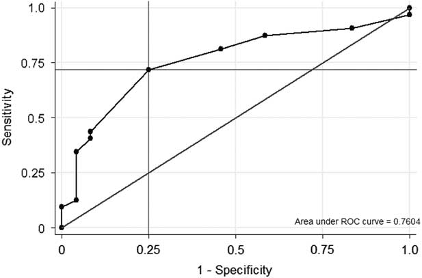
To determine the optimal cut-off point to predict which differences between band and bust would be associated with a volume ≥750 cc we conducted a ROC analysis (Figure 3). A difference of 5 inches between the band and bust circumferences showed a sensitivity and specificity of 71·9 and 75·0% to correctly determine a CT-measured breast volume of 750 cc or greater. With this cut-off the percentage of correctly classified cases was 73·2%.
Receiver operating characteristic (ROC) curve analysis for the measured difference predicting a breast volume ≥750 cc.
DISCUSSION
In this study, we describe the correlation between breast size when measured in the clinical practice and breast volume determined in a simulation CT scan performed previous to radiation therapy. We found a significant correlation coefficient of 0·61 showing that as the difference between band and bust increases, the breast volume measured in the CT scan also increases, and that 61% of the variation in breast volume can be explained from the difference between band and bust.
Breast size varies greatly in our country, with women of different areas having varying breast sizes. The population included in this study represents different areas of the country, and presented a wide range of breast sizes, with the most common being the equivalent to D and DD cup size.
We decided to measure the difference between band and bust in the clinical practice, because not all patients know their correct bra size and it is often difficult for them to find the adequate size in commercial establishments. And, as previous research has reported,Reference Coltman, MCGhee and Steele 12 breast volume can differ greatly among the same cup size. Thus, we will use the measured difference between band and bust circumferences as an indicator of breast size and volume at our clinical practice.
The variability in breast volume for the different cup sizes could be explained because some patients have breast ptosis and thus the tissue does not protrude from the thorax but hangs lower into the abdomen, making the bust measurement low while maintaining a larger volume. Other women have breasts that come from the side of the thorax, increasing the measured volume but not the cup size.
We determined that a 5-inch difference best predicts which patients will have breasts larger than 750 cc; however, this will not be the only criterion used to decide if a patient is an ideal candidate for prone breast irradiation, as some patients present smaller differences and larger volumes and other patients present larger differences but smaller volumes.
In conclusion, the difference between band and bust as measured in the clinical practice will be one of the criteria used to determine if a patient is a candidate for prone breast irradiation; however, other characteristics such as the shape of the breast will also be considered.
Acknowledgements
The authors thank Camilo Portilla, Alexandra Velez and Bladimir Gutierrez for their collaboration during data collection.
Financial support
This research received no specific grant from any funding agency, commercial or not-for-profit sectors.
Conflicts of Interest
None.