

Introduction
Numerous brain structures participate in recognising facial emotional expressions: the occipitotemporal cortices, amygdala, parietal cortices, orbitofrontal cortex, insula and basal ganglia (Adolphs, Tranel, Koenigs, & Damasio, Reference Adolphs, Tranel, Koenigs and Damasio2005). In prior research, it has been established that the basal ganglia and specifically the insula have an important role in the recognition of facial disgust (Johnson et al., Reference Johnson, Stout, Solomon, Langbehn, Aylward and Cruce2007; Phillips et al., Reference Phillips, Young, Senior, Brammer, Andrew, Calder and David1997). Nevertheless, there is no consensus regarding the different role of the left and right side of these brain structures in the process of emotion recognition. Several authors reported that exposure to disgusted facial stimuli elicits activation in the right insula and right basal ganglia (Adolphs, Damasio, Tranel, Cooper, & Damasio, Reference Adolphs, Damasio, Tranel, Cooper and Damasio2000; Anderson, Christoff, Panitz, De Rosa, & Gabrieli, Reference Anderson, Christoff, Panitz, De Rosa and Gabrieli2003; Phillips et al., Reference Phillips, Young, Senior, Brammer, Andrew, Calder and David1997, Reference Phillips, Williams, Heining, Herba, Russell, Andrew and Gray2004; Ruiz et al., Reference Ruiz, Lee, Soekadar, Caria, Veit, Kircher and Sitaram2013; Schroeder et al., Reference Schroeder, Hennenlotter, Erhard, Haslinger, Stahl, Lange and Ceballos‐Baumann2004). According to their results, it can be concluded that patients with unilateral lesions of the right insula are expected to experience serious problems with recognition of disgusted facial stimuli, whilst patients with unilateral lesions of the left insula should not experience such deficits. Partly in contrast to their reports, Sprengelmeyer, Rausch, Eysel and Przuntek (Reference Sprengelmeyer, Rausch, Eysel and Przuntek1998) demonstrated that disgusted facial expressions activated the right putamen and left insular cortex. Furthermore, Calder, Keane, Manes, Antoun and Young (Reference Calder, Keane, Manes, Antoun and Young2000) presented a patient with infarction in the left hemisphere, involving insula and basal ganglia, who showed a deficit in recognition of facial expression of disgust, even though the right basal ganglia and the right insula of this patient remained intact. Finally, some other studies demonstrated that bilateral insula lesions resulted in an impaired ability to recognise facial disgust (e.g., Adolphs et al., Reference Adolphs, Tranel, Koenigs and Damasio2005).
Additionally, basal ganglia are a part of frontal-subcortical circuit which is involved not only in motor control and emotion processing but also in cognitive and executive functions (Alexander, DeLong, & Strick, Reference Alexander, DeLong and Strick1986; Lichter & Cummings, Reference Lichter and Cummings2001; Middleton & Strick, Reference Middleton and Strick2002; Salmon, Heindel, & Hamilton, Reference Salmon, Heindel, Hamilton, Lichter and Cummings2001). Evidence that those structures are particularly important for executive functions emerges from neuropsychological studies of neurological disorders such as Huntington's and Parkinson's diseases as well as from studies of basal ganglia damage caused by stroke or traumatic brain injury (Backman, Robins-Wahlin, Lundin, Ginovart, & Farde, Reference Backman, Robins-Wahlin, Lundin, Ginovart and Farde1997; Elliott, Reference Elliott2003; Salmon et al., Reference Salmon, Heindel, Hamilton, Lichter and Cummings2001). These findings have led to the suggestion that executive function depends not on the prefrontal cortex (PFC) in isolation, but on the intact functioning of corticostriatal circuitry (Bonelli & Cummings, Reference Bonelli and Cummings2007; Elliott, Reference Elliott2003; Salmon et al., Reference Salmon, Heindel, Hamilton, Lichter and Cummings2001). The executive functions consist of those capacities that enable an individual to engage in independent, purposive, self-directed and self-serving behaviour (Lezak, Howieson, Bigler, & Tranel, Reference Lezak, Howieson, Bigler and Tranel2012). Thus, executive functions relate to various complex cognitive processes and sub-processes, such as task-switching, planning, working memory, solving novel problems, modifying behaviour, generating strategies, sequencing complex actions, etc. (Elliott, Reference Elliott2003).
The first objective of this study was to assess if the lesions of the left insula and the left part of the basal ganglia are related to impaired recognition of facial expression of emotion, especially disgust. Since the injury of our patient was almost identical to Calder et al.’s (Reference Calder, Keane, Manes, Antoun and Young2000) patient's injury, we hypothesised that he would experience the same deficits in recognition of disgusted facial expressions. The second objective was to test the patient's cognitive and executive functioning. We hypothesised that due to impaired corticostriatal connectivity our patient would show deficiency in certain cognitive and executive functions primarily related to frontal lobes.
Materials and Methods
Case Description
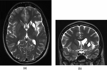
MS is a 21-year-old right-handed male attending the final year of his undergraduate study. Two years ago, MS presented to the Emergency Department with right extremities numbness and later developed choreoathetoid movements of the right arm. His mental status was normal. Brain MRI done at the time of his admission to the hospital showed an acute left cerebral hemisphere infarction involving part of the basal ganglia (putamen, caudate nucleus head, globus pallidus and claustrum), as well as left insula (Figure 1). After a month, his motor function recovered completely. On a follow-up visit done 3 months after hospital discharge his neurological examination was unremarkable. MS returned to all of his everyday activities which he used to do prior to his illness.
(a) Axial T2-weighted brain MRI shows left cerebral hemisphere post-infarction encephalomalacia involving part of the basal ganglia (putamen, caudate nucleus head, globus pallidus and claustrum), as well as left insula. (b) Coronal T2-weighted brain MRI also demonstrates post-infarction encephalomalacia of a part of the left basal ganglia and left insula.

Tests
Emotion identification test
In order to assess MS's ability to recognise emotional expressions in general and expression of disgust in particular, the Karolinska Directed Emotional Faces (KDEF) (Lundqvist, Flykt & Öhman, Reference Lundqvist, Flykt and Öhman1998) stimuli set was used. That dataset is comprised of 4900 photographs of human facial expressions, displaying anger, disgust, fear, happiness, sadness and surprise, as well as neutral expressions. The KDEF database is equivalent to Ekman and Friesen's (Reference Ekman and Friesen1976) Pictures of facial affect (PFA) database, which contains facial expressions of same six emotions. For the purpose of the present research, Calvo and Lundqvist's (Reference Calvo and Lundqvist2008) adaptations of the KDEF photographs were used. Stimuli were selected according to normative data regarding identification accuracy obtained on Spanish (Calvo & Lundqvist, Reference Calvo and Lundqvist2008) and Croatian (Milovanović, Švegar, & Kardum, Reference Milovanović, Švegar and Kardum2013) populations. Recognition of emotional expressions was examined via forced choice labelling procedure, using equivalent procedure as in Milovanović et al.’s (Reference Milovanović, Švegar and Kardum2013) study. MS was presented with a total of 70 photographs: 60 emotional (10 per each of six emotions) and 10 neutral. His task was to identify each of successively presented expressions by selecting one label from a list of seven possible expressions: anger, disgust, fear, happiness, sadness, surprise and neutral. In order to ensure higher reliability, the procedure was repeated after 14 days, with the same stimuli, displayed in a randomly selected serial order.
Neuropsychological tests
In order to assess cognitive and executive functions, we applied standard neuropsychological tests listed and described in detail below. All neuropsychological tests were translated to Croatian language and standardised for use on a Croatian population (Galić, Reference Galić2002). MS's performance on all neuropsychological tests, except the Stroop test and the Tower of Hanoi test, were compared with a normative results obtained on Croatian (Galić, Reference Galić2002) and international (Lezak et al., Reference Lezak, Howieson, Bigler and Tranel2012; Strauss, Sherman & Spreen, Reference Strauss, Sherman and Spreen2006) samples of healthy adults stratified for age and years of education. In order to evaluate the performance of MS on the Stroop and the Tower of Hanoi tests, we formed two control samples, each comprised of 20 university students. The detailed description of these samples is provided later in the Method section.
Tests for cognitive functions
Rey-Osterrieth's Complex Figure test (R-OCF) (Meyers & Meyers, Reference Meyers and Meyers1995; Strauss et al., Reference Strauss, Sherman and Spreen2006) was given in two parts: copy and memory. The subject was given a blank sheet of paper, and another sheet of paper on which the R-OCF was printed. He was instructed to copy the figure as accurately as possible, with no time restrictions. Each time a portion of the drawing was completed, the subject was given a different coloured pencil whilst the examiner was noting the order of colour used. Switching colours affords an adequate record of the subject's strategy. Following completion, the examiner removed both the original and the copied figure. Completion time was recorded. After a retention interval of 30 minutes during which the Verbal Fluency tests, Auditory Verbal Learning test and Clock Drawing test (CDT) were completed, the subject was again given a blank sheet of paper, and asked to reproduce the R-OCF from memory.
Rey-Osterrieth's Auditory Verbal Learning Test (RAVLT) (Strauss et al., Reference Strauss, Sherman and Spreen2006) was given to assess verbal learning and memory. The most commonly used version consists of 15 nouns (List A) that are read aloud (with a 1-s interval between words) for five consecutive trials, each trial followed by a free-recall test. On completion of Trial 5, an interference list of 15 words (List B) is presented, followed by a free-recall test of that list. Immediately after this, delayed recall of the first list is tested (Trial 6) without further presentation of those words. After a 20-minute delay period during which the CDT and R-OCF (memory) were completed, the subject is again required to recall words from List A.
Wechsler Memory Scale (WMS) (Strauss et al., Reference Strauss, Sherman and Spreen2006) – The Digits Forward and Digits Backward subtest was used to assess attention and working memory. The number sequences of the WMS are two to eight and two to seven digits, respectively. On hearing them, the subject's task is to repeat them in the exact order in the ‘forward situation’ and in exactly reversed order in the ‘backward situation’. The normal raw score difference between digits forward and digits reversed tends to range a little above 1 (Lezak et al., Reference Lezak, Howieson, Bigler and Tranel2012). The reversed digit span is a measure of working memory and is distinct from the more passive span of apprehension measured by digits forward (Lezak et al., Reference Lezak, Howieson, Bigler and Tranel2012).
Boston Naming Test (BNT) (Kaplan, Goodglass, & Weintraub, Reference Kaplan, Goodglass and Weintraub1983). This test consists of 60 large ink drawings of items ranging in familiarity from common (e.g., tree) at the beginning of the test to uncommon (e.g., sphinx) near its end. The subject begins with item 30 and proceeds forward. This test effectively elicits naming deficits.
CDT (Shulman, Reference Shulman2000; Strauss et al., Reference Strauss, Sherman and Spreen2006). This test is aimed to measure visual–spatial, constructional and executive function. The subject is asked to draw a clock face with all the numbers and hands and then to state the time ‘ten after eleven’. The strength and the weakness of this test lies in the number of cognitive, motor and perceptual functions required simultaneously for successful completion.
Tests for executive functions
Wisconsin Card Sorting Test (WCST) (Heaton, Chelune, Talley, Kay, & Curtiss, Reference Heaton, Chelune, Talley, Kay and Curtiss1993). The subjects is given one pack of 64 cards in the WCST. One to four instances of each of four symbols in four different colours are printed on each card (e.g., one red triangle, two green stars, three yellow crosses and four blue circles). The subject's task is to sort the cards according to a principle inferred from the examiner's responses to the subject's placement of the cards. The test involves three principles for sorting: colour, symbol or number. The examiner responds only ‘right’ or ‘wrong’ according to the subject's placement of the card, and does not answer any questions. After 10 consecutive correct placements, the examiner shifts the principle. The test continues until the subject makes six runs of ten correct placements, or when all 64 cards are placed.
Verbal Fluency Test (Galić, Reference Galić2002). A number of verbal fluency tests, modelled on Thurstone's Word Fluency Test (Lezak et al., Reference Lezak, Howieson, Bigler and Tranel2012) have been developed to assess more ‘executive’ aspects of verbal behaviour: the ability to think flexibly, switch response sets, self-regulate and self-monitor. Two variants of verbal fluency test were applied: phonemic/letter fluency and semantic/category fluency. In the first test, the subject was asked to produce as many words as possible beginning with the letters K, L and P (Croatian version of the test, according to Galić, Reference Galić2002) with the exception of proper names, numbers and words with the same suffix. One minute was allowed for each letter. In the second test, the category fluency test, the subject was asked to name all the animals he can think of in a period of 1 minute.
Design Fluency Test (Jones-Gotman & Milner, Reference Jones-Gotman and Milner1977). This test was developed as a non-verbal counterpart of Word Fluency Test. The subject is asked to invent drawings that represent neither actual objects nor nameable abstract forms and that are not merely scribbles. After being shown examples of acceptable and unacceptable drawings made by the examiner, the subject is given 5 minutes to make up as many different kinds of drawings as he or she can. Design Fluency Test has been used to assess various aspects of executive functioning.
Stroop Test. A numerical variation of the classical Stroop task paradigm was designed for the present study. Each of the 60 trials consisted of a certain number of digits (between 2 and 6), and subject's task was to quickly answer how many digits were presented. Answers were collected via keyboard. In congruent tasks, the number and the content of stimuli was equal (e.g., 4 4 4 4), whilst in incongruent trials it differed (e.g., 2 2 2 2 2 2). The performance of MS was compared to the results of 20 healthy control subjects, undergraduate psychology students (mean age: 20.25 years; SD = 1.37) who went through the same procedure. This test is a measure of cognitive control, specifically the ease with which a person can maintain a goal in mind and suppress a habitual response (Strauss et al., Reference Strauss, Sherman and Spreen2006).
Tower of Hanoi (computerised version). This test has been considered an ‘executive function’ test focused partly on planning and mostly on inhibiting a prepotent response (the goal–subgoal conflict). The object of the Tower of Hanoi test is to move all the rings from the first column to the third column. To arrive at the best solution (most direct, fewest moves) of the test, the subject must look ahead to determine the order of moves necessary to rearrange five rings of varying sizes. The rings should be moved one by one and a larger ring cannot be placed on top of a smaller ring. This test was also administered to the group of 20 healthy control subjects, male university students (mean age: 21.65 years; SD = 2.98), comprised for the present study.
Procedure
The study was conducted at the Laboratory for Experimental Psychology of the Faculty of Humanities and Social Sciences in Rijeka, Croatia. Neuropsychological assessment was carried out over three meetings lasting about 45 minutes each, 2 years and 6 months after hospitalisation. On the first meeting, the Emotion identification test, R-OCF: copy and memory, Verbal fluency tests, Auditory Verbal Learning Test and CDT were applied. On the second meeting after 2 weeks, the Emotion identification test, Tower of Hanoi, Design Fluency Test and BNT were applied and, on the last meeting a week later, the WMS, WCST and Stroop Test were applied. Over the period of testing, the patient was cooperative and motivated to complete the tasks.
The study was granted approval by the Research Ethics Committee of the Faculty of Humanities and Social Sciences at the University of Rijeka. The patient expressed his willingness to participate in the study by signing informed consent.
Results
Emotion Recognition
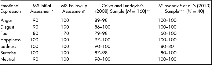
On the facial emotion recognition test no impairment was found, except some difficulties that MS had with the recognition of fearful faces, several of which he inaccurately labelled as surprised. The results of initial and follow-up assessments were similar (Table 1). However, since healthy subjects also most often misclassify fearful faces as surprised and vice versa (Calvo & Lundqvist, Reference Calvo and Lundqvist2008; Huang, Tang, Helmeste, Shioiri, & Someya, Reference Huang, Tang, Helmeste, Shioiri and Someya2001; Palermo & Coltheart, Reference Palermo and Coltheart2004), MS's ability to recognise facial emotional expressions in general and expression of disgust in particular seems to be intact. MS's results on emotion recognition test were compared to Calvo and Lundqvist's (Reference Calvo and Lundqvist2008) and Milovanović et al.’s (Reference Milovanović, Švegar and Kardum2013) participants. Calvo and Lundqvist's (Reference Calvo and Lundqvist2008) sample was comprised of 160 Spanish students (mean age: 22.2 years; SD = 3.1), whilst Milovanović et al.’s (Reference Milovanović, Švegar and Kardum2013) consisted of 58 Croatian students (mean age: 23.05; SD = 3.68) from the same university that MS attends. When the results of anxious and depressed participants are excluded from Milovanović et al.’s (Reference Milovanović, Švegar and Kardum2013) sample, 40 of 58 subjects remain, and their results together with Calvo and Lundqvist's (Reference Calvo and Lundqvist2008) subjects are compared to MS's scores in Table 1.
Recognition of Emotional Facial Expressions: Percentages of Correct Answers of MS, Calvo and Lundqvist's (Reference Calvo and Lundqvist2008) Spanish Sample of Participants and Milovanović et al.’s (Reference Milovanović, Švegar and Kardum2013) Croatian Sample

*Percentage of correct recognitions.
**Average percentage of correct recognitions per stimulus.
***Interquartile range of correct recognitions.
Cognitive Functioning
The patient successfully copied a complex drawing (R-OCF) and used an appropriate strategy for organising the sequence of the drawing process. Also, drawing from memory was successful, which reflects normal non-verbal memory and constructional abilities.
He showed normal (average and above average) performance on verbal learning and memory tasks (RAVLT), as well as on language (BNT) and visual-spatial and constructive tasks (CDT). Since these results did not differ from normal expectations we will not describe them in detail.
He showed slight difficulties in maintaining information in working memory because on the Digit Backwards subtest of the WMS, his raw score was on the lower border (four digits). According to Lezak et al. (Reference Lezak, Howieson, Bigler and Tranel2012), scores 4 to 5 can be considered ‘within normal limits’, but we should expect a better result from someone with the patient's educational background. Of course, we must be careful when interpreting a borderline result.
Executive Functions
On the WCST, MS clearly showed failure to maintain set which resulted in zero categories completed. In other words, the patient sorted correctly five cards in the first category (colour) but failed to complete this category and passed over to another sorting principle. This type of error can reflect an inability to continue using a strategy that is working successfully, or loss of train of thought (Lezak et al., Reference Lezak, Howieson, Bigler and Tranel2012). The failure to maintain set rarely occurs but is often linked to brain lesion (Galić, Reference Galić2002). Our patient obviously had some difficulties in forming and applying a principle and then continuing to use a successful strategy. This means that he could generally have problems with abstraction and flexible thinking. This inference is also supported by his results on verbal and non-verbal fluency tests.
MS expressed impaired or defective phonemic verbal fluency. His result of a total score of 22 generated words was significantly below the average result obtained by comparison group of the same age and educational level (53 words on average for healthy subjects, age range 15 to 35). His total score on the semantic/category fluency test was almost within the average range for healthy subjects. Generating the names of words beginning with particular letter is more difficult than animal naming. The former task relates to strategies of self-retrieving information from explicit memory and the latter to semantic memory. To put it more clearly, patient MS has more problems with the ability to generate words and keep track of earlier responses simultaneously, which all stand for executive aspects of verbal behaviour, than with his retrieval capacities. Although impaired verbal fluency may occur with left hemisphere damage from a variety of etiologies, imaging studies have shown that frontal damage disproportionately impairs letter fluency whilst temporal lobe damage has a greater effect on semantic fluency (Birn et al., Reference Birn, Kenworthy, Case, Caravella, Jones, Bandettini and Martin2010). Patient MS expresses a similar problem with letter fluency as patients with frontal lobe damage (Lezak et al., Reference Lezak, Howieson, Bigler and Tranel2012).
On the Design Fluency test, he achieved a low score. He successfully drew only three drawings in a 5-minute period (compared to 15 drawings in average for healthy subjects, age range 14 to 55). It has been found that patients with frontal lobe damage tended to have reduced output relative to healthy subjects and patients with posterior lesions (Lezak et al., Reference Lezak, Howieson, Bigler and Tranel2012). Design Fluency was used to assess various aspects of executive functioning, thus MS has again shown difficulties in some aspects of executive functions similar to patients with frontal lobe damage (e.g., productivity, speed, self-monitoring and self-control of own performance, remembering rules, strategy use and creative imagination).
In order to evaluate MS's performance on the Stroop test, his speed and accuracy of responding were analysed and compared to the performance of the control group. When the results of MS are transformed to Z standard values, his performance on Stroop tasks is clearly impaired. MS was slower (Z = −2.81) and made more errors (Z = −2.02) than control participants. According to Strauss et al. (Reference Strauss, Sherman and Spreen2006), patients with brain injury are typically slower to respond on all tasks and more prone to Stroop interference. Thus, we could conclude that our patient had some difficulties with cognitive control.
Performance of MS on the Tower of Hanoi test was also compared to a control group and expressed in Z standard values. Although MS did not require a lot of moves to complete the tasks (Z = 0.15), he required more time than control subjects (Z = −1.92). This could mean that he had some difficulties with response inhibition and working memory.
Results Summary
To summarise, MS's ability to recognise facial emotional expressions in general and expression of disgust in particular seems to be intact, but he has clearly showed impaired performance on subsets of the executive functions related to the elaboration of plans of actions (rule-finding, set-shifting and self-activation of cognitive strategies/cognitive control). In other cognitive tasks, he showed average or above average performance in accordance with his age and educational level.
Discussion
Analysis of MS's performance on the emotional recognition test revealed that his ability to perceive facial emotions in general and expression of disgust in particular remained intact after unilateral injury of the left insula and basal ganglia. This finding is contrary to the central finding of Calder et al. (Reference Calder, Keane, Manes, Antoun and Young2000) who demonstrated that unilateral lesions of the left insula and putamen result in deficits in recognition of facial disgust. However, the findings of the present study are in confluence with several reports that activation of the right insula is important for the recognition of disgusted faces (Anderson et al., Reference Anderson, Christoff, Panitz, De Rosa and Gabrieli2003; Phillips et al., Reference Phillips, Young, Senior, Brammer, Andrew, Calder and David1997; Phillips et al., Reference Phillips, Williams, Heining, Herba, Russell, Andrew and Gray2004; Ruiz et al., Reference Ruiz, Lee, Soekadar, Caria, Veit, Kircher and Sitaram2013; Schroeder et al., Reference Schroeder, Hennenlotter, Erhard, Haslinger, Stahl, Lange and Ceballos‐Baumann2004). Our finding is not in contradiction to the report of Adolphs et al. (Reference Adolphs, Tranel, Koenigs and Damasio2005) who claimed that bilateral insula lesions resulted in impaired ability to recognise facial disgust. When the results of Calder et al. (Reference Calder, Keane, Manes, Antoun and Young2000) are re-analysed in detail, their subject did not lose the ability to recognise disgust, since his performance on the Ekman and Friesen's (Reference Ekman and Friesen1976) PFA and Matsumoto and Ekman's (Reference Matsumoto and Ekman1988) Japanese and Caucasian Facial Expressions of Emotion (JACFEE) stimuli was above guessing by chance. On both tests, he managed to correctly recognise facial disgust in 50% of trials, whilst the expected accuracy in the case of guessing by chance would be only around 14.3–16.7% since there were six–seven possible answers in the forced-choice procedure used by Calder et al. (Reference Calder, Keane, Manes, Antoun and Young2000). Moreover, his Z-score on recognition of disgust in Ekman and Friesen's (Reference Ekman and Friesen1976) PFA stimuli equaled Z = −1.68, which means that his performance was better than about 10% of controls. Therefore, our findings support the hypothesis that the insula-striatal system is involved in recognising signals of disgust in others and that the right insula is crucial in this process.
On the other hand, MS showed preserved intellectual function in general, but impairment on subsets of the executive functions. According to Elliott (Reference Elliott2003), the executive dysfunctions associated with basal ganglia disorders provide neuropsychological evidence that frontostriatal circuitry, rather than discrete prefrontal regions, may be important in mediating executive function. This case contributes to evidence that striatal structures are important for the executive functions. Our patient has shown the similar pattern of impairment of executive functions, with emphasis on set-shifting and ability to activate or generate cognitive strategies, to that found in patients with caudate lesions (Levy & Dubois, Reference Levy and Dubois2006). Executive function deficits, therefore, appear to be a genuine concomitant of basal ganglia damage (Elliott, Reference Elliott2003). The dorsolateral PFC is tightly connected with the caudate nucleus. Electrophysiological studies focusing on the head of caudate nuclei have demonstrated patterns of neural activation similar to those observed in the dorsolateral PFC during working memory or sequencing tasks (Levy & Dubois, Reference Levy and Dubois2006). Also, in functional imaging, activation in the dorsal portion of the head of caudate nucleus was found during working memory tasks and during planning tasks (Levy & Dubois, Reference Levy and Dubois2006; Postuma & Dagher, Reference Postuma and Dagher2006). The obtained results are in line with Levy and Dubois (Reference Levy and Dubois2006) and Bonelli and Cummings’ (Reference Bonelli and Cummings2007) explanations of cognitive processing related to the dorsolateral prefrontal circuit and impairment in specific subsets of the executive functions associated with cognitive inertia, such as self-generation of rules and set-shifting as well as strategies to retrieve and organise information. Furthermore, Levy and Dubois (Reference Levy and Dubois2006) suggested that specific cognitive tasks such as the WCST (rule-finding, maintenance and set-shifting), the Tower of Hanoi task (planning) or the letter fluency task (self-activation of cognitive strategies) can be used to detect this cognitive inertia (manifested as weakened incentive or apathy). Patients with dorsolateral PFC dysfunction have difficulty focusing and sustaining attention, generating hypotheses, and maintaining or shifting sets in response to changing task demands. According to this, we could conclude that our patient, as we assumed, had impaired corticostriatal connectivity due to his brain injury.
Conclusion
MS's ability to perceive facial emotions in general and expression of disgust in particular remained intact in spite of unilateral injury of the left insula and basal ganglia. Also, MS showed preserved intellectual function in general, but he experienced difficulties on subsets of the executive functions. Impaired executive functions could lead to very serious problems such as impaired capacity to initiate activity, decreased motivation and defects in planning and carrying out activity sequences as a part of goal-directed behaviours (Lezak et al., Reference Lezak, Howieson, Bigler and Tranel2012). Because executive functions are often impaired but overlooked in patients with basal ganglia damage, it is of crucial importance to attentively define the possible impairment of executive functions and to help the patient to cope with such disabilities.
Acknowledgements
We would like to thank the participant for his time and willingness to cooperate.
Financial Support
This research was supported by the University of Rijeka grant as a part of the research project: Personality, emotions and social processes as determinants of health outcomes (Research project number: 13.04.1.2.01).
Conflict of Interest
None.
Ethical Standards
The authors assert that all procedures contributing to this work comply with the ethical standards of the relevant national and institutional committees on human experimentation and with the Helsinki Declaration of 1975, as revised in 2008.