Recent reports of infections due to multidrug-resistant bacteria attributed to contaminated duodenoscopes heighten the importance of ensuring adequate reprocessing of duodenoscopes. Outbreaks have occurred both due to deficiencies in the reprocessing protocol and in settings in which medical devices were reprocessed according to guidelines.Reference Carbonne, Thiolet and Fournier 1 – Reference Epstein, Hunter and Arwady 4 One hypothesis is that the design of the duodenoscope can lead to persistent contamination despite appropriate reprocessing.Reference Rutala and Weber 5 , Reference Rutala and Weber 6 The design of a duodenoscope is highly elaborate, consisting of a working channel (WC) with multiple internal channels and an elevator mechanism (EM) that raises components passed through the WC into the field of view to facilitate interventions during endoscopic retrograde cholangiopancreatography (ERCP). These devices can become heavily contaminated with bodily fluids and microorganisms during procedures. Moreover, the complex design of duodenoscopes makes cleaning and disinfecting them challenging. Reprocessing is a detailed, multistep process that involves manual cleaning and automated high-level disinfection (HLD) followed by rinsing, drying, and storage.Reference Petersen, Chennat and Cohen 7 , Reference Beilenhoff, Neumann and Rey 8
The current gold standard for evaluating the effectiveness of reprocessing is to obtain bacterial cultures from the endoscopes after reprocessing and prior to patient use. 9 However, the standard culture technique does not provide results in a timely manner. It typically requires sequestering instruments until proven sterile, and it is limited by the ability to grow potential pathogens on conventional culture medium.
ATP bioluminescence assays (in units of relative light units, RLU) represent a potential method for evaluating microbial contamination.Reference Rutala and Weber 5 , Reference Rutala and Weber 6 , 9 Their advantages include immediate test results and ease of use. These assays are used in the food industry as indicators of the presence of bioburden as well as indirect markers for microbial surface contamination.Reference Turner, Daugherity, Altier and Maurer 10 – Reference Poulis, de Pijper, Mossel and Dekkers 13 However, published data are limited regarding the comparison between ATP testing and microbiological culture data for evaluating the effectiveness and accuracy of bacterial contamination in the healthcare setting.Reference Batailler, Saviuc, Picot-Gueraud, Bosson and Mallaret 14 – Reference Ofstead, Wetzler and Doyle 16 Studies investigating ATP measurement and microbiologic sampling of endoscopes have provided limited analyses of the correlation between ATP and bacterial growth, and they have not been performed extensively among ERCP duodenoscopes specifically.Reference Alfa, Olson and Murray 17 – Reference Obee, Griffith, Cooper, Cooke, Bennion and Lewis 21 The Centers for Disease Control and Prevention (CDC) reports that there is insufficient data to recommend ATP testing in place of microbiologic sampling given the inadequate data demonstrating a relationship between nonculture methods and bacterial contamination. 9 The aim of this study was to quantify the correlation between adenosine triphosphate (ATP) measurements and bacterial cultures from duodenoscopes for evaluation of bacterial contamination following HLD.
METHODS
Setting
This study was conducted at Beth Israel Deaconess Medical Center, a 672-bed, tertiary referral center where approximately 1,500 ERCP procedures are performed annually. The duodenoscope reprocessing was performed in a central reprocessing unit attached to the endoscopy procedure unit. Standard ERCP duodenoscope reprocessing included a saline flush of the WC and a manual enzymatic wipe of the external duodenoscope immediately following patient procedures. Within the following hour, instruments were manually cleaned, which included the use of an EM channel brush and a WC brush. Finally, disinfection using automated endoscopic reprocessors (CU83, Custom Ultrasonic, Warminster, PA) was conducted. All duodenoscopes used were the same model (TJF-Q180V, Olympus, Center Valley, PA).
Study Design
Data were collected as part of the Duodenoscope Disinfect Study (clinicaltrial.gov ID NCT02611648), 22 during which all ERCP duodenoscopes (TJF-Q180V) used during the study period were randomly assigned to 1 of 3 disinfection arms for the duration of the study: (1) standard HLD with ortho-phthalaldehyde (OPA), (2) standard HLD with additional exposure time to disinfectant, or (3) standard HLD followed by gas sterilization with ethylene oxide (HLD/ETO). 22 The Duodenoscope Disinfect Study was reviewed and approved by the institutional review board at the study institution. In this substudy, for a convenience sample (weekday daytime hours) of ERCP duodenoscopes, we performed ATP sampling of the WC and EM sites, in addition to the samples for bacterial culture performed for the Duodenoscope Disinfect Study. A total of 18 duodenoscopes were included in the study, and data were collected from November 2015 to March 2016. Duodenoscopes acquired in 2012 were classified as “in service,” and previously unused duodenoscopes acquired immediately prior to initiation of the study in 2015 were considered “new.”
Data Collection
Sampling for ATP and bacterial contamination was performed by study investigators (A.S., R.H., L.B.O.) who received training from the manufacturer in the use of ATP sampling. To minimize environmental cross contamination, endoscopic testing was performed in a room with restricted access. Disinfectant wipes were used on surfaces, which then were covered with a sterile surgical drape prior to duodenoscope sampling. Study investigators wore hair nets, face masks, gowns, and sterile gloves. Sterile gloves, the sterile surgical drape, and sampling equipment were changed between sampling.
Sampling of ERCP duodenoscopes was performed after the completion of reprocessing and prior to patient use. The elevator mechanism (EM) and working channel (WC) of the duodenoscopes were both sampled for ATP and bacterial contamination in a contemporaneous and highly standardized fashion.
First, a dry flocked swab was used to sample the EM for bacterial culture. The swab was inserted under the EM and twirled for 5 seconds, then the swab was rubbed once on the top and bottom of the elevator, and then over the surrounding area. Second, a “flush-brush-flush” method was used to sample the WC in a series of steps: (1) Sterile water was flushed through the WC and into a sterile specimen cup. (2) A device-specific sterile brush was passed through the WC to the distal end, removed, and the brush tip was agitated in the previously collected sterile water for 10 seconds. (3) The WC was flushed again with sterile water which was captured into the same specimen cup. (4) The WC sample was divided and processed for both bacterial culture and ATP measurement.
For the ATP measurement, a swab provided with the ATP sampling kit was agitated in the WC fluid specimen for 10 seconds prior to measurement of ATP. The EM was sampled for ATP with the swab provided in the ATP sampling kit using the same technique described for the bacterial culture sample. Samples for ATP measurement were processed immediately at the time of collection according to the manufacturer’s instructions (novaLUM ATP Detection System, Charm Sciences, Lawrence, MA). ATP levels were measured in relative light units (RLU). Samples for bacterial culture were processed in the investigators’ research laboratory. Swab samples were transported in Liquid Amies media (Becton Dickinson, Franklin Lakes, NJ) and were vortexed in solution. Fluid specimens were centrifuged, and the virtual pellet was withdrawn for culture. The samples were inoculated on Mueller-Hinton agar, and growth was quantified as colony-forming units (CFU).
Statistical Analysis
The microbiological and ATP results were collected in a relational database (Microsoft Access 2010, Microsoft, Redmond, WA) and were blinded from clinician and technicians performing reprocessing. According to manufacturer recommendations for ATP sampling, a cutoff of RLU>0 was defined as significant contamination. A priori, the threshold for significant bacterial contamination was chosen as CFU>0 for both the working channel (WC) and the elevator mechanism (EM). Given the skewed distribution of ATP and CFU, medians (interquartile range [IQR]) for each were calculated.
The primary outcome of the study was the relatedness between ATP and CFU as continuous measures for both the WC and EM sites, calculated using the Spearman correlation coefficient. To explore the role of duodenoscope age and reprocessing method the duodenoscope was assigned to as potential effect modifiers of the relationship between ATP and CFU, a stratified analysis was conducted by calculating the correlation coefficient among in-service and new duodenoscopes, and among duodenoscopes assigned to each reprocessing method.
ATP and CFU agreement were analyzed by creating 2×2 contingency tables of dichotomized measurements (>0 vs 0 RLU and CFU, respectively). The discordant percentage was calculated as the number of discordant pairs (ATP>0 and CFU=0, or ATP=0 and CFU>0) divided by the total number of samples, performed separately for measurements from the WC and EM sites. Test characteristics, including sensitivity, specificity, positive predictive values (PPVs), and negative predictive values (NPVs), were calculated using bacterial culture as the reference.Reference Dawson 23 The α risk of error for all calculations was set at 0.05. Statistical analysis was performed using Microsoft Excel 2010 software (Microsoft) and SAS version 9.4 (SAS Institute, Cary, NC).
RESULTS
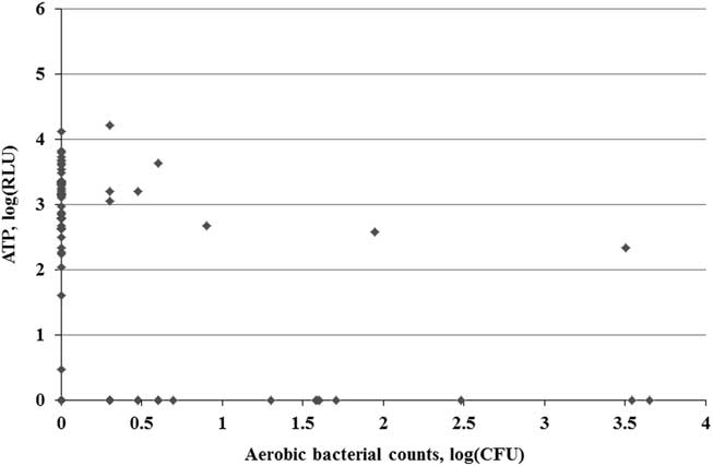
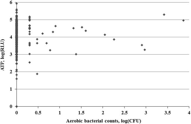
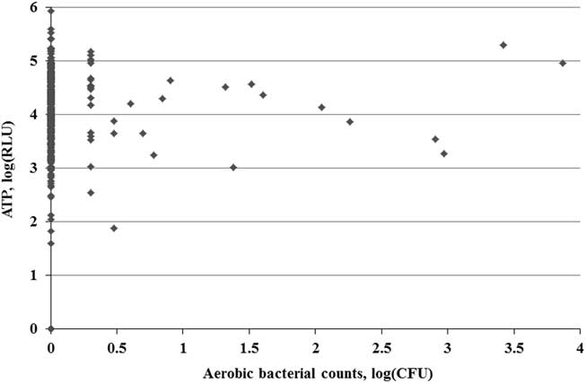
ATP RLU quantification and aerobic bacterial culture were collected for 390 duodenoscope encounters. The distribution of CFU and ATP (log-transformed) are displayed in Figure 1 for the WC sites and in Figure 2 for the EM sites. The median CFU count for the WC sites was 0 (interquartile range [IQR], 0–0) and the maximum was 4,512. The median (IQR) CFU count for the EM sites was 0 (IQR, 0–0) and the maximum was 7,352. For ATP sampling from the WC sites, the median RLU was 0 (IQR, 0–0) and the maximum RLU was 16,211. For the EM sites, the median ATP measurement was 13,836 (IQR, 3,831–42,060) and the maximum was 844,212.
Duodenoscope working channel aerobic bacterial contamination (CFU, log-transformed) versus adenosine triphosphate (RLU, log-transformed) for 390 paired observations. Abbreviations: ATP, adenosine triphosphate; CFU, colony-forming units; RLU, relative light units.

Duodenoscope elevator mechanism aerobic bacterial contamination (CFU, log-transformed) versus ATP (RLU, log-transformed) for 390 paired observations. Abbreviations: ATP, adenosine triphosphate; CFU, colony-forming units; RLU, relative light units.
The frequency of growth >0 CFU on aerobic culture for the WC duodenoscope sites was 46 (11.8%), and for the EM duodenoscope sites the frequency of growth >0 CFU was 36 (9.2%). In total, 8 (2.1%) duodenoscope samples were contaminated both at the WC and EM sites. ATP RLU measurements were >0 in 52 WCs (13.3%) and 367 EMs (94%) of the endoscopes sampled, respectively, including 51 duodenoscopes (13.1%) that were simultaneously contaminated both at the WC and the EM sites.
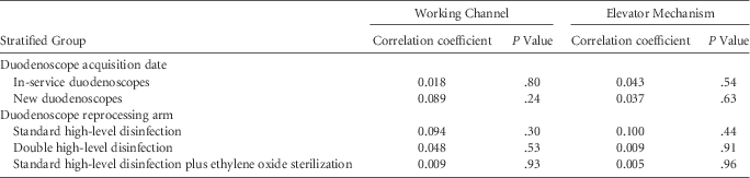
The Spearman correlation coefficient between ATP (RLU) and bacterial growth (CFU) was 0.047 for the WC sites (P=.35) and was 0.039 for the EM sites (P=.44). No significantly different distributions were detected when the data were stratified by study arm or age of the scope (Table 1).
The Correlation Coefficient Between Duodenoscope Sampling for Adenosine Triphosphate and Bacterial Contamination Among Samples Stratified by Acquisition Date of the Duodenoscope and Method of Disinfection/Sterilization Reprocessing

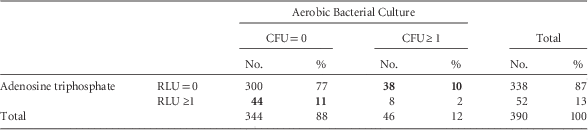
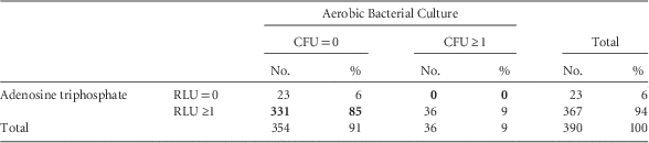
Table 2A shows the pairwise results of WC sampling and Table 2B shows the pairwise results of EM sampling for aerobic bacteria (CFU) and ATP (RLU). The results were discordant in 82 of 390 of the WC samples (21%) and in 331 of 390 of the EM samples (84.9%). For WC sites, the sensitivity was 17.4% (95% confidence interval [CI], 8.3%–32.0%), the specificity was 87.2% (95% CI, 83.1%–90.5%), the PPV was 15.4% (95% CI, 7.3%–28.6%), and the NPV was 88.8% (95% CI, 84.8%–92.0%). For EM sites, the sensitivity was 100% (95% CI, 88.0%–100%), the specificity was 6.5% (95% CI, 4.3%–9.7%), and the PPV was 9.8% (95% CI, 7.1%–13.4%). An NPV could not be calculated given the small number of measurements with no ATP detected.
Pairwise Result of Duodenoscope Working Channel Sampling for Aerobic Bacterial Culture and Adenosine Triphosphate

NOTE. CFU, colony-forming units; RLU, relative light units. Results in bold typeface represent discordant findings.
Pairwise Result of Duodenoscope Elevator Mechanism Sampling for Aerobic Bacterial Culture and Adenosine Triphosphate

NOTE. CFU, colony-forming units; RLU, relative light units. Results in bold typeface represent discordant findings.
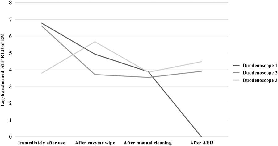
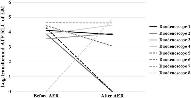
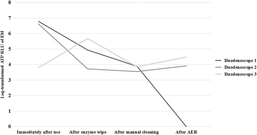
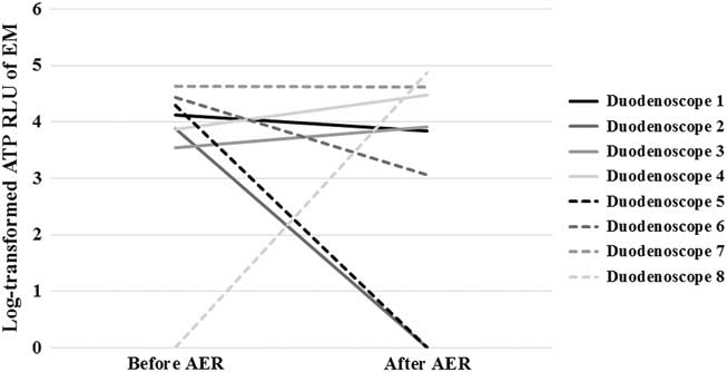
After finding the unexpected distribution of the ATP on the EM sites, we performed 3 post hoc investigations. First, to test for the adequacy of our bacterial culture technique, testing was performed on a convenience sample of several duodenoscopes immediately following the completion of an ERCP procedure to test the level of bacterial contamination and ATP RLU measurements prior to reprocessing. High levels of bacterial contamination were confirmed: On the WC sites, bacterial contamination ranged from 602 to 452,000 CFU; on the EM sites, bacterial contamination ranged from 195 to 133,000 CFU. Similarly, ATP sampling of duodenoscopes immediately after the procedure demonstrated high levels of ATP at the WC sites (ATP RLU range, 45,320–4,037,369) and the EM sites (ATP RLU range, 778,112–5,129,733). Second, we studied how the variability in ATP measurements of the EM sites changed throughout the reprocessing protocol following device use. This investigation included sampling the EM from a convenience sample of duodenoscopes (in the same fashion described in the Methods section) during 4 moments of the reprocessing cycle: (1) immediately following the procedure, (2) after bedside cleaning, (3) after complete manual cleaning in the reprocessing room, and (4) after automated endoscopic reprocessing. Test results among 3 duodenoscopes at all 4 time points are presented in Figure 3, and test results among 8 duodenoscopes before and after automated endoscopic reprocessing are presented in Figure 4. Third, we explored the theory that retained-but-dried OPA disinfectant may have affected the measurement selectively for the EM rather than the WC. We sampled the OPA disinfectant solution for ATP by pouring OPA disinfectant directly from a newly opened container into a sterile collection cup. This procdure showed results from 0 to 13,740 RLU when measured 4 times (ie, 148 and 13,740 for the first sampling in duplicate and 3,536 and 0 for repeated sampling with duplicate measurements). Notably, the sampling of 2 new and empty sterile collection cups in duplicate yielded no ATP for all 4 measurements.
Serial adenosine triphosphate measurements of the elevator mechanism during 4 moments of the reprocessing cycle: (1) immediately following the procedure, (2) after bedside cleaning, (3) after manual cleaning, and (4) after automated endoscopic reprocessing. Abbreviations: ATP, adenosine triphosphate; RLU, relative light units; EM, elevator mechanism; AER, after automated endoscopic reprocessing.
The variability in adenosine triphosphate measurements of the elevator mechanism before and after automated endoscopic reprocessing. Abbreviations: ATP, adenosine triphosphate; RLU, relative light units; EM, elevator mechanism; scope, duodenoscope.
DISCUSSION
In this study, we sought to quantify the relationship between the measurement of ATP and aerobic bacterial contamination of ERCP duodenoscopes in an effort to determine whether ATP can be used to assess ERCP duodenoscopes for bacterial contamination and reprocessing adequacy and to provide an estimation of risk of multidrug-resistant organism transmission. The main findings of our study were (1) very weak and nonsignificant relatedness between aerobic bacterial contamination (CFU) and ATP (RLU) for both the WC and EM sties on the ERCP duodenoscope; (2) marked discordance between the pairwise sampling for aerobic bacteria (CFU) and ATP (RLU), with results showing discordance in 21% of WC sites and 85% of EM sites; and (3) almost universally and markedly elevated ATP measurement from the EM sites.
Several cases of patient-to-patient transmission of infections after ERCP procedures between patients cared for using the same duodenoscope have been reported.Reference Carbonne, Thiolet and Fournier 1 – Reference Epstein, Hunter and Arwady 4 This is concerning, and it is desirable to find an efficient an accurate method of assessing the adequacy of reprocessing and bacterial contamination of duodenoscopes. Unfortunately, the literature regarding the optimal monitoring to prevent endoscopic contamination is limited. The CDC has issued an interim duodenoscope surveillance protocol, but this protocol has not yet been validated. The sensitivity, specificity, and limits in quantification or detection are not established for bacterial pathogens. 9 ATP quantification has been proposed as a surrogate for bacterial cultures to evaluate for microbial contamination of duodenoscopes. However, our study suggests that ATP bioluminescence measurement cannot be used reliably in this setting given the marked discordance and poor correlation between the pairwise sampling of both WC and EM sites for aerobic bacteria (CFU) and ATP (RLU).
Other studies that specifically evaluate the pairwise concordance and correlation between sampling for aerobic bacteria (CFU) and ATP (RLU) are scarce. A study of flexible endoscopes that compared ATP with a routine microbiological culture technique after reprocessing procedures showed that the sensitivity and specificity of the ATP compared to culture differed depending on the chosen RLU threshold. The use of a lower ATP RLU threshold was associated with lower sensitivity.Reference Hansen, Benner, Hilgenhoner, Leisebein, Brauksiepe and Popp 15 The low sensitivity of ATP measurements compared to a microbiological standard could potentially be explained by the low level of microbiologic contamination of the duodenoscopes after HLD. Studies have shown that the relationship between CFUs and RLUs is not always linear: to detect 1 ATP RLU, a sample would need to contain 103 CFU of gram-positive bacteria or 102 CFU of gram-negative bacteria.Reference Turner, Daugherity, Altier and Maurer 10 , Reference Alfa, Fatima and Olson 24 Thus, it remains unclear whether the ATP bioluminescence tests can reliably detect low levels of microbiological contamination.Reference Turner, Daugherity, Altier and Maurer 10 , Reference Aiken, Wilson and Pratten 25
An unexpected finding from this study was the consistently and markedly elevated ATP measurements from the EM. In post hoc analyses, we did demonstrate variability in ATP and did not demonstrate a significant amount of confounding from the disinfectant, which we had hypothesized might be found in higher concentrations (dried) in the EM. The ATP findings at the EM sites, therefore, likely reflect the presence of organic residue, which is difficult to clean but may be culture-negative due to the presence of nonplanktonic bacteria. Notably, however, if biofilm explains the findings in this study, the age of the duodenoscope was not associated with the presence of biofilm. Biofilm has been found on suction and biopsy channels of used endoscopes even after thorough cleaning and decontamination.Reference Pajkos, Vickery and Cossart 26 Endoscope channels with residual biofilms are subject to bacterial growth, which subsequently could be the source of bacterial contamination in outbreaks. Based on our findings, including post hoc analyses, ATP measurement of the EM may be a very useful tool for manufacturers assessing new duodenoscope designs, specifically whether they are prone to biofilm formation in complex structural areas.
Our study has several potential limitations. We used an ATP bioluminescence assay from a single manufacturer, but several other ATP measurement assays are commercially available. Omidbakhsh et alReference Omidbakhsh, Ahmadpour and Kenny 27 compared different ATP measurement assays and found that assays by different manufacturers had acceptable linearity and repeatability in their readings. Given the low level of bacterial contamination of the duodenoscopes after reprocessing. It is possible that our culture method was too insensitive to capture all pathogenic organisms or that residual disinfectant conferred bactericidal activity. This study was conducted using duodenoscopes in actual use, and the microbiologic surveillance culturing was performed according to CDC guidelines. We intentionally used a pragmatic method of culturing that institutions may employ. The current surveillance guidelines focus on aerobic bacteria and may miss microbiota. The CDC guidance allows for culture surveillance without the use of disinfectant neutralizer; its effect on the yield of duodenoscope cultures is unknown. 28 Additionally, to validate our culture method and ATP sampling method, we sampled duodenoscopes before cleaning, which demonstrated both high levels of contamination and the ability to capture the anticipated spectrum of bacterial counts and ATP RLU levels. We did not evaluate whether ATP could potentially be of use after manual cleaning prior to the duodenoscope undergoing HLD in an automated endoscope reprocessor. Using a decrease in ATP to validate the effectiveness of manual cleaning remains a potential use of ATP sampling of duodenoscopes.
In conclusion, our study investigated the relationship between ATP RLU and CFU as markers for contamination of ERCP duodenoscopes after reprocessing. In a setting of similar rates of bacterial contamination of the WC and EM, we identified both poor relatedness and poor agreement between ATP and bacterial culture methods. ATP may reflect biological material other than nonviable bacteria and should not serve as a surveillance method to detect bacterial contamination or adequacy of reprocessing of ERCP duodenoscopes.
ACKNOWLEDGMENTS
Financial support: E.B.H, A.C., B.L., M.S.S., and G.M.S. received grant support from the American Society of Gastrointestinal Endoscopy. Charm Sciences provided support (ATP testing materials) and education regarding the use of the devise and interpretation of ATP RLU results but was not involved in the design or conduct of the study, or analysis of the data.
Potential conflicts of interest: All authors report no conflicts of interest relevant to this article.