Case description
A 1.5-year-old boy presented to our outpatient department for cardiac evaluation. He was asymptomatic. An antenatal scan performed at 8 months of intrauterine life, owing to decreased foetal movements, revealed a membrane-like structure in the mid-cavitary region of the left ventricle. His perinatal course was uneventful. On examination, his vital signs and cardiovascular parameters were normal. Other systemic examinations were also normal.
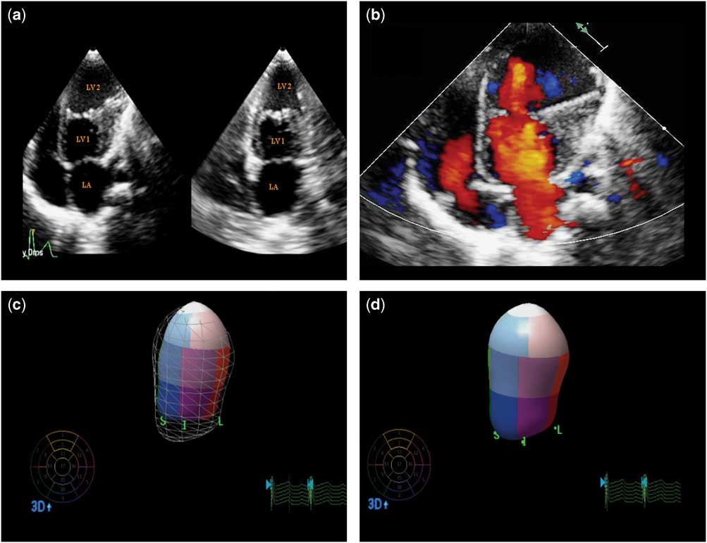
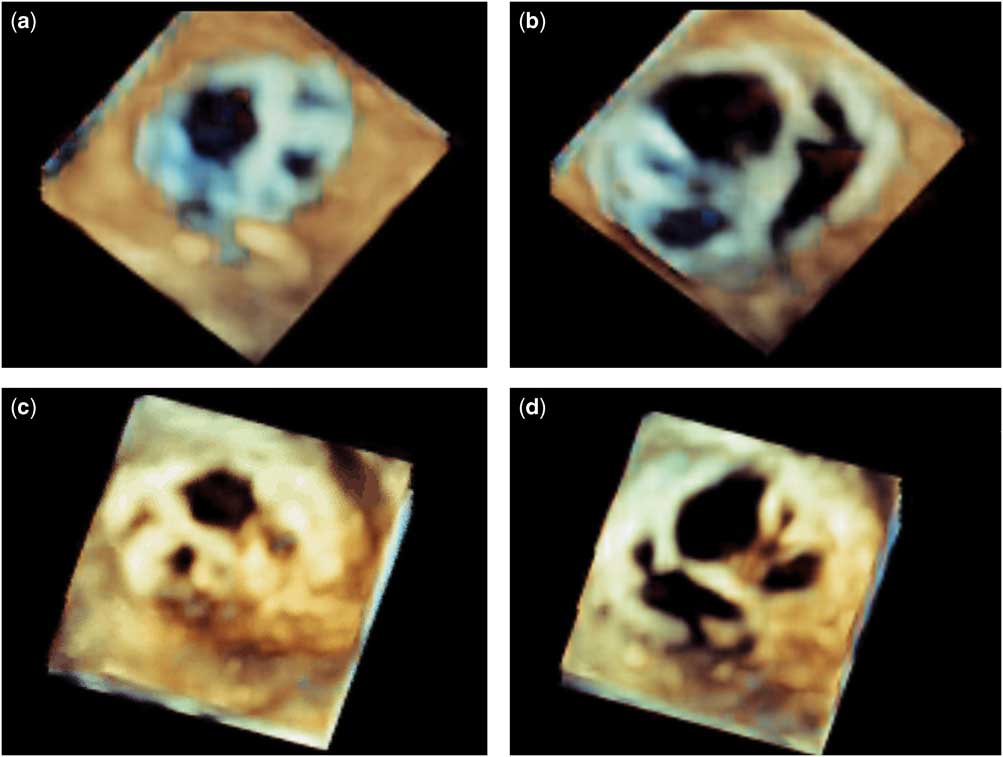
Further, two-dimensional echocardiography revealed a shelf-like membranous structure in the middle of the left ventricle (Fig 1a); two-dimensional color flow Doppler did not reveal any flow acceleration or gradient across the membrane (Fig 1b); and three-dimensional echocardiography revealed a fenestrated, muscular, membranous structure dividing the left ventricular cavity into two equal chambers. The muscular, membranous structure was seen contracting with each ventricular systole (Fig 2a–d, Supplementary videos 1 and 2). The distal chamber was dilated and had global hypokinesia, although the proximal left ventricle chamber contractility was normal; three-dimensional left ventricle ejection fraction was 33% (Fig 1c and d). Except for the presence of this muscular, membranous structure in the left ventricle and hypocontractility of the distal part of the left ventricle, other echocardiographic parameters were grossly within normal limits.
( a ) Apical four-chamber and apical two-chamber views showing a shelf-like membrane at the mid-cavitary level of the left ventricle. Color flow Doppler showing no flow acceleration at the membrane level ( b ). Three-dimensional echocardiographic images showing end-systolic and end-diastolic left ventricular volumes. The end-systolic frame revealed apical hypokinesia ( c and d ).

( a and b ) Three-dimensional echocardiographic demonstration of a muscular, membranous structure from mitral valve side during systole and diastole, respectively. ( c and d ) A muscular-membranous structure from the left ventricular apex during systole and diastole, respectively.

Discussion
Double-chambered left ventricle should be differentiated from other left ventricle outpouchings such as aneurysms, pseudoaneurysms, and diverticular and epicardial cysts. There is no clear definition of these lesions, and the terms are often used interchangeably.Reference Kraseman, Gehrmann and Fenge 1 Double-chambered left ventricle contains all three cardiac layers similar to a diverticulum and has wide communication with the left ventricle chamber, which is similar to an aneurysm. Most of the previously reported cases had a small contracting chamber attached to the main left ventricle cavity in side-by-side orientation.Reference Sanz, Rius and Kuschnir 2 – Reference Breithardt, Ropers and Seeliger 4 Although rarely the left ventricle can be divided by a fibroelastotic membrane into two chambers in series,Reference Rajiah, Thomas, Smedira and Desai 5 our case had a left ventricle cavity that was divided into two almost equal chambers by a muscular, membranous structure; three-dimensional echocardiography delineated the muscular, membranous structure in the left ventricle mid cavity with good resolution. Hence, three-dimensional echocardiography may be an ideal alternative to cardiac MRI among small children with double-chambered left ventricle. Although MRI is very sensitive in differentiating left ventricle outpouchings, definitive diagnosis is only possible with histology.Reference Rajiah, Thomas, Smedira and Desai 5
Left ventricle aneurysm is usually a saccular protrusion from the apex or the anterior part of the left ventricle. It has a wide neck (neck-to-mouth ratio usually 0.9–1.0) with the left ventricle and is dyskinetic. It contains endocardium, epicardium, and thinned-out abnormally contracting, scarred myocardium. The thinned-out, scarred myocardial area can be demonstrated on delayed enhanced MRI without pericardial enhancement, and dyskinetic movement is well appreciated in MRI.Reference Rajiah, Thomas, Smedira and Desai 5 , Reference Sharma and Kumar 6 Pseudoaneurysm formation is due to cardiac rupture. The ruptured wall is contained by the pericardium, thrombus, or the scared tissue without any viable myocardium. It has a narrow neck with a neck-to-mouth ratio of usually 0.25–0.5. Pericardial enhancement can be seen on MRI scan.Reference Rajiah, Thomas, Smedira and Desai 5 , Reference Sharma and Kumar 6 Left ventricle diverticulum is commonly congenital in origin and mostly located at the apex. It contains all three cardiac layers and contracts synchronously with the left ventricle.Reference Rajiah, Thomas, Smedira and Desai 5 , Reference Sharma and Kumar 6 It is a tubular structure with a variable neck-to-mouth ratio, although commonly associated with narrow neck, and is almost always associated with other thoracoabdominal malformations.Reference Marijon, Ou and Fremont 7 An epicardial cyst is usually multiloculated. It communicates with the ventricular cavity through narrow channels opening into intra-trabecular spaces or rarely with the coronary arteries.Reference Gerlis, Partridge and Fiddler 8 Commonly reported forms of double-chambered left ventricle have an accessory chamber, which is oriented side-by-side to the main left ventricle chamber and connected through a wide mouth. It contracts synchronously with the main left ventricle chamber and is not associated with other midline thoracoabdominal malformations.Reference Sanz, Rius and Kuschnir 2 – Reference Breithardt, Ropers and Seeliger 4 Rarely the accessory chamber can be oriented in series with the main left ventricle cavity.Reference Rajiah, Thomas, Smedira and Desai 5
The exact aetiology of the double-chambered left ventricle is still uncertain. Early reports have shown that double-chambered left ventricle appears to be related to a cardiomyopathy. Paronetto and StraussReference Paronetto and Strauss 9 and Ruttenberg et alReference Ruttenberg, Jue, Elliott, Anderson and Edwards 10 hypothesised that it was due to congenital weakness of the heart muscles. Gerlis et alReference Gerlis, Partridge and Fiddler 8 reported that associated endocardial fibroelastosis may render the main left ventricle cavity hypocontractile or non-contractile, and thus the functional portion of the left ventricle may derive from expansion of intra-trabecular myocardial sinusoids. Hence, a single aetiological explanation for double-chambered left ventricle is hard to find.
In our patient, the hypocontractile distal left ventricle chamber was considered a possible source for thrombus formations with risk of thromboembolism. Therefore, oral anticoagulation with warfarin was initiated, and parents were advised to monitor the international normalized ratio at regular intervals. Surgical options were not considered as the muscular, membranous structure was non-obstructing in nature, and the child was asymptomatic.
In conclusion, double-chambered left ventricle is a rare clinical entity in which the left ventricular cavity is divided into two by an abnormal septum or rarely a fibroelastotic membrane. Its aetiology is still uncertain and could be either congenital or acquired in nature; three-dimensional echocardiography and cardiac magnetic resonance imaging are considered inevitable tools for accurate non-invasive diagnosis of this condition.
Acknowledgement
None.
Financial Support
This research received no specific grant from any funding agency or from commercial or not-for-profit sectors.
Conflicts of Interest
None.
Ethical Standards
The authors assert that all procedures contributing to this work comply with the ethical standards of the relevant national guidelines on human experimentation (Ethical guidelines for biomedical research on human participants 2006, India) and with the Helsinki Declaration of 1975, as revised in 2008, and has been approved by the institutional committees (Christian Medical College, Vellore).
Supplementary Material
To view supplementary material for this article, please visit https://doi.org/10.1017/S1047951117000555