Introduction
Paragangliomas are slow-growing, benign, neuroendocrine tumours. The commonest site in the head and neck is the carotid bifurcation. They rarely occur in the larynx, with fewer than 90 reported cases. Surgery is the mainstay of treatment, although radiotherapy has also been used. Historically, there has been diagnostic confusion with atypical carcinoid. The main method of differentiation is immunohistochemical staining in addition to light microscopy.Reference Ferlito, Barnes, Rinaldo, Gnepp and Milroy1
Case report
A 78-year-old woman presented with a one-year history of throat irritation, against a background of passive smoking.
Flexible laryngoscopy revealed an exophytic lesion on the right aryepiglottic fold extending to the base of the epiglottis.
Light microscopy and immunohistochemical staining diagnosed a non-small cell carcinoma, with appearances favouring a poorly differentiated squamous cell carcinoma. The lesion was staged as a tumour stage 2, node stage 0, supraglottic squamous cell carcinoma.
The patient received a total of 66 Grays of radiotherapy, with resulting regression of the lesion.
However, three months after radiotherapy she was suspected of having a recurrence, with a 2 cm, right-sided mass observed on the laryngeal surface of the epiglottis.
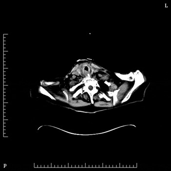
Positron emission tomography computed tomography (PET CT) showed increased uptake in the supraglottic region (Figure 1).
Axial positron emission tomography computed tomography scan showing a right-sided, supraglottic, soft tissue mass. L = left; P = posterior

Surgery was delayed due to viral gastroenteritis. The patient subsequently presented to the emergency department with acute stridor and aspiration pneumonia, requiring emergency intubation.
Panendoscopy, biopsy and tracheostomy were performed two days later. Intra-operatively, diffuse oedema of the aryepiglottic folds and false vocal folds was observed, with a fungating mass on the right laryngeal surface of the epiglottis.
Histopathological analysis of biopsies from the right vallecula, right laryngeal surface of the epiglottis and right false vocal fold showed necrotic squamous epithelium without evidence of dysplasia. However, biopsies of the left laryngeal surface of the epiglottis revealed high grade squamous dysplasia.
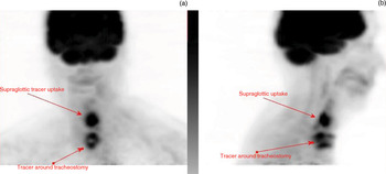
Two weeks after these biopsies, CT and repeated PET CT findings remained highly suggestive of residual malignancy (Figure 2).
(a) Coronal and (b) sagittal positron emission tomography computed tomography scans showing intense uptake of tracer in a soft tissue mass in the supraglottis, highly suggestive of recurrent carcinoma at that site. Uptake of tracer around the tracheostomy is probably inflammatory rather than neoplastic, and there are no other findings of note.

Given the histopathological and PET CT findings, and the fact that our patient had a non-functioning larynx with gross aspiration, the decision was made to proceed with total laryngectomy and bilateral selective neck dissection (i.e. levels II, III and IV).
The definitive histopathological diagnosis was paraganglioma of the laryngeal surface of the epiglottis, completely excised with no residual squamous dysplasia, despite the original biopsy finding of squamous cell carcinoma. The neck dissection was also negative for squamous cell carcinoma.
In the original biopsy, high and low molecular weight cytokeratin immunohistochemical stains were positive (pancytokeratin cocktail AE1/AE3 and cytokeratin 8), while the tumour was negative for lymphocyte markers (CD3 and CD20), thyroglobulin, synaptophysin, chromogranin, S-100 protein and Melan-A. This biopsy showed a high-grade lesion with marked cytological atypia, necrosis and abundant mitotic figures. The biopsy was reviewed by six pathologists at three different services, who all concurred with the diagnosis of squamous cell carcinoma.
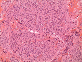
This is in stark contrast to the histopathological appearance of the resection specimen, which showed prominent ‘Zellballen’ architecture (Figure 3; note polygonal epithelioid cells in a nested arrangement). There were no infiltrative margins, no cytological atypia and no apparent mitotic figures, but the marked ‘salt and pepper’ chromatin appearance was typical of neuroendocrine neoplasia.
Photomicrograph of the tumour showing polygonal cells in a typical nested or ‘Zellballen’ arrangement. (H&E; ×200)

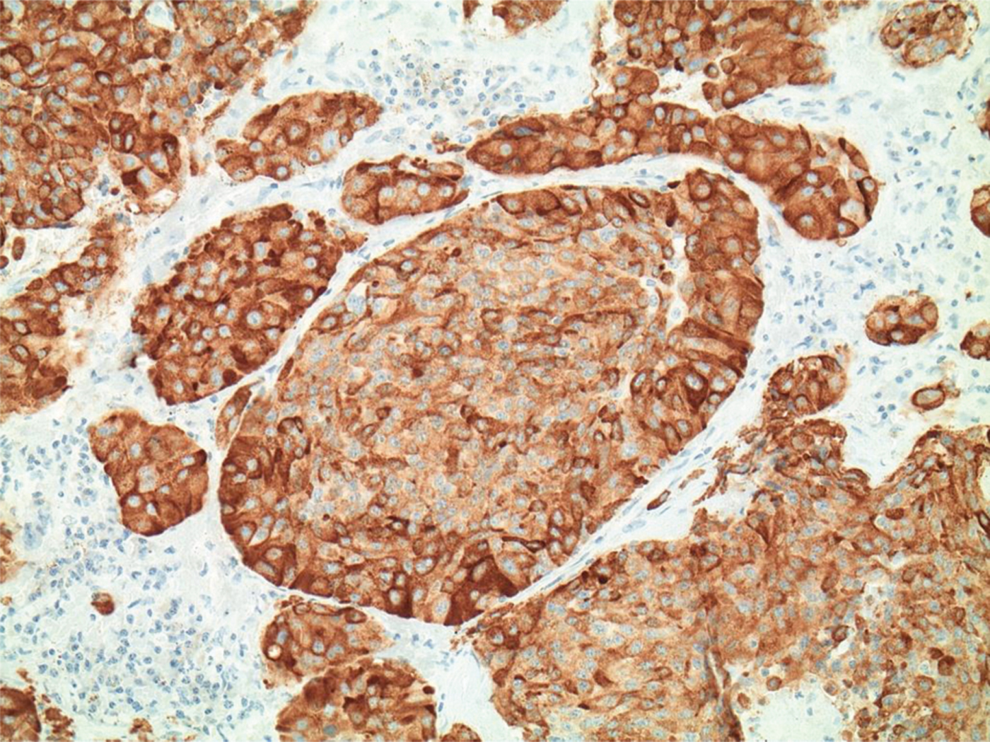
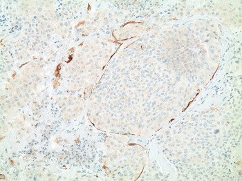
Immunohistochemical staining of the tumour cells for the neuroendocrine markers chromogranin (Figure 4) and synaptophysin showed immunoreactivity. There was positive S-100 protein staining of the sustentacular cells (Figure 5). The tumour cells were negative for cytokeratin staining. These findings supported the diagnosis of paraganglioma of the epiglottis. Extensive radiation-related change was found, but no residual squamous cell carcinoma. In retrospect, it is difficult to ascertain whether the original tumour mass was in part accounted for by a closely adjacent and coexistent submucosal paraganglioma, not yet clinically apparent.
Photomicrograph showing central chief cells stained for chromogranin. (×200)
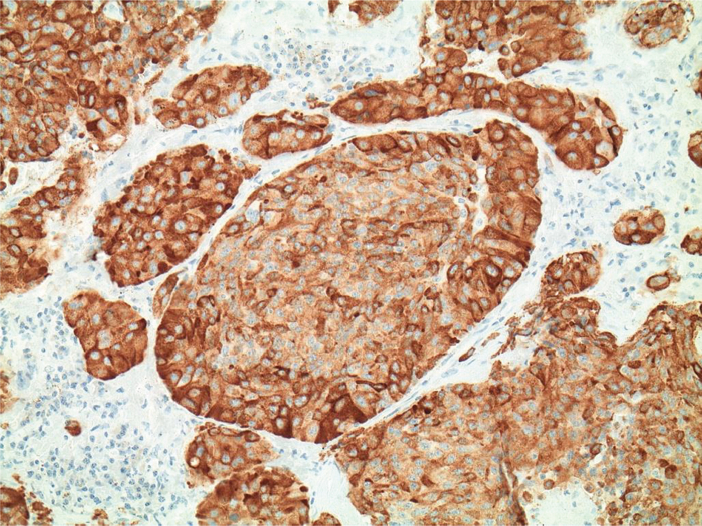
Photomicrograph showing peripheral sustentacular cells stained for S-100 protein. (×200)

Four weeks after surgery, the patient was discharged with a tracheoesophageal speaking valve. A two-month post-operative octreotide scan showed no residual tumour. At six months' follow up, there was no clinical recurrence, a good swallow and improving oesophageal speech.
Discussion
Paragangliomas are slow-growing, vascular, submucosal tumours arising from the paraganglion cells which are part of the diffuse neuroendocrine system. They are generally considered benign. In the head and neck, carotid body paragangliomas are the most common type, followed by jugulotympanic and vagal paragangliomas.Reference Snyderman, Lin and Basow2
Laryngeal paragangliomas are rare, making up less than 1 per cent of all laryngeal neoplasms.Reference Ferlito, Silver, Bradford and Rinaldo3 Since they were first described by Blanchard and Saunders in 1955,Reference Blanchard and Saunders4 there have been fewer than 90 cases reported in the literature. More than 90 per cent of laryngeal paragangliomas are supraglottic.Reference Peterson, Fu and Calcaterra5
The majority of laryngeal paragangliomas affect women (with a gender ratio of 3:1), mainly in the fourth to sixth decades of life (median age, 44 years).Reference Ferlito, Barnes and Wenig6, Reference Myssiorek, Rinaldo, Barnes and Ferlito7 Although previously thought of as a sporadic tumour, there have been two reported cases of multifocal head and neck paragangliomas in which the index lesion was a laryngeal paraganglioma.Reference Sanders, Abreo, Rivera, Stucker and Nathan8, Reference Rubin, Cheng and Bradford9 Neither of these cases underwent genetic testing; however, they raise the possibility of succinate dehydrogenase complex unit B gene mutations, which are most commonly implicated in paragangliomas of the head and neck.Reference Snyderman, Lin and Basow2 However, there is insufficient evidence to comment further on the genetic basis of paraganglioma of the larynx.
Patients typically present with dyspnoea as their primary symptom, as there is always a degree of airway obstruction. Commonly, there may also be cough, dysphagia and a foreign body sensation.
The differential diagnosis of laryngeal paraganglioma includes: neuroendocrine neoplasm of epithelial rather than neural origin; malignant melanoma; metastatic renal cell carcinoma; and medullary carcinoma of the thyroid gland. The neuroendocrine tumour that has caused the greatest diagnostic confusion with laryngeal paraganglioma is atypical carcinoid. Differentiation is important because atypical carcinoid has higher malignant potential and may require different treatment modalities.Reference Ferlito, Silver, Bradford and Rinaldo3
Macroscopically, laryngeal paragangliomas are typically well-circumscribed, tan, brown or reddish-brown, submucosal masses.Reference Barnes10 Histopathologically, they consist of two cell types – chief cells and sustentacular cells – in a Zellballen pattern which is typical, but not diagnostic, of paraganglioma. This pattern can also be found in carcinoid tumours, melanomas and medullary carcinomas of the thyroid.Reference Sanders, Abreo, Rivera, Stucker and Nathan8, Reference Barnes, Ferlito and Wenig11
The chief cells are polygonal with abundant, granular cytoplasm and round nuclei with ‘salt and pepper’ chromatin. Nuclear pleomorphism may be present, but mitotic figures are rare and tumour necrosis is absent. The sustentacular cells are spindled cells at the periphery of the nests, which are apparent on immunostaining with S-100.Reference Sanders, Abreo, Rivera, Stucker and Nathan8, Reference Ferlito, Milroy, Wenig, Barnes and Silver12
Barnes proposed that almost all alleged malignant paragangliomas are actually misdiagnosed atypical carcinoids.Reference Barnes10 Only one accepted case of metastatic paraganglioma has been reported,Reference Ferlito, Barnes, Rinaldo, Gnepp and Milroy1, Reference Barnes10 with a subsequent case published in 2010 in which the patient died of his disease.Reference Smolarz, Hanna, Williams and Kupferman13 One to three per cent of paragangliomas are considered functional, and in these cases serum and urine catecholamine levels can be measured,Reference Laudadio14 but there are no reported cases of paraneoplastic syndrome.Reference Ferlito, Silver, Bradford and Rinaldo3 There are no previous reports in the literature of paraganglioma in coexistence with squamous cell carcinoma.
Currently, the diagnostic imaging modality of choice is magnetic resonance imaging (MRI) with gadolinium enhancement (preferred over CT unless there is suspicion of cartilage destruction).Reference Myssiorek, Rinaldo, Barnes and Ferlito7, Reference Lustrin, Palestro and Vaheesan15 Octreotide scanning is also a reliable test to detect paragangliomas.Reference Myssiorek, Rinaldo, Barnes and Ferlito7 The radiological features are those of a vascular mass, usually with a dominant feeder vessel, either the superior or inferior thyroid artery.Reference Sanders, Abreo, Rivera, Stucker and Nathan8 Positron emission tomography CT has been shown to be more sensitive than MRI or plain CT in the detection of metastatic paragangliomas, but not in the detection of localising, non-metastatic paraganglioma,Reference Timmers, Chen, Carrasquillo, Whatley, Ling and Havekes16 and hence is not recommended as a first-line imaging investigation. Case reports have typically described a mass of increased uptake closely associated with a nerve;Reference Takayama, Konishi, Kishimoto, Kanazawa and Yamane17 better visualisation can be achieved in the case of catecholamine-producing paragangliomas.Reference Fiebrich, Brouwers, Kerstens, Pijl, Kema and de Jong18 Angiography has been used to assess the size, location and blood supply of paragangliomas, although they can be adequately assessed with MRI and CT.Reference Myssiorek, Rinaldo, Barnes and Ferlito7
Although these tumours are generally considered benign, the treatment of choice is surgical removal with maximal possible preservation of laryngeal function. Open surgery is preferred to endoscopic surgery because bleeding can obscure visualisation, which can result in a higher recurrence rate.Reference Myssiorek, Rinaldo, Barnes and Ferlito7 Open approaches include supraglottic laryngectomy, total laryngectomy, laryngofissure and lateral thyrotomy, depending on the tumour size and site. Neck dissection is not indicated, as laryngeal paragangliomas do not typically metastasise.
• Paragangliomas are benign, slow-growing, neuroendocrine tumours
• There are fewer than 90 reported cases of laryngeal paraganglioma
• Laryngeal paraganglioma in a previously irradiated neck, with concurrent squamous cell carcinoma (SCC), has not been reported
• A case of paraganglioma masquerading as SCC, treated with total laryngectomy in a non-functioning larynx, is reported
Radiotherapy is a well documented treatment modality in some cases of head and neck paraganglioma, as it has been shown to limit the tumour's growth and reduce its size. Experience with laryngeal paragangliomas is limited because of their rarity. Radiotherapy can be associated with airway swelling and chondronecrosis. There has only been one case report of successful tumour size reduction to 2 mm with external beam radiotherapy, in which the patient was symptom-free at five-year follow up.Reference Smolarz, Hanna, Williams and Kupferman13
The unique feature of our case is the fact the tumour occurred concurrently with squamous cell carcinoma and persisted despite irradiation, calling into question the effectiveness of radiotherapy as a treatment for laryngeal paraganglioma.
Non-laryngeal paragangliomas of the head and neck have been shown to respond to radiotherapy, with reported control rates of over 90 per cent.Reference Hinerman, Amdur, Morris, Kirwan and Mendenhall19, Reference Mendenhall, Amdur, Vaysberg, Mendenhall and Werning20 Despite this, the literature supports surgical resection as the treatment of choice for laryngeal paraganglioma.
The reported case also highlights the difficulty of paraganglioma diagnosis, not only regarding differentiation from other neuroendocrine tumours, but also regarding obtaining a positive biopsy in a previously irradiated neck with extensive radiation-related change.