Introduction
The zona pellucida (ZP), surrounded by a specialized extracellular matrix composed of glycoproteins, plays a vital role in facilitating subsequent fertilization events by allowing initial recognition and binding of the sperm to the oocyte (Zhao et al., Reference Zhao and Dean2002). Under specific physiological conditions, proteolytic enzymes gradually degrade the ZP, leading to its thinning as the embryo develops, ultimately facilitating its hatching from the ZP and successful implantation (Cohen, Reference Cohen1991). Meanwhile, a diverse array of factors regulate implantation, a complex physiological process that includes embryo invasion, attachment, localization, and hatching.
The effectiveness of cryopreservation techniques contributes to increased cumulative pregnancy and implantation rates in in vitro fertilization (IVF) procedures (Andersen et al., Reference Andersen, Goossens, Ferraretti, Bhattacharya, Felberbaum and de Mouzon2008). Cryopreservation-induced ‘ZP hardening’ has been shown to impede human embryo hatching (Kirienko et al., Reference Kirienko, Apryshko, Naumova and Yakavenko2019). Current research suggests that cryopreservation may initiate the zona hardening (Cohen et al., Reference Cohen, Alikani, Mentzer and Schimmel1999). In 1988, an additional technique called ‘assisted hatching (AH)’ was developed to address this issue. AH involves artificially disrupting the zona pellucida (ZP) (Cohen et al., Reference Cohen, Malter, Fehilly, Wright, Elsner and Kort1988). AH techniques aim to create an artificial opening or thinning of the ZP, making easy the hatching and implantation of embryos that may strive to escape the ZP otherwise (Cohen et al., Reference Cohen and Alikani1992; Hernandez-Nieto et al., Reference Hernandez-Nieto, SotoCossio and Basurta-Diaz2015).
Several techniques have been employed to facilitate embryo hatching, including chemical drilling and thinning of the zona pellucida (ZP) using acidic Tyrode’s medium, mechanical piercing known as partial zona dissection, hydrostatic pressure-induced ZP expansion, creation of piezoelectric pulses on the ZP, and laser-assisted zona drilling (Avella et al., Reference Avella, Milne, Dawood, Dawood, Tucker, Nagy, Varghese and Agrawal2019). Laser-assisted hatching (LAH) was pioneered in the early 1990s and was recognized for its accuracy, speed, and safety (Tadir et al., Reference Tadir, Neev and Berns1993). LAH can be implemented in two ways: by opening the ZP or thinning its outer wall using laser energy. Studies have compared laser-assisted opening (LAO) and control methods, as well as laser-assisted thinning techniques (Hsieh et al., Reference Hsieh, Huang, Cheng, Chang, Tsai and Lee2002; Valojerdi et al., Reference Valojerdi, Eftekhari-Yazdi, Karimian and Ashtiani2008; Kutlu et al., Reference Kutlu, Atvar and Vanlioglu2010; Frydman et al., Reference Frydman, Younes, Hesters, Frydman and Fanchin2016). Research suggests that human 8 cell embryos undergoing thinning LAH exhibit higher pregnancy rates compared to those undergoing drilling LAH (Joeng et al., Reference Jeong, Joo, Kim, Kim, Joo and Lee2018).
In this study, we evaluated the effects of two types of LAH groups and a NAC group on clinical outcomes including clinical pregnancy, ongoing pregnancy and live birth rates in frozen-thawed cleavage stage embryo transfer cycles.
Materials and methods
Patient characteristics
This retrospective study included 310 patients between 20 and 40 years old who underwent FET (frozen embryo transfer) cycles from April 2021 to December 2022 in the ART Unit of Izmir Economy University Medical Point Hospital, Izmir, Turkey and underwent frozen thawed cleavage stage embryo transfer after ‘freeze-all’ protocols and single or double embryos were transferred to the uterus. The choice of the number of embryos transferred was made following the criteria recommended by the Ministry of Health of The Republic of Turkey. Exclusion criteria were patients (i) women 41 years old, (ii) azoospermia, (iii) having partially or fully degenerated cleavage stage embryos.
The patients were categorized into 3 groups according to the LAH technique. The thinning LAH group (TAH, 100 patients), the drilling LAH (DAH, 100 patients), and non-assisted control group (NAC, 110 patients). Patients were allocated into the TAH, DAH and NAC groups consecutively according to the order of treatment cycles and laboratory availability. No specific patient characteristic (e.g., age, embryo quality, infertility diagnosis) was used to assign patients to a particular group.
Oocyte stimulation
Gonadotropins (follitropin alfa or menotropin 150–450IU) were administered starting on day 3 of the menstrual cycle, with 0.25 mg of the GnRH antagonist cetrorelix acetate (Cetrotide, Merck, Germany) on cycle day 7. Human chorionic gonadotropin was used to trigger ovulation once the dominant follicle reached 18mm and the removal of the oocytes was carried out 35 h after activation.
Criteria for selection of embryos
All embryos were cultured in Sage One Step culture medium (Origio, Denmark) in the K-system G 210 incubator (Cooper Surgical, Denmark) in an atmosphere of %5.7 CO2 and %5.0 O2. Cleaving embryos were scored and graded according to the number of blastomeres, their symmetry, and the degree of cytoplasmic fragmentation. The evaluation of each embryo was carried out according to the conventional criteria outlined by the ESHRE Istanbul Consensus Guidelines (2011), which categorizes embryos as follows.
-
Grade 1: Considered ‘good’ with less than 10% fragmentation, uniform and stage-specific cell sizes, and no multinucleation.
-
Grade 2: Rated as ‘fair’ with 10–25% fragmentation, predominantly stage-specific cell sizes, and no evidence of multinucleation.
-
Grade 3: Identified as ‘poor’ with severe fragmentation exceeding 25%, inconsistent cell sizes not aligned with the developmental stage, and multinuclear appearance.
Embryo freezing and thawing
The embryos were vitrified on day 3 and using a vitrification protocol (Irvine Scientific, USA) with an open cooling system device (Cryotec, Reprolife Inc, Japan) for FET. The SAGETM Vitrification Warming Kit (Cooper Surgical, USA) was used for the warming procedures.
In all groups, good-quality embryos in the cleavage stage were thawed on day 3 and cultured for a short period (2–4 h after thaw culture) in Sage-1 Step (Origio, Denmark). Embryos were considered to survive and are suitable for transfer if their blastomeres were fully intact after thawing. Single or double day 3 embryos were transferred in all groups.
Laser-assisted hatching procedure and embryo transfer
A computer-controlled non-contact 1480 nm diode laser (Zilos-tkTM, Hamilton Thorne Biosciences, Beverly, MA, USA) was utilized for laser hatching. The laser system was integrated with a 40X objective combination. Mounted on the objective turret of a Leica DMI3000 B inverted microscope (Leica Microsystems CMS GmBH, Germany), the Zilos-tkTM laser was configured with a pulse width of 600 µs and a standard pulse energy of 300 mW/pulse. The laser operated at 100% power. The embryos were subjected to TAH or DAH in 20 µL micro-drops containing handling medium (Origio, Denmark). Minimal laser pulses were used at the lowest feasible power setting to reduce potential damage to the blastomeres. The selection of areas with a substantial perivitelline space between the blastomere and the ZP was made to prevent blastomere damage during the procedure. TAH and DAH were performed on the day of FET after embryo thawing.
In the TAH thinning technique, the objective was to reduce the thickness of the outer layer of the protective glycoprotein coating by a specific amount. Using a laser shot size of 5 µm, thinning was carried out by administering 4–5 shots without penetrating the inner membrane, reaching a depth of 50% of the thickness of the ZP as shown in Figure-1(A). In the DAH, laser openings were created with 4–5 shots from the outer to the inner side of the ZP as shown in Figure-1(B). In the NAC, which did not receive LAH, are shown in Figure-1(C). Following laser hatching, embryos in the droplets were washed multiple times before being transferred to a four-well Nunc dish (Thermo Scientific, Denmark) containing Sage One-Step culture medium (Origio, Denmark).
The thinning laser-assisted hatching (TAH) (A), The drilling laser-assisted hatching (DAH) (B), Non- assisted control group (NAC) (C).

Embryos were transferred on the third day of development with a Sure-Pro Ultra Embryo Replacement Catheter with an oscillator (Cooper Surgical Company, The Netherlands) and a medium Sage One Step (Origio, Denmark).
Endometrial preparation for frozen embryo transfer
All patients received luteal support with daily use of a 200 mg Crinone 8% vaginal gel (Dendron Brands Limited, Watford, England) and a 200 mg Lutinus capsule (Ferring GmbH, Wittland, Kiel, Germany).
Evaluation of clinical outcomes
The pregnancy result was obtained with a blood βhCG test on day 12 after embryo transfer, and the clinical pregnancy was confirmed by detecting the gestational sac on a transvaginal ultrasound. Ongoing pregnancy was defined as the presence of a viable fetus after 12 weeks of pregnancy.
Statistical analysis
Continuous variables were summarized as mean and standard error and categorical variables as number and percentages. To compare the differences between the groups, we used the Chi-square test for categorical variables and the one-way ANOVA test for continuous variables. The student t test was performed to compare the difference among the groups. SPSS 29.0 was used in all analyzes and p values <0,05 were considered statistically significant.
Results
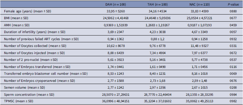
In this study, between April 2021 and December 2022, the results of 310 frozen-thawed ET cycles were evaluated. First or second cleavage stage embryos were transferred to the patients. The demographic characteristics and laboratory parameters are shown in Table 1. There were no differences in female age of the TAH, DAH, and NAC groups age (33,95 ± 5260 vs 34,16 ± 4534 vs 33,83 ± 4599; p = 880), respectively. BMI, AMH, duration of infertility, number of previous failed ART cycles (p > 0.05). Furthermore, the number of oocytes, the number of injected oocytes, and the number of fertilized oocytes were similar between the groups (p = 331, p = 672, p = 537; respectively.). Furthermore, there was no statistical difference between the groups in terms of the number of embryos cryopreserved (2,77 ± 1,58 vs 2,73 ± 1,68 vs 2,59 ± 1,48, p = 676, respectively.). As mentioned above, there were no statistical differences between the groups in terms of the mean number of transferred embryos (1,74 ± 0441 vs 1,61 ± 0490 vs 1,71 ± 0456; p = 116, respectively.). As expected, there were no statistical differences between the groups in terms of the number of blastomere cells of transferred embryos (8,53 ± 1,24 vs 8,40 ± 1,23 vs 8,16 ± 1018; p = 069, respectively.). Finally, we considered semen parameters that include semen volume, sperm concentration, and total progressive motile sperm count among the three groups and we did not find any statistical difference (p > 0,05) (Table 1).
Cycle characteristics of the participants in three groups

Values are presented as mean±standard deviation. No differences were significant (P > 0.05).
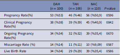
The clinical pregnancy rates (%38 vs %39 vs %40,9; p = 842), ongoing pregnancy rates (%34 vs %32 vs %35,4; p = 670,) miscarriage rates (14% vs 11% vs 9.1%; p = 587) (p > 0.05) and the live birth rates (%34 vs %29 vs %35,4; p = 586) among the groups were non-significant (Table 2).
Clinical outcomes in frozen thawed good stage cleavage embryo cycles in three groups

Discussion
The vitrification process causes the zona pellucida to harden, which can cause difficulties in embryo hatching (Zeng et al., Reference Zeng, Su and Li2018). There is a negative correlation between zona thickness and embryo implantation (Edi Osaige et al., Reference Edi-Osagie, Hooper and Seif2003). This scenario has led to the question whether LAH could improve clinical outcomes in patients whose embryos have hardened ZP. The use of LAH techniques has been shown to enhance the rates of implantation in assisted reproduction (Martins et al., Reference Martins, Rocha, Ferriani and Nastri2011). The use of LAH techniques, which involve drilling or thinning of the ZP, has shown a significant elevation of both implantation and clinical pregnancy rates (Ali et al., Reference Ali, Rahbar, Burjaq, Sultan, Al Flamerzi and Shahata2003; Zeng et al., Reference Zeng, Su and Li2018). In a large-scale retrospective cohort study conducted in 2023, which included 5779 FET cycles, LAH was associated with a significantly higher live birth rate compared to cycles without assisted hatching (34.9% vs. 31.4%; p = 0.024). However, no significant differences were observed in implantation or clinical pregnancy rates between the two groups (Yang et al., Reference Yang, Sun, Huang, Li and Hu2023). A meta-analysis by Zhang et al. (Reference Zhang, Yan, Jiali, Xinyu and Xuefeng2024), which included 2634 patients from eight studies, demonstrated that LAH significantly improved implantation rates. However, LAH was also associated with an increased risk of miscarriage in FET cycles.
Additionally, a prospective cohort study conducted between 2014 and 2016 found that zona pellucida thinning significantly improved implantation (31.2% vs. 24.6%) and clinical pregnancy rates (49.3% vs. 38.9%) in patients with previous implantation failures. However, no significant difference was observed in live birth rates between the groups (Lee et al., Reference Lee, Cha and Shin2019).
However, in a randomized study, Ng et al. did not show any advantageous impact of LAH on the clinical pregnancy rate when comparing NAC-assisted hatching after FET (Ng et al., Reference Ng, Naveed, Lau, Yeung, Chan, Tang and Ho2005). Primi et al. also failed to show any advantage of assisted hatching in frozen-thawed ET cycles (Primi et al., Reference Primi, Senn, Montag, Van der Ven, Mandelbaum, Veiga, Barri and Germond2004). A 2024 systematic review by Jin et al. analyzing nine studies found no statistically significant difference between zona drilling (D-LAH) and zona thinning (T-LAH) regarding clinical pregnancy, implantation, or live birth rates. However, D-LAH was associated with a higher singleton pregnancy rate.
It is still unclear whether the methods can be used interchangeably on day 3 FET. In this study, the clinical outcomes of frozen embryos frozen on day 3 that underwent TAH, DAH and NAC were examined. Our results have not shown any significant difference in terms of clinical pregnancy rates in the assisted hatching group based on the thickness of the ZP between the methods.
The starting point of this study was the findings of previous research. In general, these findings were similar and there were no superiority to each other in differences studies (Ali et al., Reference Ali, Rahbar, Burjaq, Sultan, Al Flamerzi and Shahata2003; Primi et al., Reference Primi, Senn, Montag, Van der Ven, Mandelbaum, Veiga, Barri and Germond2004, Ng et al., Reference Ng, Naveed, Lau, Yeung, Chan, Tang and Ho2005; Zeng et al., Reference Zeng, Su and Li2018). The blastocyst must escape from the ZP prior to implantation in the endometrium. The underlying hypothesis of LAH is that altering the ZP can enhance the rates of implantation for embryos that are incapable of hatching naturally (Cohen et al.,Reference Cohen, Malter, Wright, Kort, Massey and Mitchell1989; Cohen et al., Reference Cohen and Alikani1992). Despite the efforts of numerous researchers, there is still an interest in the results of LAH, and several studies have noted that some patients did not observe any benefits from the use of LAH (Bider et al., Reference Bider, Livshits, Yonish, Yemini, Mashiach and Dor1997; Edirisinghe et al., Reference Edirisinghe, Ahnonkitpanit, Promviengchai, Suwajanakorn, Pruksananonda, Chinpilas and Virutamasen1999; Fryman et al., Reference Fryman, Madoux, Hesters, Duvernoy, Feyereisen, Le Du, Tachdian, Frydman and Fanchin2006). Unlike other methods, LAH can be applied through an opening or thinning and has the advantage of allowing the simple removal of the ZP by approaching the embryos in a non-contact mode without mechanical or mutagenic side effects (Germond et al., Reference Germond, Nocera, Senn, Rink, Delacrétaz and Fakan1995; Mantoudis et al., Reference Mantoudis, Podsiadly, Gorgy, Venkat and Craft2001). Similar studies conducted on mouse models suggest that thinning at the 8-cell stage does not enhance or promote in vitro hatching (Uppangala et al., Reference Uppangala, D’Souza, Pudakalakatti, Atreya, Raval, Kalthur and Adiga2016). Similarly, Lee et al. investigated the impact of thinning versus opening on clinical outcomes based on maternal age in patients experiencing repeated failures of implantation with human embryos, and both groups had similar outcomes (Lee et al., Reference Lee, Cha and Shin2019). In our study, we evaluated TAH, LAH and NAC in good-quality cleavage-stage embryos and did not show any advantage in clinical outcomes after FET. Our results of frozen thawed cycles showed a trend toward lower clinical pregnancy rates and higher miscarriage rates in the TAH and DAH groups compared to NAC. However, this did not result in a statistical difference. Comparison of frozen thawed embryos DAH and LAH did not show any benefit in performing AH based on ZP hardening, and the results were similar between groups. Several previous studies have shown higher miscarriage rates following LAH groups but had indistinct outcomes on the potential risk of miscarriage due to insufficient statistical power.
In comparison to other methods, LAH stands out as the preferred and widely used technology due to several key advantages. First, it saves time and reduces the number of laser shots needed. Second, embryos spend less time outside the incubator. Lastly, the risk of temperature increase near the embryos from laser thermal shock is minimized (Wan et al., Reference Wan2014). Human embryos possess a zona pellucida (ZP) consisting of two layers: an outer layer that is easily dissolved and a denser and more resistant inner layer (Tucker et al., Reference Tucker, Biol, Wiker and Kort1993). The complete breach of ZP could increase the risk of bacterial infection and other negative environmental exposure. Furthermore, during cleavage, blastomeres can become more susceptible to separation if the ZP is completely breached (Tucker et al., Reference Tucker, Biol, Wiker and Kort1993).
In contrast, thinning the ZP by removing the outer layer while preserving the inner layer appears to minimize the risk to the embryos. Despite these considerations, the advantages of LAH, whether through drilling or thinning the ZP, remain controversial. Previous studies have suggested potential drawbacks of opening the zona pellucida in mouse embryos, including the risk of losing blastomeres during contractions of the female reproductive tract or inhibiting the natural expansion of the blastocyst (Nichols et al., Reference Nichols and Gardner1989).
Conclusion
In conclusion, our study has demonstrated that LAH techniques do not damage cleavage stage embryos compared to the non-assisted control group. However, more studies involving a larger number of embryos in a prospective randomized clinical trial design are needed to resolve these issues.
Data availability
The data that support the findings of this study are available from the corresponding author (I.P.) upon reasonable request.
This research was presented as an oral presentation at the 11th International Turkish Society of Reproductive Medicine Congress, held Nov 16-19, 2023, Antalya, Türkiye.
Acknowledgements
Ibrahim Pala for the writing of the article has nothing to disclose. Funda Gode, data analysis, proofreading, has nothing to disclose.
Author contributions
IP designed, planned and wrote the manuscript; collected data and applied the treatment in the laboratory; FG drafted the manuscript and supervised the manuscript writing. All authors read and approved the final manuscript.
Funding statement
The authors declare that no funds, grants, or other support was received during the preparation of this manuscript.
Competing interests
The authors have no relevant financial or non-financial interests to disclose.
Ethical standards
This study was carried out in accordance with the principles of the Declaration of Helsinki. The Ethics Committee of Izmir Economy University granted approval.
Consent to participate
Informed consent was obtained from all individual participants included in the study.
Consent to publish
Participants signed their informed consent to publish their data.