INTRODUCTION
Brachytherapy has historically played an important part in the treatment of cervical cancer. In the 1940’s, brachytherapy was undertaken via the manual insertion of live sources such as radium, and dosimetric techniques—such as the Manchester System were developed in tandem. The Manchester System was then adapted for use with Low Dose Rate (LDR) afterloading machines and is still widely used.Reference Radstone, Kunkler, Bomford and Kunkler1–3
In the UK, 63% of centres currently use LDR machines, and 73% of centres currently plan treatments with orthogonal x-rays.4 The Manchester System is still widely used, with dose prescribed to points based upon applicator geometry rather than tumour topography or Organs At Risk (OAR). However, the vast majority (83%) of these UK centres anticipate using image guided planning for brachytherapy within the next three years.4 Furthermore, LDR machines that have been popular in the UK are now coming to the end of their natural lives and are being phased out of production by their manufacturers.Reference Radstone, Kunkler, Bomford and Kunkler1,3 Replacement technology such as High Dose Rate (HDR) and Pulsed Dose Rate (PDR) afterloading machines offer versatility to shape dose distributions, unlike LDR machines.Reference Chajon, Dumas, Touleimat, Magné, Coulot, Verstraet, Lefkopoulos and Haie-Meder5 Therefore, there is a current opportunity for cancer centres in the UK to re-evaluate and reassess, as to how the different technological solutions available will influence future management of disease, and consider their impact upon the service.
This discussion paper will explore the impact of MRI on brachytherapy technology with specific reference to the management and treatment of cervical cancer. The role of radiotherapy in treating cervical cancer will be discussed, contextualising the use of brachytherapy, and the authors will elaborate on how this may influence the further development of brachytherapy technology and services.
Cervical cancer: A general overview and use of brachytherapy in disease management
Gynaecological cancers account for 19% of worldwide cancer, within which cancer of the cervix is the second most common.Reference Sankaranarayanan and Ferlay6 Radiotherapy is an important part of the radical management of cancer of the cervix; chemoradiotherapy is the gold standard for patients with locally advanced disease or poor prognostic indicators.Reference Patrick and Michel7,Reference Kehoe8 Brachytherapy has traditionally been a common constituent of the radiotherapy treatment, providing a local boost to compliment the external beam (EBR) treatment to the whole pelvis. Doses over 50 Gy to the whole pelvis with EBR have been associated with increased complications and lower survival in cervical cancer,Reference Logsdon and Eifel9 and hence, in combination, brachytherapy allows higher doses to be given to the primary site. However, there are still associated risks of small bowel, rectal and bladder toxicity.Reference Pötter, Dimopoulos, Georg, Lang, Waldhäusl, Wachter-Gerstner, Weitmann, Reinthaller, Knocke, Wachter and Kirisits10
Using MRI for delivering brachytherapy to cervical tumours
Image quality and target delineation
MRI has been used in staging investigations for cervical cancer to assess the volume of disease and the parametrial, bladder, or rectal invasion. Lymph node status in the obturator, internal and external iliac groups may also be assessed with MRI, and although PET imaging is more sensitiveReference Viswanathan11 it is not widely available. In EBR, MRI has demonstrated superior Gross Tumour Volume (GTV) and OAR definition when compared to portal films and CT images.Reference Khoo, Dearnaley, Finnigan, Padhani, Tanner and Leach12,Reference Barillot and Reynaud-Bougnoux13 Furthermore, in a large study of patients undergoing brachytherapy after EBR, MRI showed that the visualisation of macroscopic and partially microscopic disease, as well as areas of tumour regression, was possible.Reference Dimopoulos, Schard, Berger, Lang, Goldner, Helbich and Pötter14
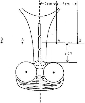
Traditionally, brachytherapy planning has relied on using orthogonal x-rays to calculate point doses, such as to point A (Figure 1) and the International Commission on Radiation Units and Measurements (ICRU) bladder and rectal points. The use of MRI images for patient-specific planning would allow a transition away from point doses to a volume-based analysis and reporting. The GEC-ESTRO recommendationsReference Haie-Meder, Pötter, Van Limbergen, Briot, De Brabandere, Dimopoulos, Dumas, Hellebust, Kirisits, Lang, Muschitz, Nevinson, Nulens, Petrow and Wachter-Gerstner15,Reference Pötter, Haie-Meder, Van Limbergen, Barillot, De Brabandere, Dimopoulos, Dumas, Erickson, Lang, Nulens, Petrow, Rownd and Kirisits16 suggest that areas identifiable on MRI as macroscopic disease, partially microscopic disease and tumour regression should be used to delineate target volumes at high or intermediate risk of local recurrence. These Intermediate-risk Clinical Target Volumes (IR-CTVs) or High-risk Clinical Target Volumes (HR-CTVs) can then be used to optimise plans to patient-specific disease characteristics.
The position of Point A, situated 2 cm superior of the vaginal ovoids and 2 cm lateral of the intrauterine sources. (Reproduced by kind permission of Elsevier from Meredith WJ (1967) ‘Radium Dosage: The Manchester System’ Second Edition. Edinburgh, E&S Livingstone Ltd, p. 48.)

Image registration and applicator reconstruction
Although no extra margins are needed for patient related uncertainties such as organ movement,Reference Haie-Meder, Pötter, Van Limbergen, Briot, De Brabandere, Dimopoulos, Dumas, Hellebust, Kirisits, Lang, Muschitz, Nevinson, Nulens, Petrow and Wachter-Gerstner15 the literature identifies a number of geometric uncertainties. Uncertainties are associated with geometric distortion from the MRI, but some studies have shown this can be limited to 2 mm in the area of treatment.Reference Anderson, Beenstock, Bryant and Chapman17 Another possible uncertainty is ‘reconstruction of the applicator’; this is the process whereby the geometry of the source channels and dwell positions are defined on three dimensional (3D) images to enable planning. Hack et al describes a method of applicator reconstruction for MRIReference Haack, Nielsen, Lindegaard, Gelineck and Tanderup18 via the visualisation of source channels using catheters containing copper sulphate.
Unlike when planning for EBR, electron density information is not necessary for brachytherapy planning; CT images are therefore not needed.Reference Huh, Park and Han19 However, applicator reconstruction using CT images followed by CT-MR fusion is still a widely reported method of planning in the literature.20,21,22,23 It has been shown that valuable information can be lost on CT in between slices and this is less likely to occur with MR reconstruction.Reference Hellebust, Tanderup, Bergstrand, Knutsen, Røislien and Olsen20 Although inter-observer variability due to image artefacts is evident in applicator reconstruction with MRI, the uncertainties in reconstruction can be minimised as part of a stringent quality assurance program,Reference Haack, Nielsen, Lindegaard, Gelineck and Tanderup18 and particular care must be taken when using conformal loading patternsReference Berger, Dimopoulos, Georg, Georg, Pötter and Kirisits24.
Patient specific planning for cervical brachytherapy: what is the evidence base?
The GEC-ESTRO recommendations for target volume definition and dose reporting have been adopted by the Royal College of Radiologists Working Party,4 and are widely used in the literature to describe both planning studies and clinical results.
Planning studies are often descriptive rather than comparative, and are commonly used to report the use of a new technique, giving a synopsis of the dosimetric results. However, some of these planning studies have compared new techniques against old institutional practices.Reference De Brabandere, Mousa, Nulens, Swinnen and Van Limbergen25,Reference Lindegaard, Tanderup, Nielsen, Haack and Gelineck26 Planning studies also may be limited as they have small sample sizes (between one and twenty two patients in the surveyed studies) and it is therefore debatable that the results are transferable to the general patient population. Nevertheless they do provide some useful indications for future work, and importantly identify some issues that can be followed up in future studies. One study identified strategies for treating large HR-CTV and OAR close to the GTV with interstitial needles and/or adapting source dwell times.Reference Kirisits, Lang, Dimopoulos, Berger, Georg and Pötter22 Another identified a subgroup of patients that were problematic to plan (severely antiflexed uterus), and subsequently developed two contrasting treatment plans that either optimised target coverage or improved organ sparingReference Duan, Kim, Elassal, Lin and Shen21.
Two papers were identified that directly compared standardised loading/prescription to point A against conformal loading using MRI images.Reference De Brabandere, Mousa, Nulens, Swinnen and Van Limbergen25,Reference Lindegaard, Tanderup, Nielsen, Haack and Gelineck26 Although both studies found that mean D90 (minimum dose delivered to 90% of the volume) to the GTV increased slightly when using MRI, mean rectal, bladder and small bowel D1cc (dose to 1cc of most irradiated tissue) and 2 cc results were equivocal. However, De Brabandere et al.Reference De Brabandere, Mousa, Nulens, Swinnen and Van Limbergen25 emphasised that the reporting of mean dose across a cohort hides the specific advantages that conformal loading brings to individual plans. For example, when altering a standardised plan, the planner would sometimes reduce a high D90 to the GTV in order to reduce dose to critical structures. Similarly, doses to critical structures were sometimes increased (within tolerance) to increase D90 coverage of the GTV.
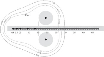
Both studies used natural modifications of existing techniques to allow MRI imaging and non-conformal loading. MRI compatible versions of traditional applicators (the ring, and tandem and ovoids, as used throughout UK centres4 were used and the ‘pear shape’ dose distribution was the common starting point for planning (Figure 2). The plans produced by three different types of applicator adapted for MRI (tandem and ovoids, individual moulds and Vienna ring) have been compared and have shown relative equivalence, with differences in dose distribution quality attributed to patient anatomy.Reference Langa, Nulensb and Briotc27 Additional modifications of traditional applicators have been developed, such as the insertion of interstitial needles through guide holes in the ring.Reference Kirisits, Lang, Dimopoulos, Berger, Georg and Pötter22 Even when dose distributions have been deliberately kept close to the classic ‘pear shape’, interstitial needles enabled larger GTVs to be treated to higher doses without increasing OAR doses.Reference Kirisits, Lang, Dimopoulos, Berger, Georg and Pötter22,Reference Lindegaard, Tanderup, Nielsen, Haack and Gelineck26
A cross-sectional view, through an intrauterine tube and vaginal ovoids, showing caesium pellet positions, and the resulting dose distribution. The pear shape dose distribution is representative of a Manchester System treatment, which uses a standardised pellet loading pattern.

Clinical Results
Reporting of clinical results of MRI guided planning for gynaecological brachytherapy are often limited to case series reports. A series from Vienna compares a cohort of consecutive patients treated at a single institution before and after a switch to MRI assisted planning.Reference Pötter, Dimopoulos, Georg, Lang, Waldhäusl, Wachter-Gerstner, Weitmann, Reinthaller, Knocke, Wachter and Kirisits10 Data suggests that the use of MRI guidance leads to both an overall reduction of toxicity and an increase in local control and survival in a subset of patients with tumours greater than 5 cm at implant.Reference Pötter, Dimopoulos, Georg, Lang, Waldhäusl, Wachter-Gerstner, Weitmann, Reinthaller, Knocke, Wachter and Kirisits10 The authors reported that interstitial needles allowed them to treat large tumours, which they would have normally treated with an EBR boost, but no comparison of outcomes between these two options is given. In Nayaran et al.Reference Narayan, van Dyk, Bernshaw, Rajasooriyar and Kondalsamy-Chennakesavan28 a survival difference was not observed after a switch from LDR to MRI-planned HDR, but a significant decrease in bowel and bladder symptoms is reported.
What are the possible alternatives to MR guided brachytherapy?
CT brachytherapy planning
In the UK, only one institution has reported on the use of 3D imaging with cervix brachytherapy.Reference Tan, Coles, Hart and Tait29 This series from Addenbrooke’s NHS Trust reported an increase in local control and decrease in complications using CT planning when compared to standard loading and prescribing to point A. The use of CT images rather than MRI was necessary due to the impracticality of scheduling MR scans to coincide with intracavitary insertions at that institution. In the UK in 2008, only 4% of centres had access to brachytherapy planning using MRI, and it is likely that many other centres will see CT planning as an alternative.4 Data suggests that CT planning leads to larger target volumes than MRI,Reference Viswanathan, Dimopoulos, Kirisits, Berger and Pötter23,Reference Tan, Coles, Hart and Tait29 and this series reports a cautious use of dose optimisation as a result, with V90 for patients limited by OAR tolerance.
The Addenbrooke’s series utilised a ring applicator, but does not use the additional capability of the interstitial needles, and it reported a lower increase in local control in tumours greater than 5 cm compared to the Vienna series. Addenbrooke’s relies more on giving an EBR boost to patients with bulkier tumours, and so the number of patients in their series with tumours greater than 5 cm was much lower than in the Vienna series.Reference Tan, Coles, Hart and Tait29 The use of a conventional EBR boost when brachytherapy is not possible is increasing at other UK centres, and good survival and complication rates can be seen using this technique.Reference Barraclough, Swindell, Livsey, Hunter and Davidson30
What is the feasibility of the MRI-guided EBR boost for cervix carcinoma?
A number of planning case studies have been undertaken to understand the possible role of an MR-guided IMRT boost in the treatment of cervix carcinoma. Dosimetric comparison between IMRT and brachytherapy is difficult due to the dose inhomogeneity inherent in brachytherapy, but there is evidence to suggest that IMRT plans struggle to provide comparable V90 and V95 doses without compromising normal tissue tolerances.Reference Georg, Kirisits, Hillbrand, Dimopoulos and Pötter31,Reference Assenholt, Petersen, Nielsen, Lindegaard and Tanderup32 One planning case study reported that conventionally loaded or optimised brachytherapy in combination with a concomitant IMRT boost was superior to optimised brachytherapy alone; this was particularly profound in cases where the uterus was severely antiflexed.Reference Duan, Kim, Elassal, Lin and Shen21
It must be noted that the above studies assume that as the CTV is immobilised using a stereotactic applicator, PTV margins are minimal. Implementation of IMRT after applicator insertion would require quick turnaround of planning and verification, which is logistically challenging.Reference Duan, Kim, Elassal, Lin and Shen21 Without stereotactic immobilisation, PTV margins surrounding the cervix and uterus are greatly affected by internal organ motion, and recent studies have tried to quantify this problem in cervical cancer using MRI.Reference Huh, Park and Han19,Reference van de Bunt, Jurgenliemk-Schulz, de Kort, Roesink, Terteeg and van der Heide33 These studies in particular have identified the influence of tumour regression and OAR filling has on the position of the GTV, and it is suggested that non-uniform margins and interfractional imaging are required if the aim is to achieve a reduction in the PTV margin to reduce OAR toxicity.
Implementing a change in treatment delivery for cervical cancer brachytherapy
A change from LDR to PDR or HDR machines requires a significant change in practice regardless of an institution’s decision to use MRI, as new dose and fractionation regimes would need to be determined to re-create the radiobiological effects of LDR treatment using the linear quadratic model.Reference Haie-Meder, Pötter, Van Limbergen, Briot, De Brabandere, Dimopoulos, Dumas, Hellebust, Kirisits, Lang, Muschitz, Nevinson, Nulens, Petrow and Wachter-Gerstner15,Reference Petereit, Sarkaria, Potter and Schink34 Although this model has been used previouslyReference Petereit, Sarkaria, Potter and Schink34 and is now well accepted within the radiotherapy community,3 it must be emphasised that if a large number of treatments are undertaken, any small inaccuracies in the model are more likely to have a detrimental clinical effect. So, although MR optimisation may have mitigatory role, clinical results must be closely monitored.
The introduction of MRI assisted planning introduces an additional step into a treatment process that is arguably simple and effective. The maintenance of expertise in brachytherapy has been recognised as a problem,35 and MRI planning will add to the challenge of educating and training practitioners as well as maintaining those skills. To date there has been no specific research that has explored staff training needs, or evaluated the extra resources needed to plan with MRI. In the literature, the time reported to produce a plan ranged from 20 to 40 minutes per insertion,Reference Dimopoulos, Kirisits and Petric36 with around 15–20 minutes allocated for patient contouring.Reference Tan, Coles, Hart and Tait29 HDR treatments may require multiple insertions, which could result in extra time needed for contouring. The use of interstitial needles would require the use of supplementary theatre time and the development of new skills; scheduling MRI guidance would also require additional co-ordination between infection control experts, nurse, anaesthetists, diagnostic radiographers and radiologists. CT planning would also require additional staff training, quality assurance procedures to be put in place and the appropriate scheduling of CT planning time. The limited availability of MRI to all brachytherapy service providers in the UK may require referral to neighbouring cancer centres with MRI facilities and available capacity for specific categories of patient.4
Although inverse planning solutions for brachytherapy have been discussed in the literature,Reference Dewitt, Hsu, Speight, Weinberg, Lessard and Pouliot2,Reference Chajon, Dumas, Touleimat, Magné, Coulot, Verstraet, Lefkopoulos and Haie-Meder5 the use of the ‘pear-shaped’ dose distribution is still widely accepted as an important starting point for optimisation, and severe deviations from it using inverse planning were not apparent in the literature. Therefore, manual optimisation may suffice, as no indication is given in the literature that inverse planning will reduce planning times. However, this could be a future area of exploration that would require the close collaboration between the oncologist, the dosimetrist and the rest of the planning and treatment delivery team, during the early stages of implementation.
CONCLUSION
Brachytherapy is an important part of the treatment of cervical cancer with radiotherapy, and the use of MRI in planning and delivering treatment allows the definition of macroscopic and partially microscopic disease even after initial EBR. HDR and PDR machines can simulate the current techniques, but they also facilitate the use of image guided brachytherapy planning using MRI. Uncertainties in applicator reconstruction and image fusion can be minimised by developing appropriate quality assurance systems, which address training and resource issues.
Planning studies show the advantages of using MRI to conform dose to the target volumes identified by the GEC-ESTRO guidelines. The majority of evidence favours the approach of MR imaging with patient specific loading compared to standardised loading and prescribing the use of the Manchester System. However, the reviewed clinical results for MRI dose optimisation were limited and further clinical trials will be needed to explore this further. Although optimising plans using CT imaging work on the same principles, the limited clinical evidence so far does suggest that MRI is superior for brachytherapy, especially in patients with larger tumours. Currently those patients with large tumours or patients that cannot have brachytherapy frequently have a fractionated EBR boost instead, which may be improved in the future from the use of IMRT and MRI-CT fusion for planning. Further studies into the definition of organ movement and PTV reduction may improve outcomes for this already well-established and clinically acceptable technique. Although the skill base in IMRT is already well established, planning case studies in the literature and current logistical difficulties with dose verification would suggest that IMRT boosts cannot be used as a direct alternative to MR brachytherapy.
The process of dose optimisation throughout the literature is still reliant upon the classic ‘pear-shape’ for a starting point. Dose constraints for volumes are as yet unproven in clinical trials, so this could be an area for further exploration. However, emphasis should still be placed on ensuring that good clinical outcomes are maintained.
Evidence suggests that there is no superiority in using a particular type of applicator and it would be therefore acceptable to continue using the current styles of applicator if a switch to MRI dose optimisation was to occur. Although no formal assessment of skills and time needed to implement a change to MR brachytherapy has been published, the use of interstitial needles, particularly in conjunction with MRI guidance, would require a new set of skills across professions, as well as additional time and new logistical solutions. Dedicated staff across disciplines should be appointed to co-ordinate learning during the introduction of MRI planning, and education and training should be offered along with the dissemination of knowledge to the wider professional team.35 These staff, dependent on their scope of practice could be at consultant/advanced practice level,35 and further role extension and development of radiographers could be utilised more, for example in vaginal vault insertions.35