Implications
Mortality is increased in low birth weight offspring, and surviving offspring often exhibit poor growth performance. Identifying dietary supplements that mitigate low birth weights in the sheep industry would improve food animal production. This research article describes maternal and fetal nutrient availability in ewes supplemented with dietary melatonin during a compromised pregnancy. In addition, supplementing feeds with melatonin during pregnancy is economically feasible due to the low amount of melatonin provided per day. The alteration in fetal nutrient availability following maternal melatonin supplementation implicates melatonin as a partial mediator of fetal development.
Introduction
Several animal models of fetal and placental growth restriction (e.g. maternal nutritional plane, maternal age, heat stress, hypoxic stress, and fetal number) have been developed to better unravel the relationship between uteroplacental blood flow, placental vascularity, and nutrient delivery to the fetus (Kwon et al., Reference Kwon, Ford, Bazer, Spencer, Nathanielsz, Nijland, Hess and Wu2004; Reynolds et al., Reference Reynolds, Borowicz, Vonnahme, Johnson, Grazul-Bilska, Redmer and Caton2005). In sheep, increasing uterine blood flow during the last half of gestation is vital for maintaining a continual delivery of sufficient oxygen and nutrients to the exponentially growing fetus (Ford, Reference Ford1995; Redmer et al., Reference Redmer, Wallace and Reynolds2004; Reynolds et al., Reference Reynolds, Caton, Redmer, Grazul-Bilska, Vonnahme, Borowicz, Luther, Wallace, Wu and Spencer2006). Maternal nutrient restriction causes aberrations in uterine blood flow and placental vascularity, which is highly associated with the magnitude of fetal growth restriction. In addition, compromised pregnancies exhibit a decrease in fetal plasma total α-AA concentrations and umbilical cord blood flow (Rigano et al., Reference Rigano, Bozzo, Ferrazzi, Belloti, Battaglia and Galan2001; Kwon et al., Reference Kwon, Ford, Bazer, Spencer, Nathanielsz, Nijland, Hess and Wu2004).
Melatonin supplementation has been associated with improved oxidative status and altered cardiovascular function (Paulis and Simko, Reference Paulis and Simko2007). Recently our laboratory observed an increase in umbilical artery blood flow in ewes supplemented with dietary melatonin (Lemley et al., Reference Lemley, Meyer, Camacho, Neville, Newman, Caton and Vonnahme2012). Therefore, supplementing melatonin may improve fetal development by increasing placental blood perfusion and/or uteroplacental nutrient transport capacity. Apart from characterizing uteroplacental hemodynamics during melatonin supplementation, a paucity of information exists on the relationship, if any, between uteroplacental nutrient transport and melatonin secretion. We hypothesized that dietary melatonin supplementation would increase uteroplacental nutrient flux during a compromised pregnancy. Using a mid- to late-gestation model of intrauterine growth restriction (IUGR), the objectives were to examine uterine, fetal, and uteroplacental flux of glucose and AA during dietary melatonin supplementation.
Material and methods
Animal care and use were according to protocols approved by the North Dakota State University Institutional Animal Care and Use Committee #A10071.
Animals and experimental design
The animal management, breeding, and experimental design were previously published (Lemley et al., Reference Lemley, Meyer, Camacho, Neville, Newman, Caton and Vonnahme2012). Briefly, nulliparous western whiteface ewe lambs were transported to the Animal Nutrition and Physiology Center (ANPC; Fargo, ND, USA) in September of 2010 and fed to meet nutrient requirements during early gestation. Initially 64 ewe lambs were placed on pasture with ad libitum access to hay and water with two rams of proven fertility fitted with crayon marking harnesses. Mating was recorded every 12 h. Breeding dates were intentionally distributed over a large range to decrease the number of dams receiving surgery at day 130 of gestation. These breeding dates ranged from 24 September 2010 to 2 December 2010. At day 28 of gestation, ewes were moved from pasture into a temperature-controlled facility (14°C; ANPC) with a 12 : 12 light: dark cycle with lights on at 0700 h and off at 1900 h for the remainder of the study. Singleton pregnant ewes were identified via ultrasonography between day 28 and 35 of gestation and all dams were stratified by maternal weight and breeding date. On day 45 of gestation, singleton pregnant ewes were acclimated to a common pelleted diet (Lemley et al., Reference Lemley, Meyer, Camacho, Neville, Newman, Caton and Vonnahme2012; CP of 15.8%, ME of 2.66 Mcal/kg), provided trace mineral salt blocks (American Stockman, Overland Park, KS, USA), and ad libitum access to water. Melatonin and nutritional plane treatments began on day 50 of gestation. Ewes were supplemented with 5 mg/day of melatonin (MEL; Spectrum Chemical Mfg. Corp., Gardena, CA, USA) or no melatonin (CON) and provided 100% (ADQ; adequate diet) or 60% (RES; restricted diet) of nutrient recommendations for the remainder of the experiment (2 × 2 factorial design). A total of 32 singleton pregnant ewes were selected for the study; however, one ewe was removed due to a perforated esophagus, which existed before dietary treatment. This resulted in four treatment groups consisting of CON-ADQ (n = 7), MEL-ADQ (n = 8), CON-RES (n = 8), or MEL-RES (n = 8). Nutritional requirements were based on NRC (2007) recommendations for a 60-kg body weight pregnant ewe lamb during mid- to late-gestation so that adequate ewes were fed to receive adequate nutrients and energy for maintenance, maternal growth, and fetal growth. This mid- to late-gestation maternal undernutrition model has been previously described in our laboratories (Meyer et al., Reference Meyer, Reed, Neville, Taylor, Hammer, Reynolds, Redmer, Vonnahme and Caton2010; Lemley et al., Reference Lemley, Meyer, Camacho, Neville, Newman, Caton and Vonnahme2012). All ewes were fed and/or supplemented with melatonin-enriched pellets at 1400 h (5 h before the start of the dark cycle at 1900 h). Melatonin powder was dissolved in 95% ethanol at a concentration of 5 mg/ml. The day before feeding, a subset of the control pellet (100 g) was placed into a small plastic bag and top dressed with 1 ml of melatonin solution. The ethanol was left to evaporate overnight at room temperature with no exposure to light. All MEL ewes consumed treatments in ∼5 min. The remainder of control pellet was given to all groups shortly after consumption of melatonin-treated pellets. Description of peripheral serum concentrations of melatonin, maternal body condition, fetal weight, and fetal organ development were described previously (Lemley et al., Reference Lemley, Meyer, Camacho, Neville, Newman, Caton and Vonnahme2012).
Intraoperative measurements and blood sampling
On day 130 of gestation, ewes were weighed and anesthesia was induced with 3 mg/kg of body weight sodium pentobarbital (Fort Dodge Animal Health, Overland Park, KS, USA) at 0800 h. Anesthesia was maintained by intermittent infusion of sodium pentobarbital via a jugular catheter. A catheter was placed into the maternal saphenous artery and advanced to the iliac artery via the femoral artery. The uterus was exposed via a midventral laparotomy, covered with warm surgical towels, and liberal amounts of saline (37°C) were applied to the uterus every 5 min. Uterine and umbilical blood flow were assessed using a duplex B-mode (brightness mode) and D-mode (Doppler spectrum) program of the color Doppler ultrasound instrument (Aloka SSD-3500; Aloka America, Wallingford, CT, USA) fitted with a 7.5 MHz finger transducer (Aloka UST-995) using our previously published techniques (Lemley et al., Reference Lemley, Meyer, Camacho, Neville, Newman, Caton and Vonnahme2012).
Following measurements of uteroplacental blood flow, concurrent blood samples were collected from the catheterized maternal saphenous artery and the gravid uterine vein. Approximately 30 ml of blood was collected using a 10 ml syringe (BD Diagnostics, Franklin Lakes, NJ, USA) attached to a three-way stopcock. Immediately following collection of maternal blood, the gravid uterine horn was dissected. The umbilical cord was located and concurrent blood samples were collected from the umbilical vein and umbilical artery. Similar to the maternal blood samples, ∼30 ml of umbilical blood was collected using a 10 ml syringe (BD Diagnostics) attached to a three-way stopcock with a 20 G needle. Blood was equally divided between BD vacutainer serum tubes, placed on ice, allowed to clot and later spun at 2000 × g for 20 min. Serum was stored at −20°C until further analysis for glucose and AA profiles. The fetus was removed while under general anesthesia of sodium pentobarbital, which crosses the maternal fetal circulation through the placenta. Fetal liver was removed, weighed, snap frozen in supercooled isopentane (submerged in liquid nitrogen) and stored at −80°C.
Amino acid (AA) analysis
Serum AA profiles were determined using an Ultra Performance Liquid Chromatograph (UPLC). Two-hundred fifty microliters of serum was deproteinized with 250 μl of 10% sulfosalicylic acid to which 250 μM norvaline was added as an internal standard. This mixture was vortexed and centrifuged for 5 min at 16 000 × g. Twenty microliters of supernatant was added to 60 μl of borate buffer and sodium hydroxide solution as well as 20 μl of MassTrac Amino Acid Analysis derivating reagent. The samples were then capped, mixed, and heated in a digestion block at 55°C for 10 min. Samples were then injected into the UPLC. This method utilizes the MassTrac Amino Acid Analysis system for the full profile of AA in physiological fluids. Derivatization chemistry for physiological samples is a precolumn method and is based on a derivatizing reagent, 6-aminoquinolyl-N-hydroxysuccinimidyl carbamate, which converts both primary and secondary AA to stable chromophores for UPLC detection.
Blood glucose and liver glycogen analysis
Maternal and fetal serum glucose concentrations and liver glycogen content were determined using a colometric assay, following Lekatz et al. (Reference Lekatz, Ward, Borowicz, Taylor, Redmer, Grazul-Bilska, Reynolds, Caton and Vonnahme2010, Reference Lekatz, Wu, Caton, Taylor, Reynolds, Redmer and Vonnahme2011). For serum glucose concentrations the intraassay and interassay CV were 4.6% and 5.4%, respectively. For liver glycogen content the intraassay and interassay CV were 7.9% and 8.8%, respectively. Total fetal liver glycogen was calculated by multiplying fetal liver glycogen concentration by liver weight.
Uteroplacental nutrient flux calculations
Evaluation of transplacental exchange in vivo was determined using equations from Reynolds and Redmer (Reference Reynolds and Redmer1995), which are based on the Fick principal. Gravid uterine flux of nutrients is equal to uterine blood flow multiplied by the maternal arterial − venous concentration difference (maternal saphenous artery − gravid uterine vein) of any given substance. Fetal flux of nutrients is equal to umbilical blood flow multiplied by the fetal venous − arterial concentration difference (umbilical vein − umbilical artery) of any given substance. Uteroplacental flux of nutrients is calculated as the difference between gravid uterine flux (which consists of the uteroplacental and fetal compartments) and fetal flux. Moreover, positive flux of nutrients represents tissue uptake, and negative flux of nutrients represents tissue release (Reynolds and Redmer, Reference Reynolds and Redmer1995).
Statistical analysis
Blood flow was determined and serum samples collected in all 31 ewes and their respective fetuses (n = 31) consisting of the following experimental units for each treatment group: CON-ADQ (n = 7), MEL-ADQ (n = 8), CON-RES (n = 8), or MEL-RES (n = 8). However, AA analysis from the maternal artery was not determined in three of the ewes, which resulted in the following experimental units for maternal artery AA concentrations, maternal arterial − venous difference, uterine flux, and uteroplacental flux: CON-ADQ (n = 7), MEL-ADQ (n = 7), CON-RES (n = 7), or MEL-RES (n = 7). The effects of diet on dependent variables were tested with the MIXED procedure of SAS (SAS software version 9.2, SAS Institute Inc., Cary, NC, USA). The model statement included: melatonin treatment, nutritional plane, fetal sex, and melatonin treatment × nutritional plane interaction. Fetal sex was removed from the model statement if P > 0.25 was observed. For each treatment group male and female fetuses were distributed as: CON-ADQ (n = 3, n = 4), MEL-ADQ (n = 2, n = 6), CON-RES (n = 5, n = 3), and MEL-RES (n = 6, n = 2), respectively. Means were separated for melatonin treatment, nutritional plane and melatonin treatment × nutritional plane interaction using the PDIFF option of the LSMEANS statement. For each dependent variable homogeneity v. heterogeneity of variance was tested in the MIXED procedure and the best-fit model was selected based on Bayesian Information Criteria. Breeding date was used as a covariant due to the range of breeding dates from early September (13.5 h of daylight) to late December (8 h of daylight); however breeding date was removed from the model if P > 0.25 was observed. Least square means and s.e. are reported. Statistical significance was declared at P ⩽ 0.05. Main effects are discussed in the absence of a significant interaction (P > 0.06).
Results
Total α-AA concentrations and uteroplacental uptake
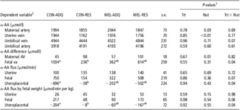
Total α-AA concentrations and uteroplacental flux of total α-AA at day 130 of gestation are illustrated in Table 1. Total α-AA in maternal artery were decreased (P < 0.05) by 8% in RES v. ADQ fed ewes (1851 v. 2019 ± 52 μmol/l). Similarly, total α-AA in the gravid uterine vein were decreased (P < 0.01) by 10% in RES v. ADQ fed ewes (1759 v. 1960 ± 48 μmol/l). Umbilical vein and umbilical artery total α-AA concentrations were not different across all treatment groups (P > 0.06). Maternal arterial − venous difference in total α-AA showed a main effect of nutritional plane (P < 0.05), where RES ewes had a 96% increase in arterial − venous concentration difference v. ADQ fed ewes (100 v. 51 ± 13 μmol/l). In contrast, uterine uptake of total α-AA was not different (P > 0.40) across all treatment groups. There was a melatonin treatment × nutritional plane interaction (P < 0.05) in fetal venous − arterial difference of total α-AA, where concentration differences in total α-AA were decreased in fetuses from CON-RES v. CON-ADQ ewes, while melatonin supplementation in nutrient restricted ewes (MEL-RES) increased total α-AA to the level of CON-ADQ ewes. Fetal uptake of total α-AA was not different across all treatment groups (P ⩾ 0.07). Uteroplacental release of total α-AA showed a melatonin treatment × nutritional plane interaction (P < 0.05), where total α-AA uteroplacental release was decreased in CON-RES v. CON-ADQ ewes, while melatonin supplemented ewes were intermediate. Similar results were observed when uterine, fetal, and uteroplacental fluxes of total α-AA were analyzed relative to fetal weight (Table 1).
Total α-AA concentrations, maternal AV difference, fetal va difference, uterine flux, fetal flux, and uteroplacental flux at day 130 of gestation from ewes supplemented with 5 mg of MEL or CON and provided 100% (ADQ diet) or 60% (RES diet) of nutrient recommendations beginning on day 50 of gestation

AV = arterial-venous; va = venous-arterial; MEL = melatonin; CON = no melatonin; ADQ = adequate; RES = restricted; AA = amino acids.
a,bLeast square means with different letter superscripts depict differences (P ⩽ 0.05).
1Main effect of melatonin supplementation (Trt), main effect of nutritional plane (Nut), or the interaction between melatonin supplementation × nutritional plane (Trt × Nut). Least square means and s.e.m. for main effects are reported in the text.
2Fetal sex had no influence on any of the dependent variables for total α-AA (P > 0.25).
Branched-chain AA (BCAA) concentrations and uteroplacental uptake
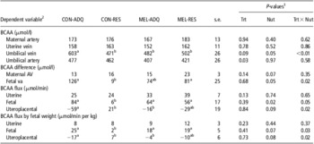
Total BCAA concentrations and uteroplacental flux of BCAA at day 130 of gestation are illustrated in Table 2. BCAA concentrations in the maternal artery and gravid uterine vein were not different (P < 0.40) across all treatment groups. Umbilical vein concentrations of BCAA showed a melatonin treatment × nutritional plane interaction (P < 0.01), where CON-RES, MEL-ADQ, and MEL-RES ewes had decreased BCAA compared with CON-ADQ. In addition, umbilical artery concentrations of BCAA showed a main effect of melatonin treatment (P < 0.05), where BCAA were decreased in the umbilical artery of MEL v. CON ewes (414 v. 469 ± 18 μmol/l). Maternal arterial − venous difference in BCAA and uterine flux of BCAA was not different (P > 0.06) across all treatment groups. There was a melatonin treatment × nutritional plane interaction (P < 0.05) in fetal venous − arterial difference of BCAA, where concentration differences in BCAA were decreased in fetuses from CON-RES v. CON-ADQ ewes, while melatonin supplementation in nutrient restricted ewes (MEL-RES) increased BCAA to the level of CON-ADQ ewes. There was a melatonin treatment × nutritional plane interaction (P = 0.05) for fetal uptake of BCAA, which was decreased in CON-RES v. all other treatment groups. Uteroplacenta flux of BCAA showed a melatonin treatment × nutritional plane interaction (P < 0.05), where BCAA were released from the uteroplacental of CON-ADQ ewes and taken up by the uteroplacental of CON-RES ewes, while MEL-RES ewes were intermediate. Similar results were observed when uterine, fetal, and uteroplacental fluxes of BCAA were analyzed relative to fetal weight.
BCAA concentrations, maternal AV difference, fetal va difference, uterine flux, fetal flux, and uteroplacental flux at day 130 of gestation from ewes supplemented with 5 mg of MEL or CON and provided 100% (ADQ diet) or 60% (RES diet) of nutrient recommendations beginning on day 50 of gestation

BCAA = branched-chain AA; AV = arterial-venous; va = venous-arterial; MEL = melatonin; CON = no melatonin; ADQ = adequate; RES = restricted.
a,bLeast square means with different letter superscripts depict differences (P ⩽ 0.05).
1Main effect of melatonin supplementation (Trt), main effect of nutritional plane (Nut), or the interaction between melatonin supplementation × nutritional plane (Trt × Nut). Least square means and s.e.m. for main effects are reported in the text.
2Fetal sex had no influence on the dependent variables for BCAA in umbilical vein, maternal AV difference, fetal va difference, fetal flux, and fetal flux by fetal weight (P > 0.25). Fetal sex was included in the model statement for maternal artery, uterine vein, umbilical artery, uterine flux, uteroplacental flux, uterine flux by fetal weight, and uteroplacental flux by fetal weight (P < 0.25).
Individual AA data
Maternal and fetal concentrations of individual AA at day 130 of gestation are illustrated in Supplementary Table S1 through Supplementary Table S4. Maternal arterial − venous difference and fetal venous − arterial difference of individual AA are illustrated in Supplementary Table S5 and Supplementary Table S6, respectively. Uterine, fetal, and uteroplacental flux of individual AA are illustrated in Supplementary Table S7 through Supplementary Table S9.
Glucose concentrations and uteroplacental uptake
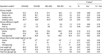
Glucose concentrations and uteroplacental utilization of glucose at day 130 of gestation are illustrated in Table 3. There was no effect (P ⩾ 0.09) of treatment on glucose concentrations in either the maternal artery or uterine vein. Umbilical vein concentrations of glucose were decreased (P < 0.01) in fetuses from RES v. ADQ fed ewes (37.4 v. 50.6 ± 3.0 mg/dl). Conversely, there was a melatonin treatment × nutritional plane interaction (P < 0.05) on umbilical artery glucose concentration, where fetuses from MEL-RES ewes had decreased concentrations compared with all other treatment groups. Maternal arterial − venous difference of glucose was not different (P ⩾ 0.06) across all treatments, while fetal venous − arterial difference was decreased (P = 0.05) in RES v. ADQ fed ewes (2.7 v. 7.9 ± 1.7 mg/dl). Uterine uptake of glucose was not different (P ⩾ 0.12) across all treatment groups. Fetal uptake of glucose was decreased in RES v. ADQ fed ewes (6.3 v. 72.7 ± 15.0 mg/min), while uteroplacental uptake of glucose was increased (P ⩽ 0.05) in RES v. ADQ fed ewes (107.2 v. 22.2 ± 28.3 mg/min). Uterine uptake of glucose relative to fetal weight showed a melatonin treatment × nutritional plane interaction (P = 0.05), where MEL-RES had an increased uptake compared with all other treatment groups. Fetal uptake of glucose relative to fetal weight was decreased in RES v. ADQ fed ewes (1.8 v. 21.9 ± 4.8 mg/min per kg). Uteroplacental uptake of glucose relative to fetal weight was increased in RES v. ADQ fed ewes (34.7 v. 10.9 ± 8.3 mg/min per kg). Fetal concentration of liver glycogen was similar across all treatment groups, while total liver glycogen was decreased (P = 0.05) in fetuses from RES v. ADQ fed ewes (2.8 v. 4.0 ± 0.3 g). This was primarily due to a decrease (P < 0.01) in absolute liver weight in fetuses from RES v. ADQ fed dams (Lemley et al., Reference Lemley, Meyer, Camacho, Neville, Newman, Caton and Vonnahme2012).
Glucose concentrations, maternal AV difference, fetal va difference, uterine uptake, fetal uptake, and uteroplacental uptake of glucose and fetal concentrations of liver glycogen and total liver glycogen at day 130 of gestation from ewes supplemented with 5 mg of MEL or CON and provided 100% (ADQ diet) or 60% (RES diet) of nutrient recommendations beginning on day 50 of gestation

AV = arterial-venous; va = venous-arterial; MEL = melatonin; CON = no melatonin; ADQ = adequate; RES = restricted.
a,bLeast square means with different letter superscripts depict differences (P ⩽ 0.05).
1Main effect of melatonin supplementation (Trt), main effect of nutritional plane (Nut), or the interaction between melatonin supplementation × nutritional plane (Trt × Nut). Least square means and s.e.m. for main effects are reported in the text.
2Fetal sex had no influence on the dependent variables for glucose in the uterine vein, umbilical vein, umbilical artery, fetal va difference, uterine flux, and fetal flux by fetal weight (P > 0.25). Fetal sex was included in the model statement for maternal artery, maternal AV difference, uterine flux, uterine flux by fetal weight, and uteroplacental flux by fetal weight (P < 0.20).
3Absolute fetal liver weight taken from Lemley et al. (Reference Lemley, Meyer, Camacho, Neville, Newman, Caton and Vonnahme2012).
Discussion
Previous data from our laboratory has shown that maternal body weight, body condition, loin muscle area, and internal fat mass were decreased in our model of IUGR, while no main effects or interactions of melatonin supplementation were observed for variables pertaining to maternal body weight or condition throughout gestation (Lemley et al., Reference Lemley, Meyer, Camacho, Neville, Newman, Caton and Vonnahme2012). We previously reported a decrease in fetal weight at day 130 of gestation in RES v. ADQ fed ewes. Moreover, a tendency (P < 0.07) for a melatonin treatment × nutritional plane interaction showed a greater difference in fetal weight among MEL ewes v. CON ewes, which was not due to a difference in fetal weight between CON-RES and MEL-RES ewes, but rather an increase in fetal weight between MEL-ADQ v. CON-ADQ ewes. Therefore, the current melatonin supplementation model did not rescue fetal growth restriction during maternal nutrient restriction, but rather increased fetal growth in ADQ fed ewes. In addition, we observed an increase in abdominal girth and ponderal index in fetuses from MEL-ADQ ewes v. all other treatment groups (Lemley et al., Reference Lemley, Meyer, Camacho, Neville, Newman, Caton and Vonnahme2012). Therefore, melatonin supplementation during gestation may be a beneficial model of developmental programming for animals born with disproportionate body growth.
Discussions on uteroplacental and fetal uptake of AA can be primarily divided into two areas of interest focusing on either metabolic pathways or transport systems (Battaglia and Regnault, Reference Battaglia and Regnault2001; Jansson, Reference Jansson2001; Fowden et al., Reference Fowden, Ward, Wooding, Forhead and Constancia2006). From a metabolic standpoint, previous research has shown consistent uterine and fetal uptakes of essential AA (Chung et al., Reference Chung, Teng, Timmerman, Meschia and Battaglia1998). Contradictory to previous research we observed an increased uptake for the majority of AA in the fetus compared with the uterus, which resulted in many AA being released from the uteroplacenta. These differences compared with the previous literature may be due to the timing of sampling in relation to feeding (18 h post-feeding), or stress during the surgery and anesthesia procedures, which could have led to an increased placental release of essential AA due to protein breakdown, or both. It is important to note that in the current study all animals underwent the same procedures, so therefore the majority of the discussion will focus on treatment differences v. direct comparisons to previously published ovine placenta AA characterization studies.
In the current data set maternal arterial − venous differences in total α-AA was dramatically increased (96%) in RES v. ADQ fed ewes; however, uterine flux of total α-AA was similar across nutritional plane. The lack of a nutritional plane effect on uterine total α-AA flux is due partly to the previously observed decrease in uterine artery blood flow (∼20%) in RES v. ADQ fed ewes (Lemley et al., Reference Lemley, Meyer, Camacho, Neville, Newman, Caton and Vonnahme2012). This relationship highlights the importance of both sufficient AA transport and sufficient uterine blood perfusion during late gestation. Moreover, nutrient-restricted ewes, which showed a decrease in total α-AA concentrations in both the maternal artery and gravid uterine vein during late gestation, may have compensated by utilizing and/or extracting a greater proportion of AA as measured by the increased maternal arterial − venous difference in RES v. ADQ fed ewes. However, this compensation in AA uterine uptake did not rescue fetal weight in our maternal nutrient restriction model (Lemley et al., Reference Lemley, Meyer, Camacho, Neville, Newman, Caton and Vonnahme2012).
BCAA have been shown to regulate mammalian target of rapamycin (mTOR), which has been implicated as a nutritional sensor, that regulates cell growth and protein synthesis via rates of gene transcription and mRNA translation (Lynch et al., Reference Lynch, Halle, Fujii, Vary, Wallin, Damuni and Hutson2003; Wullschleger et al., Reference Wullschleger, Loewith and Hall2006). Moreover, supplementation of BCAA to dams on a protein deficient diet can restore fetal growth and minimize the decreases in fetal organ mass and carcass fat, which is associated with increased mTOR signaling (Teodoro et al., Reference Teodoro, Vianna, Torres-Leal, Pantaleao, Matos-Neto, Donato and Tirapegui2012). For the current study, a decreased fetal uptake of BCAA was observed in fetuses from nutrient restricted ewes, while maternal melatonin supplementation improved BCAA uptake. This increased fetal uptake cannot be entirely dependent upon maternal blood concentrations of BCAA, which were not different in maternal artery or gravid uterine vein. Therefore, placental nutrient transport capacity of BCAA, accomplished primarily via sodium independent transporter system I (Battaglia and Regnault, Reference Battaglia and Regnault2001; Jansson, Reference Jansson2001), may be decreased during maternal nutrient restriction and rescued by melatonin supplementation. This would account for the dramatic differences in maternal and fetal concentrations of BCAA across our treatment groups. This alteration in AA release across the placenta due to melatonin supplementation may be independent of direct melatonergic pathways and could be related to the antioxidant properties of melatonin (Poeggeler et al., Reference Poeggeler, Reiter, Tan, Chen and Manchester1993; Juaniaux et al., Reference Juaniaux, Poston and Burton2006; Reiter et al., Reference Reiter, Melchiorri, Sewerynik, Poeggeler, Barlow-Walden, Chuang, Ortiz and AcunaCastroviejo2007), although further investigation is needed in this area of research.
In the current study, maternal nutrient restriction decreased umbilical vein glucose concentrations, with no effect on maternal glucose concentrations. In addition, we observed a limited glucose concentration gradient between maternal v. fetal circulation. This alteration in umbilical vein glucose concentrations may depict a decrease in placental glucose transporter density. Although we observed similar uterine uptake of glucose across treatment groups, uteroplacental uptake was increased in RES v. ADQ fed ewes. Several models of IUGR brought about by placental insufficiency or decreased placental mass, such as heat stress and maternal overnutrition, have decreased glucose uptake in IUGR v. control dams (Wallace et al., Reference Wallace, Bourke, Aitken, Leitch and Hay2002; Limesand et al., Reference Limesand, Rozance, Smith and Hay2007). In the hyperthermia ovine model, reduced glucose transport capacity occurs due to a smaller placenta size and reduced concentrations of glucose transporters per gram of placenta (Thureen et al., Reference Thureen, Trembler, Meschia, Makowski and Wilkening1992; Limesand et al., Reference Limesand, Rozance, Smith and Hay2007). In the overnutrition ovine model, reduced glucose transport capacity occurs primarily due to a decreased placental surface area and placental size, whereas glucose transporter density remains similar across treatment groups (Hay, Reference Hay2006; Wallace et al., Reference Wallace, Luther, Milne, Aitken, Redmer, Reynolds and Hay2006). Similar to our maternal nutrient restriction model, which decreased fetal weight with no change to placental weight, Leury et al. (Reference Leury, Bird, Chandler and Bells1990) observed a decrease in both fetal and uteroplacental glucose uptake. In the current study, melatonin supplementation and maternal nutrient restriction did not alter placental mass at day 130 of gestation (Lemley et al., Reference Lemley, Meyer, Camacho, Neville, Newman, Caton and Vonnahme2012); therefore, changes in umbilical vein glucose concentrations may be dependent on placental glucose transporter density. However we cannot rule out the possibility of altered placental surface area within the placentome, as this was not measured.
In conclusion, maternal nutrient restriction from mid- to late-gestation altered maternal AA concentrations and uterine flux of AA. Dietary melatonin supplementation did not alter maternal artery, uterine vein, or uterine flux of AA; however, dietary melatonin did alter fetal venous − arterial differences of total α-AA as well as fetal and uteroplacental flux of BCAA. Similar maternal-fetal results were observed in our previously published report on the effects of dietary melatonin supplementation on uteroplacental hemodynamics, whereby maternal nutrient restriction decreased uterine artery blood flow and melatonin supplementation increased umbilical artery blood flow. Therefore, melatonin supplementation may improve umbilical hemodynamics and umbilical vein concentrations of AA even in the presence of increased uterine artery resistance and decreased uterine blood flow. Future studies will address the relationship between melatonin amplitude and placental nutrient transporter density.
Acknowledgments
The authors acknowledge support from USDA Postdoctoral Fellowship grant no. 2011-67012-30683 from the National Institute of Food and Agriculture to C.O. Lemley. The authors would like to thank the employees of the NDSU Animal Nutrition and Physiology Center.