Implications
The results of this study show that the feeding of an ovine serum immunoglobulin (Ig) influenced gut mucins in the growing rat as evidenced by increased mucin gene expression and digesta mucin protein concentrations as well as an increased goblet cell count. This is the first report regarding the effects of serum Igs on mammalian mucin secretion. Supplementation of diets with serum Ig may have a role in supporting the gut mucus barrier of animals in times of stress, such as post-weaning.
Introduction
The mucosal surface of the gastrointestinal tract (GIT) is made up of a complex organization of epithelium, immune cells and resident microflora (McCracken and Lorenz, Reference McCracken and Lorenz2001). Gut epithelial cells are continually exposed to numerous macromolecules including chemical irritants, digested foods, toxins, resident bacteria and intestinal pathogens and their products (Moncada and Chadee, Reference Moncada and Chadee2002). The mucus coat separates and defends gastric and intestinal cells from the exterior environment, allows lubrication of the cell surface and regulates ion fluxes (Forstner and Forstner, Reference Forstner and Forstner1994). Mucin-producing goblet cells along with antimicrobial peptides, luminal immunoglobulins (Ig), lysozymes and resident microflora collectively contribute to innate immunity and provide the front line of defence against pathogens. A well-established function of mucins is their role in preventing entry of enteric pathogens via the gut tissue. As mucins play an important role in protecting the underlying epithelium, any quantitative alteration in mucus secretion may change this defensive barrier and have important physiological implications (Linden et al., Reference Linden, Sutton, Karlsson, Korolik and McGuckin2008). Gastrointestinal diseases and intestinal stressors modify the mucus layer both quantitatively and qualitatively (Corfield et al., Reference Corfield, Myerscough, Longman, Sylvester, Arul and Pignatelli2000). Crohn's disease, colitis and gastric carcinogenesis associated with Helicobacter pylori alter mucin secretion and reduce the number of goblet cells that can lead to degradation of the mucus layer (Corfield et al., Reference Corfield, Myerscough, Longman, Sylvester, Arul and Pignatelli2000; Babu et al., Reference Babu, Jayanthi, Devaraj, Reis and Devaraj2006).
Orally administered Ig present in spray-dried animal plasma (SDAP) have been shown to confer multiple benefits in terms of health and growth in farm animals (Gatnau et al., Reference Gatnau, Paul and Zimmerman1989; Pierce et al., Reference Pierce, Cromwell, Lindemann, Russell and Weaver2005). The mucus layer is affected by numerous dietary components and the reported beneficial effects of dietary Ig on gut health may be due, at least in part, to effects on the mucus layer (Claustre et al., Reference Claustre, Toumi, Trompette, Jourdan, Guignard, Chayvialle and Plaisancie2002; Dock-Nascimento et al., Reference Dock-Nascimento, Junqueira and Aguilar-Nascimento2007; Han et al., Reference Han, Deglaire, Sengupta and Moughan2008; King et al., Reference King, Morel, Pluske and Hendriks2008). Recently, we have reported that feeding a freeze-dried ovine serum Ig preparation selectively improves growth performance, the weight of some digestive organs and gut morphology in the growing rat (Balan et al., Reference Balan, Han, Rutherfurd, Singh and Moughan2009). Dietary Ig preserved a higher degree of immunological activity and selectively altered the composition of the gut microbiota (upregulation of Lactobacillus species) in normal and in Salmonella enteritidis challenged growing rats (Balan et al., Reference Balan, Han, Rutherfurd-Markwick, Singh and Moughan2010, Reference Balan, Han, Rutherfurd-Markwick, Singh and Moughan2011a and Reference Balan, Han, Rutherfurd, Singh and Moughan2011b). There are no reports in the literature, however, regarding the effects of ovine Ig on gut mucins in animals. Therefore, the objective was to investigate whether orally administered ovine serum Ig affects gut mucin in the growing rat. Comparison of goblet cell count, mucin gene expression and digesta mucin concentrations was made between a casein-based control diet (CON) and a similar test diet containing freeze-dried ovine Ig (FDOI).
Material and methods
Preparation and quantitation of ovine serum Ig
An ovine serum Ig fraction was prepared according to the procedures described in an earlier study, using an ammonium sulphate precipitation method (Balan et al., Reference Balan, Han, Rutherfurd, Singh and Moughan2009). The IgG content in the ovine Ig fraction was determined in quadruplicate by direct ELISA using a rabbit anti sheep IgG (AbD Serotec, Kidlington, Oxford, UK) as described by Balan et al. (Reference Balan, Han, Rutherfurd, Singh and Moughan2009). The IgA and IgM contents in the ovine Ig fraction were determined as for IgG but using rabbit anti sheep IgA and rabbit anti sheep IgM (AbD Serotec). The respective protein, IgG, IgA and IgM concentrations of the FDOI were 94.0%,73.0%, 15% and 5%. There was no residual ammonium sulphate present in the ovine Ig fraction.
Animal study
This study was approved by the Massey University Animal Ethics Committee (MUAEC 06/132) and procedures complied with the New Zealand Code of Recommendations and Minimum Standards for the Care and Use of Animals for Scientific purposes (New Zealand Animal Welfare Advisory Committee, 1995). Fourteen Sprague–Dawley male rats (140 to 160 g body weight) were housed singly in wire-bottomed stainless steel cages and had free access to water in a room maintained at 22 ± 2°C with a 12 h light/dark cycle.
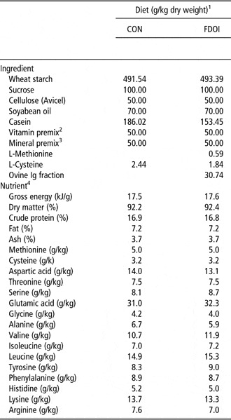
The rats were given 1 week to acclimatize to their surroundings during which time they consumed a basal diet ad libitum. After acclimatization, the rats were randomly allocated to two diets (Table 1, n = 7 per diet) including a control diet (CON) and a test diet (FDOI) and underwent a 3-week study.
Ingredient composition and determined nutrient and energy contents of the CON and FDOI diets

CON = control diet; FDOI = freeze-dried ovine immunoglobulin; Ig = immunoglobulin.
1All diets were formulated to meet or exceed the nutrient requirements of the growing rat.
2The mixture supplied – mg/kg diet: retinol acetate 5.0, dl-α-tocopheryl acetate 200, menadione 3.0, thiamin hydrochloride 5.0, riboflavin 7.0, pyridoxine hydrochloride 8.0, d-pantothenic acid 20, folic acid 2.0, nicotinic acid 20, d-biotin 1.0, myo-inositol 200, choline chloride 1500; μg/kg diet: ergocalciferol 25, cyanocobalamin 50.
3The mixture supplied – g/kg diet: calcium hydrogen phosphate 6.29, magnesium oxide 1.06, potassium chloride 5.24, sodium chloride 1.97; mg/kg diet: chromic potassium sulphate 1.97, Cupric carbonate 10.7, ferric citrate 424, manganese sulphate 78.0, zinc oxide 48.2; μg/kg diet: cobalt chloride 29.0, potassium iodate 151, ammonium molybdate152, sodium selenite 151.
4values for nutrients were determined and are given on an ‘as-fed’ basis. Methionine and cysteine were calculated values.
The estimated respective IgG and Ig concentrations in the FDOI diet were 2.2% and 2.9% (as-fed).
The diets were formulated (with methionine plus cysteine as the first limiting amino acid (AA)) to meet or exceed National Research Council (NRC, 1995) recommendations for the growing rat for the major nutrients (Table 1). The FDOI fraction was included in the FDOI diet at 3.07%. The resultant amount of ovine Ig in the FDOI diet was equal to the amount of porcine IgG contributed by spray-dried porcine plasma diets used in other studies (Pierce et al., Reference Pierce, Cromwell, Lindemann, Russell and Weaver2005; Balan et al., Reference Balan, Han, Rutherfurd, Singh and Moughan2009). Food was given in a powdered form ad libitum.
Chemical analysis
Dietary dry matter, ash, crude protein, total fat and gross energy were determined according to methods described by the Association of Official Analytical Chemists (AOAC, 2003). The AA contents of the diets were determined in duplicate after hydrolysis (6 mol/l HCl for 24 h at 110°C) and using ion exchange HPLC with ninhydrin after column derivatization. Methionine, cysteine and tryptophan were not determined.
Post-mortem procedure
At day 21 of the experiment, rats were anaesthetized (∼10 h) by intra-peritoneal injection (0.1 ml/100 g live weight) of a mixture containing two parts acepromazine maleate BP (0.2%, w/v, acepromazine maleate), five parts ketamine (10%, w/v), one part xylazine (10%, w/v) and two parts sterile water. Rats were then killed by exsanguination.
The stomach of each rat was removed and its contents emptied. The tissues were then dried with absorbent paper and weighed. The entire small intestine from the pylorus to the ileocaecal valve was removed, dissected free of mesenteric attachments, with the duodenum (upper 12% from the pylorus end) being isolated and with the rest being divided into two parts (equal in length), designated to approximate the jejunum (centre of the small intestine) and the ileum (anterior to the ileocaecal valve). The measures of total gut mass and gut histology are reported elsewhere (Balan et al., Reference Balan, Han, Rutherfurd, Singh and Moughan2009). The contents of each part (gastric, intestinal and colonic tissues) were flushed out using phosphate-buffered saline (PBS) and stored at −20°C. The tissues were immediately frozen on dry ice and then stored at −80°C for subsequent analysis of mucin gene expression. For goblet cell count, lengths of tissues (2 cm) were collected from the middle of the duodenum, jejunum, ileum and colon, and stored immediately in 10% buffered formalin for 24 h.
Intact and cavitated goblet cell count
Each tissue fixed with formalin was excised, dehydrated and embedded in paraffin wax. From each tissue, transverse sections were cut, stained with haemotoxylin, eosin and alcian blue (pH 2.5), and the goblet cells were examined under a light microscope. Goblet cells were enumerated in intact villi (from the tip of the villus to villus-crypt junction) and crypts (from villus-crypt junction to the base of the crypt). Positive staining with alcian blue indicated goblet cells containing acid mucins (van Sandick et al., Reference van Sandick, van Lanschot, van Felius, Haringsma, Tytgat, Dekker, Drillenburg, Offerhaus and ten Kate2002). For duodenum, jejunum and ileum, the five longest and straightest villi, and their associated crypts were taken from each transverse tissue section, and goblet cells were enumerated. For colon tissue, goblet cells in crypts were enumerated from five elongated and straight crypts from each transverse tissue section. All goblet cells were also examined for the presence of cavitation of their apical membrane. A goblet cell with clear apical indentation into the intracellular store of mucus granules was taken as showing signs of the cavitation that accompanies recent compound exocytotic activity (Barcelo et al., Reference Barcelo, Claustre, Moro, Chayvialle, Cuber and Plaisancie2000).
RNA extraction and quantitative real-time PCR
The procedure followed the method described by Han et al. (Reference Han, Deglaire, Sengupta and Moughan2008). Six rats from each group were used to determine the expression of mucin genes, Muc2, Muc3, Muc4 for ileum and colon, Muc5Ac for stomach, (seven rats per treatment and control were not used here and for the mucin anlaysis as one set of sample was misplaced). Total RNA was extracted from stomach, ileum and colon tissue with Trizol reagent (Invitrogen New Zealand Limited, Auckland, New Zealand) according to the manufacturer's guidelines. Total RNA was digested by RNA-free DNase I (In Vitro Technologies, Auckland, New Zealand) to ensure that there was no contamination by genomic DNA.
Briefly, the first strand of cDNA was synthesized utilizing the transcriptor first-strand cDNA synthesis kit (Roche Diagnostics New Zealand Limited, Wellington, New Zealand). DNase treated RNA (1 μg), anchored-oligo(dT)18 primer (2.5 μM), transcriptor reverse transcriptase (10 U), deoxynucleotide mix (0.001 mol/l each), RNase inhibitor (20 U) and reaction buffer (0.008 mol/l MgCl2) were mixed to yield a final volume of 20 μl, and incubated at 50°C for 60 min and then at 85°C for 10 min. The real-time PCR measures were performed in duplicate using the LightCycler 480 system with the LightCycler 480 probes master kit (Roche Diagnostics). Primers and probes for mucin genes and an internal reference β-actin gene were designed with the assistance of Roche Universal ProbeLibrary Assay Design Center (https://www.roche-applied-science.com/sis/rtpcr/upl/adc.jsp; Table 2, Han et al., Reference Han, Deglaire, Sengupta and Moughan2008). The reaction mixture contained 2 μl of diluted cDNA (1 : 5), forward and reverse primers (0.4 μM each), probe (0.1 μM) and 10 μl of LightCycler 480 probes master in a final volume of 20 μl. The cycling conditions of PCR were as follows: preincubation at 95°C for 10 min, followed by 45 amplification cycles (95°C, 10 s, and 55°C, 45 s), and cooling (40°C, 10 s). Real-time monitoring was achieved by measuring the fluorescence at the end of the elongation phase. The target genes (Muc2, Muc3, Muc4 and Muc5AC) were normalized based on the β-actin gene expression of each sample. Comparison was made using LightCycler 480 release 1.5.0 SP3 software between CON- and FDOI-fed groups.
Primers and probes for quantitative real-time PCRFootnote 1

Muc = mucin.
1 Han et al. (Reference Han, Deglaire, Sengupta and Moughan2008).
Enzyme-linked lectin assay (ELLA) for mucin
Stomach chyme, Ileal and colonic digesta of six rats from each group were freeze-dried and assayed for glycosylated mucin by an ELLA using wheat germ agglutinin (WGA) as the lectin, as described by Trompette et al. (Reference Trompette, Blanchard, Zoghbi, Bara, Claustre, Jourdan, Chayvialle and Plaisance2004) with some modification. Porcine gastric mucin (Sigma, Auckland, New Zealand) was used as the standard. Briefly, various dilutions of standards and samples were prepared in a carbonate buffer (0.5 mol/l, pH 9.6) and coated on 96-well microtiter plates (NUNC microplates, New York, USA). After incubating overnight at 4°C, the plates were washed three times with PBS Tween (PBS-T, pH 7.2). Microplates were then blocked with 300 μl of a PBS-T solution containing bovine serum albumin (BSA, 5 g/l; PBS-T-BSA) and incubated for 1 h at 37°C. Plates were washed again, and 100 μl of biotinylated WGA (sigma) in PBS-T-BSA was added and the plates were incubated for 1 h at 37°C. Plates were washed and incubated with 100 μl of avidine-peroxidase conjugate (Sigma) for 1 h at room temperature. After washing, 100 μl O-phenylenediamine dihydrochloride solution (Sigma) were added to each well and the plates incubated in the dark room at a temperature of 24°C for 10 min. The reaction was stopped by adding 50 μl of sulphuric acid (1 mol/l). Absorbance was read at 492 nm using an ELISA reader (Anthos 2010, Lagerhausstr, Salzburg, Austria). Mucin concentration in samples was calculated with reference to a porcine gastric mucin standard curve. Data were expressed as μg or ng mucin per g freeze-dried digesta matter. The ovine Ig fraction and formulated experimental diets were not checked for possible cross-recognition by the ELLA test.
Statistical analysis
Results were given as mean ± s.e.m. Normality was assessed using the Kolmogorov–Smirnov test. Intact and cavitated goblet cell numbers were compared across groups using the Mann–Whitney t-test. Mucin gene expression and mucin protein contents were analyzed using the SAS/PROC t-test (SAS, version 9.1, SAS Institute Inc., Cary, NC, USA). Statistical significance was accepted at P < 0.05.
Results
Food intake and growth rate
Food intake and growth rate over the 21-day trial were not affected by the dietary treatments (CON and FDOI). Mean (±s.e.) food intake was 22 (±0.65) g/d and mean growth rate was 9 (±0.36) g/d across the diets. On the day of slaughter, mean (±s.e.) food intake was 21 (±0.80) g/d across the diets.
Intact and cavitated goblet cell count
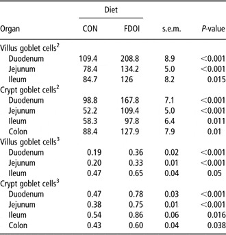
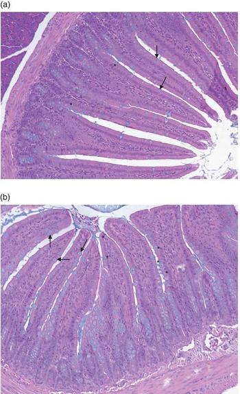
When compared to the CON diet, rats consuming the FDOI diet had a higher (P < 0.05) number of goblet cells in the villi of duodenum, jejunum and ileum. Crypt goblet cells were also higher in number (P < 0.05) for the FDOI diet in the duodenum, jejunum, ileum and colon (Table 3, Figure 1).
Goblet cell counts in the small intestine and colon for rats fed a diet containing ovine Ig for 21 daysFootnote 1

CON = control diet; FDOI = freeze-dried ovine immunoglobulin diet.
1 Values are mean ± s.e.m. (n = 7).
2 Number of goblet cells per five complete villi and their associated crypts.
3 Number of goblet cells per μm for five complete villi and their associated crypts.
Representative transverse sections of rat ileum. Section was stained with haemotoxylin, eosin and alcian blue. Mucus in goblet cells is stained with alcian blue. Arrows indicate examples of stained goblet cells. Stained mucus cells exhibit deep apical membrane cavitations from which streams of stained mucus (asterisk) emerged to join the mucus blanket in the ileal lumen. (a) Section from a rat fed with control diet and (b) freeze-dried ovine immunoglobulin diet.

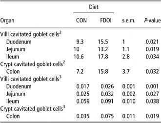
In duodenal, jejunal, ileal and colonic (only crypt) segments, histological analysis indicated that rats fed the FDOI diet had significantly (P < 0.05) more cavitated goblet cells (in villi and crypts) when compared to the rats fed the CON diet (Table 4, Figure 1).
Cavitated goblet cell counts in the small intestine and colon for rats fed a diet containing ovine immunoglobulin for 21 daysFootnote 1

CON = control diet; FDOI = freeze-dried ovine immunoglobulin diet.
1 Values are means ± s.e.m. (n = 7).
2 Number of goblet cells per five complete villi and their associated crypts.
3 Number of goblet cells per μm for five complete villi and crypts.
Mucin gene expression
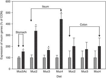
Muc5Ac mRNA expression in the stomach was significantly (P = 0.021) higher for rats fed the FDOI diet compared to rats fed the CON diet (Figure 2). Further, Muc2, Muc3 and Muc4 mRNA expression in the ileum was higher (P < 0.05) for rats given the FDOI diet (Figure 2). There was no significant (P > 0.05) difference between the two groups for mucin gene expression in the colon.
Expression of mRNA levels of mucin genes (Muc5Ac, Muc2, Muc3 and Muc4) in the stomach, ileum and colon of rats fed a diet containing ovine immunoglobuline for 21 days. Values are mean ± s.e. (n = 6). Asterisk denotes that expression of mRNA levels of mucin genes of the indicated diet group is significantly (P < 0.05) different from the other group ( = control diet;
 = freeze-dried ovine immunoglobulin diet).

Quantification of Mucin protein
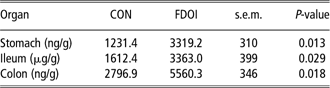
Mucin protein content was significantly (P < 0.05) higher in stomach chyme, and ileal and colonic digesta for rats fed the FDOI diet compared with rats fed the CON diet (Table 5).
Mucin protein concentrations in stomach chyme and ileal and colonic luminal digesta for rats fed a diet containing ovine immunoglobulin for 21 daysFootnote 1
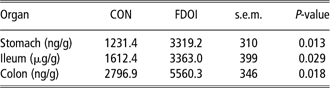
CON = control diet; FDOI = freeze-dried ovine immunoglobulin diet.
1 Values are means ± s.e.m. (n = 6) and were expressed per gram of freeze-dried digesta matter.
Discussion
SDAP included in the post-weaning diet of animals has been shown in a number of studies to enhance growth performance and reduce levels of gastrointestinal infection (Pierce et al., Reference Pierce, Cromwell, Lindemann, Russell and Weaver2005). The primary bioactive component of dried animal plasma is Ig.
Igs are the main anti-infective components of body fluids. These specialized proteins protect the body from harmful pathogens by either binding to them or by formation of an encapsulating barrier (Kraehenbuhl and Neutra, Reference Kraehenbuhl and Neutra1992; Bosi et al., Reference Bosi, Casini, Finamore, Cremokolini, Merialdi, Trevisi, Nobili and Mengheri2004). It is known that orally administered Ig is able to retain its activity in the GIT (Hammarstrom et al., Reference Hammarstrom, Smith and Hammarstrom1993). Rodriguez et al. (Reference Rodriguez, Blanch, Romano, Saborido, Rodenas and Polo2007) showed that porcine Ig partially resisted the digestion process in the GIT of adult dogs and cats fed diets containing SDAP or concentrated Ig derived from pig blood. Bovine Ig resists digestion in the upper GIT of humans and retains toxin-binding and neutralizing activity (Warny et al., Reference Warny, Fatimi, Bostwick, Laine, Lebel, LaMont, Pothoulakis and Kelly1999).
One of the important functions of the gut is to prevent lumen pathogenic bacteria and toxins from reaching the systemic circulation, organs and internal tissues (Allori et al., Reference Allori, Aguero, de Ruiz Holgado, Nader and Perdigon2000). The gut mucins afford the first line of defence against bacteria and are one of the main constituents of the gut barrier (Allori et al., Reference Allori, Aguero, de Ruiz Holgado, Nader and Perdigon2000). Mucins are produced in the gut by specialized goblet cells and an enhanced presence of goblet cells may lead to an improved synthesis of mucus that may help to protect the epithelial surface. The number of goblet cells in gut tissue may be increased by dietary interventions such as probiotic bacteria (Dock-Nascimento et al., Reference Dock-Nascimento, Junqueira and Aguilar-Nascimento2007). Other investigators (Sherman et al., Reference Sherman, Forstner, Roomi, Khatri and Forstner1985) have shown that chronic protein depletion or protein-energy undernutrition decrease the number of goblet cells and mucin synthesis in the proximal small intestine of rodents and piglets. Recently, King et al. (Reference King, Morel, Pluske and Hendriks2008) reported that feeding pigs with either colostrum or dried plasma resulted in an increase of villus and crypt goblet cells.
In the present study, the rats fed the FDOI diet had a significantly higher number of goblet cells per villus and associated crypt in the duodenal, jejunal, ileal and colonic segments when compared to rats fed the CON diet. Moreover, when compared to rats fed the CON diet, rats consuming the FDOI diet had a higher (P < 0.05) number of goblet cells per μm of villi and associated crypts in the duodenum, jejunum and ileum (Table 3). On the basis of visual examination of the histological preparations, rats fed the FDOI diet also appeared to have more numerous villi per length of intestinal tissue than the control animals. Taken together, these results indicate that feeding FDOI appears to have resulted in more goblet cells in the entire intestinal segments. This is consistent with our previous findings (Balan et al., Reference Balan, Han, Rutherfurd, Singh and Moughan2009) in which we measured gut tissue mass and intestinal villus and crypt architecture and found that the villus length, crypt depth (except in the duodenum and jejunum), villus : crypt ratio and individual villus surface area were significantly greater (P < 0.05) in rats receiving the FDOI diet.
Probiotic bacteria such as lactic acid bacteria have been shown to stimulate mucus production in the small intestine (Hecht, Reference Hecht1999). Animal plasma or colostrum (a major source of Ig) fed pigs have been shown to have a higher number of intestinal goblet cells (King et al., Reference King, Morel, Pluske and Hendriks2008). Recently, we have reported that rats receiving FDOI had a relative increase in probiotic bacteria such as Lactobacillus species in the gut digesta (Balan, Reference Balan2011). It appears that Ig may directly or indirectly (by probiotic bacteria) induce an increase of goblet cells, but how these effects are mediated remains to be elucidated.
Intestinal goblet cells may increase their discharge of mucin in response to mucin secretagogue (such as prostaglandin E2, cholera toxin, interleukin (IL)-4, IL-6, interferon-γ, tumor necrosis factor-α) stimulation by two processes and through the action of various secondary messengers such as intracellular Ca2+, cAMP and diacylglycerol, which activates protein kinase C. In many mucus cells, a phenomenon of compound exocytosis is induced, resulting in deep cavitation of the apical membrane surface of mucus cells (Specian and Oliver, Reference Specian and Oliver1991; Forstner, Reference Forstner1995; Dharmani et al., Reference Dharmani, Srivastava, Kissoon-Singh and Chadee2009). Some intestinal goblet cells may also react to stimulation by way of a second process that decreases the intracellular store of mucus granules but without cavitation (Specian and Oliver, Reference Specian and Oliver1991; Forstner, Reference Forstner1995). The latter mechanism results in a total loss of mucus granules and goblet cells are no longer stained by alcian blue and appear identical to other epithelial cells. A higher luminal mucin protein content and more intact and cavitated goblet cells per villus as observed here suggest that the FDOI diet fed to the rats induced more mucus discharge than for the rats fed the CON diet.
In the present study, stomach Muc5Ac, ileal Muc2, Muc3 and Muc4 gene expression increased by 154%, 337%, 166% and 441%, respectively, in the rats fed the FDOI diet when compared to rats fed the CON diet. The mRNA levels in the stomach tissue were highly correlated with chyme mucin protein concentrations (r = 0.83; P < 0.005). Similarly in the ileum, mRNA levels of Muc2, Muc3 and Muc4 were significantly correlated (r = 0.97, P < 0.001; r = 0.95, P < 0.001; and r = 0.96, P < 0.001) with digesta mucin protein contents. Conversely, in the colon, mRNA levels of Muc2, Muc3 and Muc4 were not significantly correlated (r = 0.62; P > 0.05, r = 0.61; P > 0.05 and r = 0.63; P > 0.05) with mucin protein. The observed upregulation of mucin genes (mainly stomach Muc5AC and ileal Muc2) may have resulted in the observed increase in luminal mucus in the stomach and intestine, although this has not been confirmed in the present study. A lack of correlation between mucin gene expression and colonic luminal mucin content may reflect the microbial degradation of mucins in the colon (Barcelo et al., Reference Barcelo, Claustre, Moro, Chayvialle, Cuber and Plaisancie2000). Further study is warranted to demonstrate the causal links between mucin gene expression and digesta mucin protein content to explain regional variations in the expression of these genes along the gut. The FDOI diet did not lead to a statistically significant upregulation of mucin gene expression in the rat colon, but our data were highly variable.
Other studies have also reported an effect of specific dietary components on gut mucins. Claustre et al. (Reference Claustre, Toumi, Trompette, Jourdan, Guignard, Chayvialle and Plaisancie2002) found that milk protein hydrolysates (casein and lactalbumin hydrolysates) and β-casomorphin-7 induced mucin release in rat jejunum and Han et al. (Reference Han, Deglaire, Sengupta and Moughan2008) showed that feeding rats with hydrolyzed casein upregulated Muc3 expression in the gut. Growing rats fed with dietary fibers with increasing viscosities were associated with an increase in ileal endogenous nitrogen flow and mucoprotein (sialic acid) secretion (Larsen et al., Reference Larsen, Wilson and Moughan1994). In addition, Barcelo et al. (Reference Barcelo, Claustre, Moro, Chayvialle, Cuber and Plaisancie2000) reported that dietary fibers (alginate, ulvan), uronic acids (glucuronic acid, galacturonic acid), and short-chain fatty acids may all stimulate the secretory activity of intestinal goblet cells. Caballero-Franco et al. (Reference Caballero-Franco, Keller, De Simone and Chadee2007) studied three bacterial groups (Lactobacilli, Bifidobacteria and Streptococci) contained in a probiotic formula and reported that the Lactobacillus species were the strongest potentiator of mucin secretion in vitro. In the present study, mucin protein was found to be significantly higher in the luminal contents of the stomach, ileum and colon for the FDOI-fed rats when compared to the CON-fed rats. Previously, we have observed that feeding rats an FDOI containing diet results in an increase in the Lactobacillus species in both ileal and colonic digesta (Balan, Reference Balan2011). Thus, feeding the FDOI diet may have led to an increase in Lactobacillus species that might have potentiated the mucin secretion in vivo. The present study has not directly addressed the mechanisms leading to higher amounts of mucin in the gut luminal contents. However, on the basis of the present results and our earlier findings (Balan et al., Reference Balan, Han, Rutherfurd, Singh and Moughan2009), it appears that Ig do have a trophic effect on intestinal cells and stimulate the mRNA expression of genes responsible for mucin synthesis.
In conclusion, orally administered FDOI selectively altered gut mucin gene expression and digesta mucin protein contents as well as the number of tissue goblet cells per length of villus in the growing rat. This may be associated with an enhanced mucus layer overlying the epithelial lining of the gut which may in turn enhance GIT protection, but this remains to be demonstrated.
Acknowledgements
Riddet Institute Doctoral Scholorship financially supported Prabhu Balan for his PhD studies.