Ventricular tachycardia may be associated with complex congenital heart diseases, especially following surgical ventriculotomies; Reference Zeppenfeld, Schalij and Bartelings1 however, it is rarely documented in Ebstein’s anomaly. Reference Walsh2,Reference Moore, Shannon and Gallotti3 Histological studies of the atrialised right ventricle have demonstrated a decreased myocardial fibre concentration and large areas of fibrous tissue Reference Walsh2 . Such features might be relevant to the genesis of ventricular tachycardia. We report a successful point ablation of a macro-reentrant ventricular tachycardia in a case with Ebstein’s anomaly and pulmonary atresia without a history of a ventriculotomy.
Case report
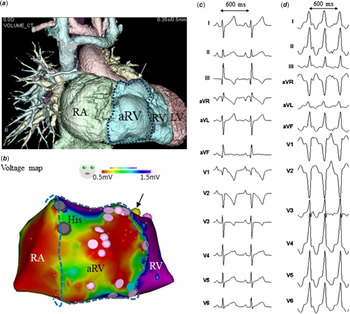
A female patient with Ebstein’s anomaly and pulmonary atresia had the first episode of ventricular tachycardia during percutaneous transluminal angioplasty of the left pulmonary artery at the age of 4 months, which required a 5 joule cardioversion. Further, ventricular tachycardia and fibrillation occurred repeatedly during a bidirectional Glenn operation at the age of 5 months, which required a transvenous infusion of amiodarone and, after the surgery, subsequent oral amiodarone. Since a total cavopulmonary connection was planned for a hypoplastic tricuspid valve, large atrialised right ventricle, and hypoplastic right ventricle, we performed an electrophysiological study before the surgery at the age of 17 months. The 3D mapping (Ensite-NavX™ system, St. Jude Medical, St. Paul, MN, USA) was focussed on the area of interest, exhibiting low voltage areas (Fig 1b, red zones) and multiple fragmented potentials (Fig 1b, pink tags) in the atrialised right ventricle; however, the bipolar electrograms in the hypoplastic right ventricle were more than 1.5 mV (Fig 1b, purple zones).
( a ) 3D computed tomographic view of the large atrialised right ventricle and hypoplastic right ventricle. ( b ) 3D mapping shows low voltage areas (red zones) and multiple fragmented potentials (pink tags) in the atrialised right ventricle; however, the bipolar electrograms in the hypoplastic right ventricle are more than 1.5 mV (purple zones). aRV = atrialised right ventricle; LV = left ventricle; RA = right atrium; RV = right ventricle. ( c ) 12-lead electrocardiograms during sinus rhythm. ( d ) 12-lead electrocardiograms during ventricular tachycardia.

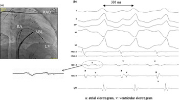
The 12-lead electrocardiograms during sinus rhythm and the induced ventricular tachycardia are shown (Fig 1c and d). A 4-Fr quadripolar electrode catheter was positioned in the right atrium and a 5-Fr bipolar electrode catheter in the left ventricle via an atrial septal defect (Fig 2a). The clinical ventricular tachycardia was reproducibly induced by programmed electrical stimulation in the left ventricle with a left bundle branch block QRS morphology and inferior axis (Fig 1d). An area with diastolic continuous fractionated potentials (Fig 2b) was found between the atrialised right ventricle and hypoplastic right ventricle (Fig 1a and b) and exhibited concealed entrainment and a post pacing interval equal to the cycle length of the ventricular tachycardia, suggesting the mechanism of the tachycardia was macro-reentry. The ventricular tachycardia became non-inducible after a single radiofrequency application using a 7-Fr ablation catheter at that point (Fig 1a white arrow, Fig 1b yellow tag, and Fig 2a). She underwent a total cavopulmonary connection procedure for 8 months after the successful ablation and has experienced no ventricular tachycardia for more than 3 years.
( a ) Catheter position at the successful catheter ablation site in the right anterior oblique view (RAO). ABL = ablation catheter; LV = left ventricle; RA = right atrium. ( b ) Intracardiac electrograms at the successful catheter ablation site. A = atrial electrogram; V = ventricular electrogram.

Discussion
Ventricular tachycardia is rarely documented in Ebstein’s anomaly. Reference Walsh2–Reference Shivapour, Sherwin and Alexander4 Shivapour et al reported that only 3 of 74 patients (4%) with an Ebstein’s anomaly undergoing a Cone procedure had sustained monomorphic ventricular tachycardia. Reference Shivapour, Sherwin and Alexander4 Ventricular tachycardia may be associated with complex congenital heart diseases, especially following surgical ventriculotomies. Reference Zeppenfeld, Schalij and Bartelings1 Reentry circuit isthmuses of ventricular tachycardia are often located within anatomically defined isthmuses bordered by unexcitable tissue, such as a patch in the right ventricular outflow tract or tricuspid valve. Reference Zeppenfeld, Schalij and Bartelings1 Linear ablation of the isthmus is necessary to eliminate the ventricular tachycardia in this situation. On the other hand, the point catheter ablation technique for macro-reentrant ventricular tachycardia has rarely been reported and drawing an entire activation map of the ventricular tachycardia could be avoided. Reference Shoda, Kasanuki and Ohnishi5 Moving a mapping catheter from the systolic phase to the diastolic phase is useful to detect the area of diastolic fragment potentials, which is an appropriate ablation target. If the phenomena of concealed entrainment and a post pacing interval equal to the cycle length of the ventricular tachycardia are defined, the point ablation technique could be utilised. Reference Shoda, Kasanuki and Ohnishi5
A prior histologic evaluation of the atrialised right ventricle in Ebstein’s anomaly has demonstrated clusters of cardiomyocytes isolated by fibrotic networks. Reference Anderson and Lie6,Reference Frescura, Angelini and Daliento7 Circuits of ventricular tachycardia are likely to be intrinsic to the congenitally abnormal atrialised right ventricle or hypoplastic right ventricle and are a consequence of degenerative remodelling. Moore et al demonstrated that ventricular arrhythmia substrates were localised to the atrialised right ventricle in un-operated patients with Ebstein’s anomaly. Reference Moore, Shannon and Gallotti3 They also reported focal ventricular tachycardia in eight patients and macro-reentrant ventricular tachycardia in four; however, the efficacy of the point ablation for macro-reentry was not well described. In our case, the tachycardia circuit was eliminated by a single point catheter ablation at an area with diastolic fractionated potentials. The boundary between the atrialised right ventricle and hypoplastic right ventricle can be identified with 3D substrate mapping. Recorded potentials in the atrialised right ventricle differ from those in the hypoplastic right ventricle.
Ebstein’s anomaly is characterised by apical displacement and dysplasia of the septal leaflet of tricuspid valve with a right ventricular atrialisation compromising its function. The functional part of the right ventricle is often small and ill-equipped to maintain a full cardiac output, especially in patients with Ebstein’s anomaly with pulmonary atresia. The choice of surgery for patients with a small functional right ventricle might be a single-ventricle repair. Reference Shinkawa, Polimenakos and Gomez-Fifer8 After a total cavopulmonary connection operation, access to the right ventricle is difficult because it should be approached using a conduit puncture or transaortic procedure. Hence, ventricular tachycardia substrates should be eliminated before a total cavopulmonary connection.
We reported a successful point ablation in a case with Ebstein’s anomaly and pulmonary atresia associated with a sustained macro-reentrant ventricular tachycardia and history of a ventriculotomy. The ventricular tachycardia substrate and slow conduction zone existed in a low voltage area between the atrialised right ventricle and hypoplastic right ventricle.
Financial support
This research received no specific grant from any funding agency or from the commercial or not-for-profit sectors.
Conflicts of interest
None.
Ethical standards
The author asserts that all procedures contributing to this study comply with the ethical standards of the relevant guidelines on human experimentation and with the Helsinki Declaration of 1975, as revised in 2008, and has been approved by the institutional committees.