Introduction
Trichinellosis is a serious zoonotic disease (Al-Attar et al., Reference AL-Attar, El-Kersh, Sadek, Harba, Osheiba and Brakat2020) that is mainly transmitted by the ingestion of raw or undercooked pork meat (Rayia et al., Reference Rayia, Saad, Ashour and Oreiby2017). Currently, more than 11 million individuals in the world are infected by Trichinella spiralis (Muñoz-Carrillo et al., Reference Muñoz-Carrillo, Maldonado-Tapia, López-Luna, Muñoz-Escobedo, Flores-De La Torre and Moreno-García2018). The risks of trichinellosis outbreaks have been continuously increasing in rural areas of Africa, Central and South America, and Asia (Bruschi, Reference Bruschi2012). After consumption of undercooked meat from infected animals, it takes approximately 17 days for the larvae to enter the striated muscles, where intact encapsulations will be formed on day 30. The capsule wall consists of collagen fibres secreted by the host fibroblasts, which escape the humoral and cellular immune responses. These muscle larvae can survive and persist within the host for its entire life (Ren et al., Reference Ren, Qin, Zhang, Zheng, Dai and Wu2018).
Fever, muscle fatigue and eyelid oedema are symptoms of trichinosis (Park et al., Reference Park, Kang, Jo, Baek, Yu, Choi, Cha and Ock2018). Severe cases of trichinosis-related myocarditis can even lead to death (Bruschi & Dupouy-Camet, Reference Bruschi and Dupouy-Camet2014). An inflammatory response aiming for the elimination of the parasite is elicited during the course of infection. However, several studies have confirmed that the host is threatened by this inflammatory response (Muñoz-Carrillo et al., Reference Muñoz-Carrillo, Muñoz-Escobedo, Maldonado-Tapia, Chávez-Ruvalcaba and Moreno-García2017).
Conventional treatment with benzimidazole derivatives, such as mebendazole and albendazole, is frequently used against trichinellosis. However, in addition to their imperfect action against the encapsulated larvae and the emerging resistance against it, their restricted absorption creates a clear impediment to the overall treatment (Prichard, Reference Prichard2007). Furthermore, many of these medications are contraindicated in pregnant women and children aged <3 years, and others have a high risk of carcinogenicity (Yadav & Temjenmongla, Reference Yadav and Temjenmongla2012). Therefore, there is a strong need for novel antihelminthic preparations, particularly those derived from herbal sources that are well tolerated with minimal side effects (Gilleard & Beech, Reference Gilleard and Beech2007). Pharmaceutical plants have been used to control parasites for decades and have demonstrated sufficient potency (Bauri et al., Reference Bauri, Tigga and Kullu2015).
Citrus limon (lemon) is a small tree of the Rutaceae family, which originated in Asia and now grows globally in tropical, semitropical and warm temperate countries (Youmsi et al., Reference Youmsi, Tsouh Fokou, Menkem, Bakarnga-Via, Keumoe, Nana and Boyom2017). Citrus limon has a broad range of activities, including anti-inflammatory, antimicrobial, anticancer and antiparasitic (Klimek-Szczykutowicz et al., Reference Klimek-Szczykutowicz, Szopa and Ekiert2020). Capsicum frutescens is a small bush of the Solanaceae family and is known as ‘the malagueta pepper’ (Vasconcelos et al., Reference Vasconcelos, Martins, de Oliveira and Duarte2014). It grows in tropical climates because it requires a wet and humid atmosphere (Anthony et al., Reference Anthony, Ese and Lawrence2013). In addition to its antibacterial and antioxidant properties, C. frutescens produces saponins, flavonoids and diterpenoids together with phenolic compounds that are antiparasitic (Madhumathy et al., Reference Madhumathy, Aivazi and Vijayan2007; Otunola et al., Reference Otunola, Afolayan, Ajayi and Odeyemi2017).
In the present study, we aimed to identify novel and safe therapeutic agents that reduce the morbidity and mortality of trichinellosis. We assessed the antiparasitic and anti-inflammatory effects of C. limon and C. frutescens on murine trichinellosis and compared them with those of albendazole and prednisolone. The efficacy of the drugs was evaluated using parasitological, biochemical, histopathological and ultrastructural assays. To the best of our knowledge, this is the first study to evaluate the therapeutic effects of C. limon and C. frutescens on murine trichinellosis.
Material and methods
Animals and parasites
Fifty parasite-free, 5-week-old Swiss albino male mice with an average weight of 20–25 g were used in this study. The mice were obtained from the animal house of Theodor Bilharz Research Institute, Giza, Egypt, and maintained in accordance with institutional and national guidelines.
Trichinella spiralis was isolated and maintained by repetitive passage in rats at the Medical Parasitology Department laboratory, Faculty of Medicine, Zagazig University, Egypt. Each mouse was orally infected with 200–250 T. spiralis larvae (Shoheib et al., Reference Shoheib, Shamloula, Abdin and El-Segai2006).
Ethics guidelines
The protocol of this study was approved by the ethics committee of the Faculty of Medicine, Zagazig University (approval number: 4002).
Experimental design
Five groups of mice, with ten mice each, were included in our study. Group 1: negative control (uninfected mice); group 2: positive control (infected mice which did not receive any treatment); group 3: infected mice treated with albendazole and prednisolone; group 4: infected mice treated with C. limon extract and group 5: infected mice treated with C. frutescens extract.
Assessment of samples
At 7 days post infection (dpi), five mice from each group were sacrificed and blood samples were collected to determine serum tumour necrosis factor (TNF)-α levels. The small intestine was removed, longitudinally opened and washed. Approximately 1 cm of the mid-intestinal region was placed in 10% formalin for histopathological examination. The rest of the intestine was used for counting T. spiralis adult worms. The adult worms were then preserved in a fresh fixative for ultrastructural study. At 35 dpi, the remaining five mice from each group were sacrificed and blood samples were collected to determine serum TNF-α levels. The peritoneum was opened and the diaphragm was carefully dissected for histopathological and ultrastructural studies. The remaining muscle samples were digested to obtain the total larval count.
Drug and plant preparations
Albendazole (Alzental suspension, EIPICO) was orally administered at a dose of 50 mg/kg/day for 3 successive days, starting from 3 dpi (Attia et al., Reference Attia, Mahmoud, Farrag, Makboul, Mohamed and Ibraheim2015).
Prednisolone (Predsol suspension, BORG PHARMACEUTICAL) was orally administered at a dose of 0.7 mg/kg/day for 3 successive days, starting from 3 dpi (Manzur et al., Reference Manzur, Kuntzer, Pike and Swan2008).
Fresh C. limon peel (300 g) and C. frutescens fruit (500 g) were cut into small pieces and separately soaked in 90% ethanol for 10 days, which was replaced every 3 days. A viscous filtrate of crude extracts was obtained using a filter paper and rotatory evaporator at 50°C under reduced pressure. This was followed by lyophilization, which produced 25 and 55 g of powdered extracts of C. limon and C. frutescens, respectively (Zhang et al., Reference Zhang, Lin and Ye2018).
The suspensions of lyophilized extracts were prepared for oral administration using 0.5% Tween-80 (ADWIC, Egypt) in normal saline. The concentration was adjusted so that each 0.1 ml of the prepared suspension contained 0.3 mg of the plant extract. Extracts were orally administered to mice at a dose of 100 mg/kg/d for C. limon (Tag et al., Reference Tag, Kelany, Tantawy and Fahmy2014) and 150 mg/kg/d for C. frutescens from 1 dpi until the day of sacrifice (Zimmer et al., Reference Zimmer, Leonardi, Zimmer, Kalinine, Onofre de Souza, Portela and Gosmann2012).
Parasitological assays
Isolation and counting of adult worms
After sacrifice, the small intestine was removed, washed, opened longitudinally, divided into small pieces and incubated in saline for 3–4 h at 37°C. The samples were then shaken and washed with saline, and the adult worms were allowed to sediment for 30 min. The supernatant was removed and the sediment was poured into a Petri dish with 3–5 drops of physiological saline, and the adult worms were examined and counted under a dissecting microscope (Basyoni & El-Sabaa, Reference Basyoni and El-Sabaa2013).
Estimation of total larval burden in muscles
Mice were dissected, cut into small pieces and digested in 1% pepsin hydrochloride prepared in distilled water for 2 h at 37°C with continuous stirring using an electromagnetic stirrer. The digested mixture was filtered through a 50-mesh/cm² sieve, then through a 200-mesh/cm² sieve, and then washed with tap water. The supernatant was discarded and the larvae in the sediment were counted microscopically using a McMaster counting chamber (Mayer-Scholl et al., Reference Mayer-Scholl, Pozio, Gayda, Thaben, Bahn and Nöckler2017).
Determination of serum TNF-α levels
Blood samples were collected and allowed to clot for 2 h at room temperature, before centrifugation at 2000×g for 20 min. Serum was separated and stored at ≤−20°C. Serum TNF-α levels were determined at 7 and 35 dpi using Mouse TNF-alpha Quantikine ELISA Kit (R&D Systems, Cat. No. MTA00B), following the manufacturer's instructions.
Histopathological study
Haematoxylin and eosin staining
The collected intestinal and muscular tissue samples were fixed in 10% formalin for 24 h, washed in water for 12 h, and then dried out in ascending ethanol concentrations (70% ethanol for 120 min, then 90% ethanol for 90 min, and finally 100% ethanol (two cycles) for 1 h per cycle). Samples were then cleared by immersing them in a mixture of 50% ethanol and 50% xylene for 1 h, followed by pure xylene for 1.5 h. Next, the samples were embedded in paraffin wax. Paraffin sections (4–5 μm) were stained with haematoxylin and eosin (Kiernan, Reference Kiernan1999). The extent of inflammatory cell infiltrates within the core of the intestinal villi and submucosa and surrounding the larval capsule was evaluated. Five histological sections per mouse were examined. The average score of ten low-power fields (100×) from each of the examined sections was then calculated (+1 = mild reaction; +2 = moderate reaction; and +3 = intense reaction) (Elgendy et al., Reference Elgendy, Othman and Hasby2020).
Masson's trichrome staining
Muscle sections were deparaffinized and rehydrated using descending concentrations of ethanol (100%, 95% and 70%). The sections were then washed in distilled water, fixed in Bouin's solution and rinsed in running tap water for 5–10 min. Next, they were stained in Weigert's iron haematoxylin solution for 10 min, rinsed and washed in distilled water. Then, they were placed in Biebrich scarlet acid fuchsin solution for 10–15 min before washing in distilled water. They were then differentiated in phosphomolybdic–phosphotungstic acid solution for 10–15 min, transferred directly to aniline blue solution for 5–10 min and rinsed briefly in distilled water. Sections were differentiated in 1% acetic acid solution for 2–5 min, washed in distilled water, and dehydrated very quickly in 95% and 100% ethanol. They were then cleared in xylene and mounted with DPX (dibutylphthalate polystyrene xylene) (Suvarna et al., Reference Suvarna, Layton and Bancroft2013). The content of collagen and fibroblast around the encysted larvae was evaluated by selecting eight separate views from each sample (400×) and calculating the ratio of the area occupied by collagen fibres to the total area (Loos et al., Reference Loos, Dávila, Rodrígues, Petrigh, Zoppi, Crocenzi and Cumino2017; Chen et al., Reference Chen, Peng, Wang, Hu, Shen, Xu, Yin, Liu and Cao2019; Zeng et al., Reference Zeng, Wang and Yang2019). The analysis was performed using ImageJ software (Schneider et al., Reference Schneider, Rasband and Eliceiri2012).
Ultrastructural evaluation (transmission electron microscopy)
Adults
Adult worms were processed as described by Sukontason et al. (Reference Sukontason, Chaiwong, Chaisri, Kurahashi, Sanford and Sukontason2011) and Karunovsky (Reference Karunovsky1965). They were collected at 7 dpi and centrifuged at 7000 rpm for 1 min and then resuspended in fresh fixative agent (2.5% glutaraldehyde) at 4°C for 24 h. After 3 days, the fixative was removed and the parasites were post-fixed for 1 h in 2% osmium tetroxide and then dehydrated in a graded ethanol series. The specimens remained in each ethanol concentration for 12 h, followed by 100% ethanol. Next, the specimens were placed in acetone for 2 h, after which they were transferred into ratios of resin to acetone of 1:3 for 24 h, 1:1 for 24 h and 3:1 for 24 h, sequentially. Specimens were then treated twice with pure resin for 3 h each time. After that, the material was embedded in the epoxy resin Epon 812 according to Luft's method (Luft, Reference Luft1961) and incubated at 70°C for 24 h. After hardening, semithin sections (0.5 μm) were prepared and stained with toluidine blue. Appropriate specimens were selected using light microscopy. Ultrathin sections (90 nm) were then made from the same blocks. Serial sections were collected onto copper slot grids and poststained in 8% uranyl acetate for 10 min and 1% lead citrate for 5 min. After drying for 15 min, ultrathin sections were examined using a JEOL JEM 2100 transmission electron microscope at 160 kV.
Muscle samples
Very small muscle samples (1 mm³) were used and fixed within <5 min. Muscle samples were then processed as described above for adults.
Statistical analysis
Statistical analysis was performed using SPSS version 18.0 (IBM, Armonk, USA). Results were expressed as means ± standard deviation (SD). Data were analysed using one-way ANOVA followed by Tukey's post hoc test for multiple comparisons between groups. The chi-squared test was used for histopathological scoring. Differences were considered statistically significant at P < 0.05 and highly significant at P < 0.01.
Results
Parasitological assessments
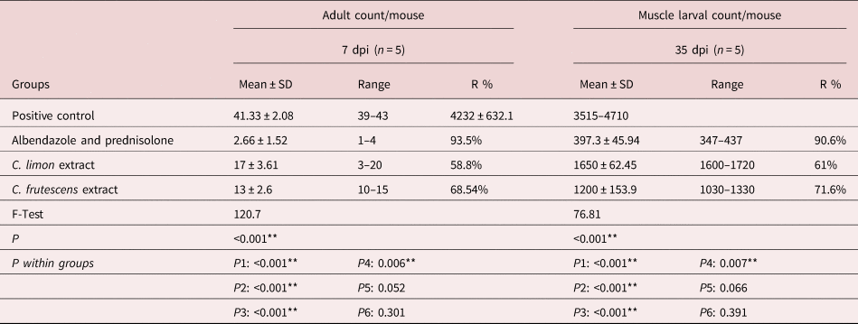
All treatment groups showed a significant reduction in the mean number of adult T. spiralis compared with the positive control group. The highest reduction was seen in the mice receiving albendazole and prednisolone, followed by those receiving C. frutescens and C. limon (93.5%, 68.54% and 58.8% respectively). Moreover, the muscles of the treatment groups showed a significant decrease in the mean larval count compared with the positive control group. Mice treated with albendazole and prednisolone showed the most significant reduction (90.6%), followed by those treated with C. frutescens and C. limon, with reduction percentages of 71.6% and 61%, respectively (table 1).
Mean T. spiralis adult count in the small intestine and mean larval count in the muscles.

n = number of mice in each group; SD = standard deviation; P = probability.
** Highly significant difference
P1: positive control group vs. albendazole and prednisolone–treated group.
P2: positive control group vs. C. limon extract-treated group.
P3: positive control group vs. C. frutescens extract-treated group.
P4: albendazole and prednisolone–treated group vs. C. limon extract-treated group.
P5: albendazole and prednisolone–treated group vs. C. frutescens extract-treated group.
P6: C. limon extract-treated group vs. C. frutescens extract-treated group.
Serum levels of TNF-α
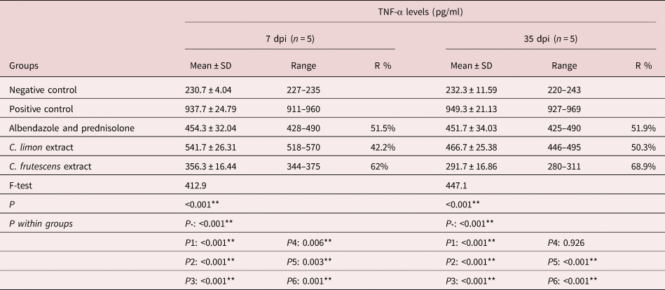
Treatment with herbal extracts significantly reduced serum TNF-α levels at 7 and 35 dpi, with C. frutescens showing the highest reduction percentages during both phases (62% and 68.9%, respectively), followed by albendazole and prednisolone (51.5% and 51.9%, respectively) and C. limon (42.2% and 50.3%, respectively; table 2).
TNF-α levels in the different groups.

n = number of studied mice in each group; SD = standard deviation; P = probability.
** Highly significant difference
P *: negative control group vs. positive control group.
P1: positive control group vs. albendazole and prednisolone–treated group.
P2: positive control group vs. C. limon extract-treated group.
P3: positive control group vs. C. frutescens extract-treated group.
P4: albendazole and prednisolone–treated group vs. C. limon extract-treated group.
P5: albendazole and prednisolone–treated group vs. C. frutescens extract-treated group.
P6: C. limon extract-treated group vs. C. frutescens extract-treated group.
Histopathological findings
Haematoxylin and eosin staining
Small intestine
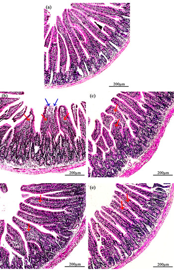
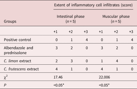
Histopathological examination of the positive control group showed dense inflammatory cell infiltrate mainly in the core of the villi and extending into the submucosa. Flattening of villi and sloughing of villous tips were also detected. Cross-sections of adult T. spiralis could be observed. The C. frutescens group showed a significant reduction in inflammation intensity and elongated villi. On the other hand, mice treated with albendazole and prednisolone showed mild to moderate inflammation within the core of the intestinal villi. The C. limon group showed moderate inflammation (fig. 1). The extent of inflammatory cell infiltrates within the core of the intestinal villi and submucosa is shown in table 3.
Histopathological findings of the small intestine sections at 7 dpi. (a) Negative control group showing intestinal villi with normal architecture and length. (b) Positive control group with obvious inflammatory cell infiltrate (red arrows) in submucosa and the core of the villi and flat sloughed villous tips (blue arrows). (c) Albendazole and prednisolone-treated group showing a decrease in inflammatory infiltrate in the core of villi (arrows) and reconstitution of the intestinal villous structure. (d) Citrus limon extract-treated group showing moderate inflammatory infiltrate in the core of villi (arrows). (e) C. frutescens extract-treated group with an evident reduction in inflammation intensity (arrows) and improvement of intestinal villi architecture and length (200×).

Extent of inflammatory cell infiltrates in the small intestine and diaphragm .

n = number of mice in each group; P = probability; *significant difference.
Skeletal muscles
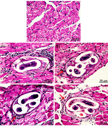
Histopathological examination of skeletal muscles of the positive control group showed encysted T. spiralis larvae surrounded by nurse cells, a collagen capsule and dense inflammatory cell infiltrate. The C. frutescens group showed a marked reduction in deposited larvae with a significant decrease in inflammatory cell infiltrate around the larvae. Moreover, the C. limon group showed moderate inflammation around the larvae (fig. 2). The extent of the inflammatory cell infiltrates in the skeletal muscle sections is presented in table 3.
Histopathological findings of the muscle sections at 35 dpi (H&E). (a) Negative control group with normal diaphragm muscle fibres. (b) Positive control group showing (1) T. spiralis larva, (2) nurse cell, (3) collagen capsule and (4) marked inflammatory infiltrate. (c) Albendazole- and prednisolone-treated group showing mild inflammatory infiltrate (arrow) around T. spiralis larva. (d) C. limon group showing larval deposition surrounded by moderate inflammatory infiltrate (arrow). (e) C. frutescens group showing T. spiralis larva with mild inflammatory infiltrate (arrow) (400×).

Masson's trichrome stain
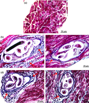
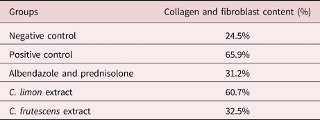
Masson's trichrome stain showed blue collagen fibres and black nuclei against a red background. The positive control group showed large number of encysted larvae surrounded by an intense inflammatory reaction with associated fibroplasia. The C. frutescens group showed a few encysted larvae surrounded by a mild inflammatory reaction and fibroplasia. The C. limon group showed larval deposition surrounded by a moderate inflammatory reaction with fibroplasia (fig. 3). ImageJ software was used to evaluate collagen and fibroblast content (table 4). They represented 31.2% of the total cellular content in albendazole and prednisolone-treated group, 32.5% in the C. frutescens group and 60.7% in the C. limon group.
Diaphragm sections at 35 dpi (Masson's trichrome stain). (a) Negative control group with normal diaphragm muscle fibres and no inflammatory reaction or mesenchymal cell proliferation. (b) Positive control group showing T. spiralis larva surrounded by an intense inflammatory reaction with associated fibroplasia (arrows). (c) Albendazole- and prednisolone-treated group showing minimal mesenchymal and inflammatory cell reaction around the larva (arrow). (d) C. limon group showing a moderate inflammatory reaction with associated fibroplasia (arrows) (e) C. frutescens group showing T. spiralis larva surrounded by a mild inflammatory reaction with associated fibroplasia (arrows) (400×).

Collagen and fibroblast content of murine muscles .

Transmission electron microscopy
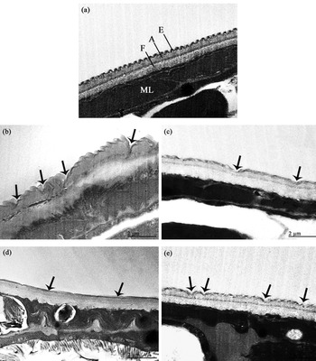
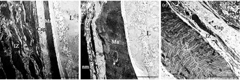
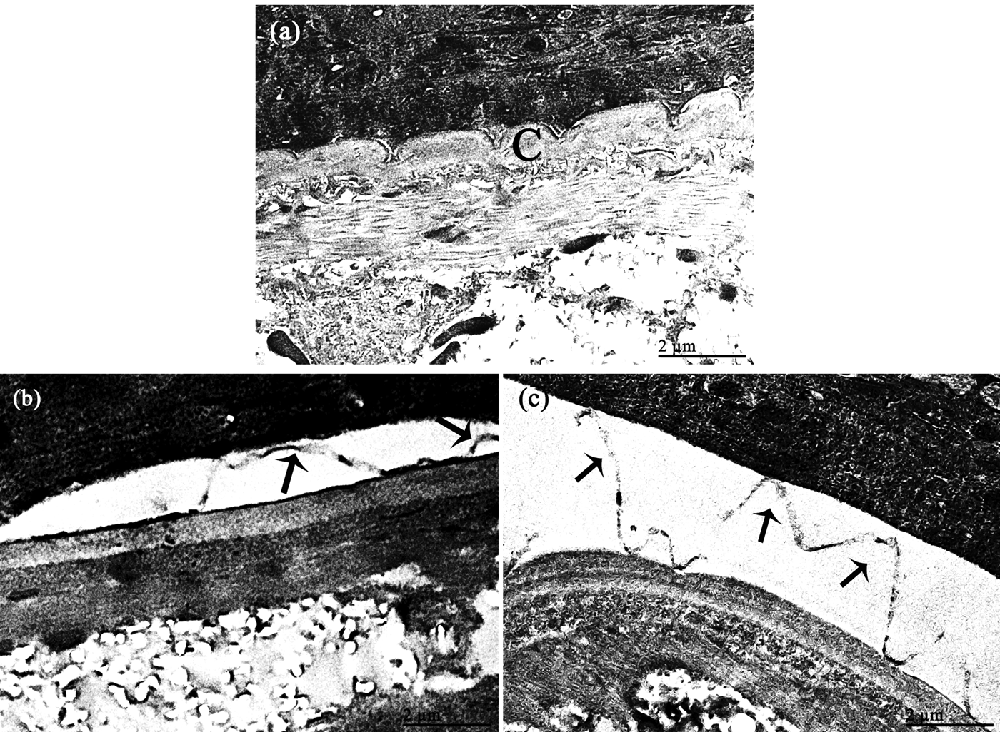
Adult T. spiralis showed epicuticle blunting, extensive loss of epicuticular corrugation, zones of depression and disturbed continuity in the C. limon and C. frutescens groups. The albendazole- and prednisolone-treated groups showed cuticular deformity with deep grooves (fig. 4). Transmission electron microscopy examination of muscle samples of the groups treated with C. frutescens and C. limon displayed a reduction in the inflammatory zone. The cuticle of the larvae showed degenerative changes, including cuticular blebbing and separation or detachment of superficial layers of the cuticle (figs. 5 and 6).
Transmission electron micrographs of T. spiralis adult at 7 dpi. (a) Positive control group with the characteristic cuticular structure being formed the amorphous layer (A), the fibrillar layer (F) and the wavy covering epicuticle (E). ML refers to the somatic muscles of the body wall. (b) Albendazole and prednisolone group showing deformity and deep grooves in the cuticle (arrows). (c) Citrus limon group displaying depressed areas in the cuticle (arrows). (d) Citrus limon group showing loss of epicuticular waviness and lack of discrimination of cuticle layers (arrows). (e) Capsicum frutescens group showing multiple zones of cuticular damage and depressions (arrows) (2000×; bar 2 μm).

Transmission electron micrographs of the diaphragm at 35 dpi. (a) Positive control group showing T. spiralis larva (L) enclosed by the matrix (Ma), the capsule (cap) and wide inflammatory zone (IZ) displaying the loss of structural integrity of sarcomere, disorganization of contractile function and disappearance of light and dark bands. (b) Citrus limon group displaying decrease in the IZ. (c) Capsicum frutescens group showing extensively destructed T. spiralis larva (L), distinct decrease in the IZ and appearance of normal muscle (nm) with regular light and dark bands (600×; bar 10 μm).

Transmission electron micrographs of the diaphragm at 35 dpi with higher magnification. (a) Positive control showing the larval cuticle (c). (b) Citrus limon group showing obvious separation and blebbing of the superficial layers of the cuticle (arrows). (c) Capsicum frutescens group showing separation of the superficial layers of the cuticle in wide area (arrows) (2000×; bar 2 μm).

Discussion
Although benzimidazole derivatives are the drug of choice to treat trichinellosis, they have limited effects against the muscular phase (Basyoni & El-Sabaa, Reference Basyoni and El-Sabaa2013). Universally prescribed steroidal anti-inflammatory drugs possess many adverse effects that include increasing the muscular parasite burden (Alvarado et al., Reference Alvarado, Meza, García, Saldívar and Moreno1996; Piekarska et al., Reference Piekarska, Michalski, Obminska-Mrukowicz and Gorczykowski2010). Therefore, there is an urgent need for safe products with strong antiparasitic and anti-inflammatory effects. The use of natural products has been recommended by several studies, as synthetic compounds have many adverse effects and some may even be carcinogenic. Consequently, a safe and effective natural alternative is needed for the treatment of both enteral and parenteral stages of T. spiralis (Shalaby et al., Reference Shalaby, Moghazy, Shalaby and Nasr2010). In this context, we assessed the efficacy of two herbal remedies, C. frutescens and C. limon.
Our study showed a significant decrease in the counts of adult worms and larvae after using C. frutescens (68.54% and 71.6%, respectively) and C. limon (58.8% and 61%, respectively). The reduction percentages in the albendazole and prednisolone-treated group were 93.5% and 90.6%. Our results are in agreement with those of Shalaby et al. (Reference Shalaby, Moghazy, Shalaby and Nasr2010) and Shoheib et al. (Reference Shoheib, Shamloula, Abdin and El-Segai2006). Both reported a reduced efficacy of albendazole against encysted muscle larvae with the number of larvae reduced only by 26.4% and 65.2%, respectively.
The effect of albendazole on T. spiralis larvae was lower than that on adults, due to the low water solubility and bioavailability of oral administration (Caner et al., Reference Caner, Döşkaya, Değirmenci, Can, Baykan, Üner, Başdemir, Zeybek and Gürüz2008). Interestingly, C. limon and C. frutescens extracts had a better effect on larvae than on adults. Therefore, using the same concentrations for longer periods may produce a better biological activity and increased parasite mortality.
The antiparasitic effects of C. frutescens could be explained by the fact that the plant fruits contain abundant amounts of the active compounds, capsaicinoids (Kurian, Reference Kurian2007; Vinayaka et al., Reference Vinayaka, Nandini, Rakshitha, Ramya, Shruthi, Shruthi, Prashith and Raghavendra2010; Nascimento et al., Reference Nascimento, Nascimento, Ramos, Silva, Câmara, Silva, Moreira and Porto2013). In accordance with our results, Neves et al. (Reference Neves, Freitas, Coutinho, Dallemole-Giaretta, Fabry, Dhingra and Ferraz2009) reported a strong nematicidal effect of chloroformic and cetonic extracts of C. frutescens.
Upadhyaya (Reference Upadhyaya2018) investigated the antiparasitic effects of C. limon extract and reported a significant activity against the Indian earthworm Eicinia foetida, which is anatomically and physiologically similar to the intestinal roundworm of humans. These findings are consistent with our results. The antiparasitic effect of C. limon could be due to its limonene content (52.6% of lemon composition) (Gomes et al., Reference Gomes, Cardoso, Soares, Batista, Machado, Andrade, de Azeredo, Resende and Rodrigues2014). The mode of action of limonene is still unknown; however, it is thought to be responsible for the anthelmintic effect of lemon (Rosskopf Erin et al., Reference Rosskopf Erin, Therrien Leonard, Adkins Scott, Iriarte, Foster Joyce and Kokalis-Burelle2008; Squires et al., Reference Squires, Foster, Lindsay, Caudell and Zajac2010). Previous studies have suggested that it inhibits nematode development, enzymes and plasma membrane pumps, and affects metabolic pathways (Kaur et al., Reference Kaur, Jain, Kaur and Jain2009; Squires et al., Reference Squires, Foster, Lindsay, Caudell and Zajac2010).
In the small intestine, the C. frutescens group showed elongation of villi and a significant reduction in inflammation intensity compared with positive control group that showed dense inflammatory cell infiltrate observed mainly in the core of the villi and extending into the submucosa. The therapeutic effect of C. frutescens extract was superior to that of C. limon extract, which showed moderate inflammation.
Compared with the positive control group, the C. frutescens group showed a marked reduction in the larvae deposited in muscles, with a significant decrease in inflammatory cell infiltrate around the larvae. On the other hand, the C. limon group exhibited a moderate decrease in muscle larval count with moderate inflammation around the larvae.
Collagen and fibroblast content in muscles was markedly decreased in the C. frutescens group (32.5%) and the albendazole- and prednisolone-treated groups (31.2%), compared with the positive control group (65.9%). On the other hand, the collagen and fibroblast content in the C. limon group was 60.7%.
Furthermore, our results showed that C. frutescens reduced TNF-α levels at 7 and 35 dpi (62% and 68.9%, respectively), more significantly than C. limon (42.2% and 50.3%, respectively). The albendazole and prednisolone group showed reduction percentages of 51.5% and 51.9%, respectively.
Previous studies have verified the detrimental outcome of the inflammatory response driven by TNF-α (Muñoz-Carrillo et al., Reference Muñoz-Carrillo, Muñoz-Escobedo, Maldonado-Tapia, Chávez-Ruvalcaba and Moreno-García2017). We believe that the capsaicin component of C. frutescens inhibited TNF-α production. Lee et al. (Reference Lee, Lee, Yon, Oh, Baek, Nahm, Lee, Yun and Nam2011) reported that TNF-α levels in the brain can be decreased by capsaicin. Moreover, Shang et al. (Reference Shang, Amna, Amina, Al-Musayeib, Al-Deyab and Hwang2017) showed that capsaicin inhibited the release of TNF-α in myoblasts. In the C. limon extract-treated group, the significant decrease in TNF-α level could be due to limonene or naringin. Yu et al. (Reference Yu, Yan and Sun2017) reported that limonene can lower TNF-α levels in serum, and Kawaguchi et al. (Reference Kawaguchi, Kikuchi, Hasegawa, Maruyama, Morita and Kumazawa1999) reported that naringin, a flavanone glycoside, significantly reduced the lipopolysaccharide-induced TNF-α levels.
Kisiel & Kaszuba (Reference Kisiel and Kaszuba2011) showed that glucocorticoid treatment for immediate hypersensitivity associated with trichinosis inhibited cellular immune response (mainly lymphocytes) and reduced production of cytokines, including TNF-α. These findings explain the reduction of TNF-α levels in the albendazole- and prednisolone-treated groups.
Ultrastructural examination showed an evident blunting of the epicuticle and degenerative changes in the cuticle of adult worms. Moreover, the thickness of the inflammatory zone was reduced in muscle samples. The larvae showed cuticular blebbing and shedding of superficial layers in the groups treated with the herbal extracts. C. frutescens extract-treated group displayed more evident effect compared with the C. limon extract-treated group. Vinayaka et al. (Reference Vinayaka, Nandini, Rakshitha, Ramya, Shruthi, Shruthi, Prashith and Raghavendra2010) suggested that capsaicinoids are responsible for the anthelminthic activity of C. frutescens. Therefore, the ultrastructural improvement may be due to the capsaicinoids in C. frutescens.
We conclude that the ethanolic extracts of C. frutescens and C. limon have therapeutic and anti-inflammatory effects in T. spiralis infection, with C. frutescens showing a superior therapeutic effect. Therefore, these extracts may be promising alternative treatments for trichinellosis. In our future studies, we plan to assess the therapeutic effectiveness of the active constituents of these extracts, to calculate the appropriate doses and to design treatment schedules for trichinosis infection.
Financial support
Nil.
Conflicts of interest
No conflict of interest.