INTRODUCTION
With a worldwide focus on northern latitudes, the bulk of fish parasitic species are yet to be discovered and described in Africa. The ectoparasitic monogenean flatworms have generated most of the volume of research done on fish parasites in Africa (Paperna, Reference Paperna1979, Reference Paperna1996; Khalil and Polling, Reference Khalil and Polling1997). Research undertaken on monogenean parasites of fishes in Africa has mainly focussed on taxonomy of tilapiine parasites such as Cichlidogyrus Paperna, 1960 (Pariselle and Euzet, Reference Pariselle and Euzet1998, Reference Pariselle and Euzet2003) because cichlids comprise a major source of food. A small number of studies have also looked at systematics and host-parasite coevolution (Pouyaud et al. Reference Pouyaud, Desmarais, Deveney and Pariselle2006), pathology (Arafa et al. Reference Arafa, El-Naggar and El-Abbassy2009) and faunistics (Khalil and Polling, Reference Khalil and Polling1997; Barson et al. Reference Barson, Bray, Ollevier and Huyse2008).
Viviparous monogeneans of the genus Macrogyrodactylus Malmberg, 1957 are common skin and gill parasites of African fishes. Currently 9 species are recognized from 4 fish families (Khalil and Polling, Reference Khalil and Polling1997; Přikrylová and Gelnar, Reference Přikrylová and Gelnar2008). The taxonomy of species belonging to the genus Macrogyrodactylus Malmberg, 1957 was previously based on haptoral morphology (Paperna, Reference Paperna1979), host and geographical range (Khalil and Polling, Reference Khalil and Polling1997), and structure of the excretory system (Malmberg, Reference Malmberg1998). The recent development of molecular markers for gyrodactylids (Matějusová et al. Reference Matějusová, Gelnar, McBeath, Collins and Cunningham2001) resulted in the first and only sequence of the ITS region of the rDNA of Macrogyrodactylus polypteri Malmberg, 1957 from the bichir, Polypterus senegalus Cuvier, 1829 (Matějusová et al. Reference Matějusová, Gelnar, Verneau, Cunningham and Littlewood2003). DNA sequence data on Macrogyrodactylus species, and African flatworm species in general, are still very rare. By virtue of their monoxenic life cycle and host specificity, monogenean parasites are excellent candidates for host-parasite coevolutionary studies. In cases where co-speciation occurs, the parasite can provide extra information on the evolution of the host (Pouyaud et al. Reference Pouyaud, Desmarais, Deveney and Pariselle2006; Šimková and Morand, Reference Šimková and Morand2008).
The purpose of this study was to identify the species of Macrogyrodactylus infesting African clariid catfishes using molecular and morphological characteristics, and to compare the morphology and genetics of these species in southern Africa (Zimbabwe), eastern Africa (Kenya) and western Africa (Senegal). These 3 regions coincide with the broad phylogeographical units of the host fish (Giddelo et al. Reference Giddelo, Arndt and Volckaert2002). The study follows up on morphological work on Macrogyrodactylus species from Zimbabwe and Senegal (Barson et al. Reference Barson, Bray, Ollevier and Huyse2008; Přikrylová and Gelnar, Reference Přikrylová and Gelnar2008). We investigated an unexpected hybridization event between Macrogyrodactylus spp. from allopatric host species to determine the historical distribution patterns of clariid catfish in Africa. We also present the ITS rDNA and partial cox1 mtDNA sequences of Macrogyrodactylus clarii Gussev, 1961, M. heterobranchiiN'douba et Lambert, Reference N'Douba and Lambert1999, M. congolensis Prudhoe, 1957 and M. karibaeDouëllou et Chishawa, Reference Douëllou and Chishawa1995 for the first time, construct their molecular phylogeny and discuss their relationships.
MATERIALS AND METHODS
Parasite collection
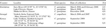
Catfish (Clarias gariepinus (Burchell, 1822) Valenciennes, 1840) were collected from the Mukuvisi River and Lake Chivero near Harare, Zimbabwe in March 2008, and from the Msaize River, a tributary of the Save River, southeastern Zimbabwe in March 2006 and February 2008 (Barson et al. Reference Barson, Bray, Ollevier and Huyse2008). Monogenean specimens were also collected from C. anguillaris L., 1758 in the Niokolo Koba National Park, Senegal (West Africa) and from C. gariepinus in Lake Turkana, Kenya (East Africa) (Table 1). Monogenean parasites, all of them Macrogyrodactylus spp., were removed from the gills of the fish and stored in 96% ethanol. In the laboratory, the opisthaptor was separated from the rest of the body enabling simultaneous morphological and molecular analyses to be carried out. The body was placed in 5 μl of milli-Q water and stored at −20°C, while the opisthaptor was digested with proteinase K solution (30 mg/ml) for 5 min at 65°C. The morphometry of Macrogyrodactylus spp. collected in Senegal has already been published (Přikrylová and Gelnar, Reference Přikrylová and Gelnar2008) and thus only the material for molecular analysis was included.
Localities, collection period and number of Macrogyrodactylus spp. analysed in the present study
(N=number of specimens in morphometrical study, n=number of specimens analysed by molecular methods.)

Morphometric analysis
The sclerites were observed under an Olympus BX51 phase-contrast microscope, and measurements of the different parts were taken according to the method of Přikrylová and Gelnar (Reference Přikrylová and Gelnar2008). The marginal hooks on the posterior part of the haptor were selected for measuring. Species identification was based on Douëllou and Chishawa (Reference Douëllou and Chishawa1995), Khalil and Mashego (Reference Khalil and Mashego1998), and Přikrylová and Gelnar (Reference Přikrylová and Gelnar2008). Drawings were made with the aid of a drawing tube and also by comparison with the drawings and measurements carried out on type material obtained from the Natural History Museum London (NHM) (M. congolensis – 3 paratypes, no. 1960.5.27.1-15; M. clarii – specimen no. 2006.9.1.1 and M. karibae – no. 2006.9.1.3), from the Muséum National d'Histoire Naturelle Paris (MNHN) (M. heterobranchii – paratype no. MNHN 574 HF, slide Tk 91 and M. congolensis karibae – paratype no. MNHN 143, slides Tg 14) and from the Swedish Museum of Natural History (M. polypteri – paratype no. 6976, 6977). Voucher specimens of each species will be deposited in the Natural History Museum, London.
DNA extraction and amplification
DNA was extracted from remainders of the bodies using the NucleoSpin ® Tissue kit (Macherey-Nagel, Germany, 2007) according to the manufacturer's protocol. The DNA was eluted in 60 μl. The Internal Transcribed Spacers (ITS) 1 and 2 were amplified using the primers ITS1A (5′-GTAACAAGGTTTCCGTAGGTG-3′) and ITS2 (5′-TCCTCCGCTTAGTGATA-3′) (Matějusová et al. Reference Matějusová, Gelnar, McBeath, Collins and Cunningham2001), and partial 18S was amplified using S1 (ATTCCGATAACGAACGAGACT) that anneals in the terminal region of the 18S gene (Sinnappah et al. Reference Sinnappah, Lim, Rohde, Tinsley, Combes and Verneau2001) and ITSR3A (GAGCCGAGTGATCCACC) annealing in the 5·8S gene (Matějusová et al. Reference Matějusová, Gelnar, McBeath, Collins and Cunningham2001), in a GeneAmp thermocycler (Applied Biosystems). Each amplification reaction contained 5 μl of DNA, 2·5 μl of 10×PCR buffer (Eurogentec), 2·5 μl of 2 mm dNTPs (Amersham Pharmacia Biotech), 1 μl of each primer (20 μm) (Eurogentec), 1 mm MgCl2 (Eurogentec), 0·2 μl of Taq polymerase (5 U/μl) and milli-Q water to a total volume of 25 μl. The reaction cycle was as follows: 3 min at 96°C followed by 36 cycles of 50 s at 95°C, 50 s at 50°C, and 50 s at 72°C; a final elongation time of 7 min at 72°C and finally cooling down at 4°C. The PCR products were visualized on an ethidium bromide-stained agarose gel (1%). The amplified product was approximately 865 base pairs long. Part of the cytochrome c oxidase I gene (cox1) was amplified (in 25 μl reaction volumes) using the newly developed primers Mono_cox1L and Mono_cox1H (unpublished), spanning a region of 705 bp. The PCR solution and cycle conditions were as above, except for the annealing temperature which was 54°C.
Nucleotide sequencing and phylogenetic analysis
The PCR products were purified using the GFX purification system (Healthcare). Both DNA strands were sequenced using the same PCR primers and Big Dye Chemistry Cycle Sequencing Kit (version 3.1) in a 3130 DNA Analyzer (Applied Biosystems). Sequences were submitted to GenBank (Accession numbers: GU252711–GU252721). Comparative sequences of M. polypteri were obtained from GenBank (Accession number AJ567672). The sequences were visually inspected and assembled in Sequencher™ 4.5 (GeneCodes Corporation) and aligned in MacClade 4.08 (Maddison and Maddison, Reference Maddison and Maddison2008). Phylogenetic analyses were conducted using PAUP* v. 4.01b (Swofford, Reference Swofford2003) and MEGA v. 4 (Tamura et al. Reference Tamura, Nei and Kumar2007). The GenBank sequence of M. polypteri was assigned as outgroup. ModelTest v. 3.06 (Posada and Crandall, Reference Posada and Crandall1998) was used to estimate the optimal model of evolution in a likelihood-testing framework. The optimal model was the General Time Reversible model with gamma-distributed rates. Neighbour-joining (NJ) and maximum likelihood (ML) searches were conducted using the selected model and the trees were statistically tested using bootstrap searches (1000 pseudoreplicates). Maximum parsimony (MP) trees were inferred with the branch and bound algorithm; gaps were treated as missing data and all sites were equally weighted.
RESULTS
Morphological and morphometric analysis
Clarias gariepinus from Lake Chivero and the Save River, Zimbabwe, were infested by M. karibae and M. clarii, and a specimen morphologically resembling both M. heterobranchii and M. clarii was additionally recorded from the Save River. The same host from Kenya was infested by M. congolensis and M. clarii, which are new records of these parasites in East Africa, as well as 2 specimens bearing intermediate features between M. heterobranchii and M. clarii. Clarias anguillaris from Senegal were infested by M. congolensis and M. heterobranchii. None of the recovered specimens resembled M. polypteri.
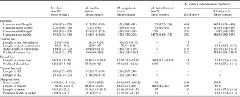
Morphometrics of haptoral sclerites of Macrogyrodactylus species from Zimbabwe, Kenya and Senegal
(All measurements are in micrometers.)

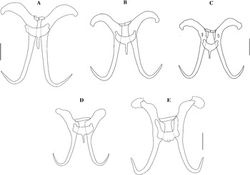
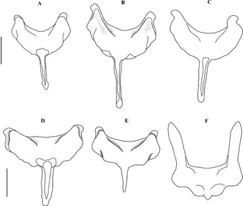
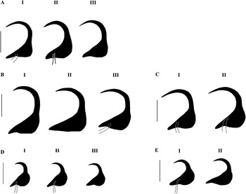
The size and shape of haptoral sclerites were used to delineate species (Table 2; Figs 1–3). The haptors of M. clarii from Zimbabwe were the largest of all species, and also larger than M. clarii specimens from the other regions. The shape of the haptors of M. clarii, M. karibae and M. congolensis were quite distinct (cf. Fig. 2 A and B), while M. clarii×heterobranchii (Ken & Zim) showed characters intermediate between those of M. clarii and M. heterobranchii. Macrogyrodactylus heterobranchii is smaller and its marginal hook sickle is more open than in M. clarii where the curve is more rounded and a little bit more closed (Fig. 3 A and B).
Haptoral sclerites of the Macrogyrodactylus spp. (A) M. clarii (Zim). (B) M. clarii (Ken). (C) M. heterobranchii (Sen), modified from Přikrylová and Gelnar (Reference Přikrylová and Gelnar2008). (D) M. karibae (Zim). (E) M. congolensis (Ken). Scale bars=100 μm.

Ventral bars of Macrogyrodactylus spp. from clariid catfish. (A) M. heterobranchii (Zim). (B) M. clarii (Zim). (C) M. clarii (Ken). (D) M. clarii×heterobranchii (Zim). (E) M. karibae (Zim). (F) M. congolensis (Ken). Scale bars=50 μm.

Marginal hook sickles of the Macrogyrodactylus spp. (A) M. heterobranchii specimens, I – Kenyan specimen, II – Senegalese specimen (Přikrylová and Gelnar, Reference Přikrylová and Gelnar2008), III – paratype specimen (MNHN 574 HF, Tk 91). (B) M. clarii specimens, I and II specimens from Zimbabwe, III – Kenyan specimen. (C) M. clarii×heterobranchii, I and II specimens from Kenya. (D) M. karibae specimens, I and II specimens from Zimbabwe, III – paratype specimen (MNHN 143, Tg 14). (E) M. congolensis specimens, I – specimen from Kenya, II – co-type specimen (1960.5.27.1-15). Scale bars=10 μm.

Molecular analysis and link with morphological results
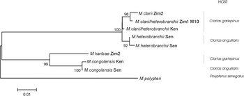
All phylogenetic methods produced the same tree; all clades are supported by high bootstrap values although the position of M. clarii×heterobranchii from Kenya remains unresolved (Fig. 4). There are 2 main groups, the first comprising M. karibae and M. congolensis, and the other group consists of M. heterobranchii, M. clarii, and the species that are intermediate between these 2 species both in genotype and in morphotype.
Neighbour-joining phylogram of African Macrogyrodactylus species inferred from the ITS1 and ITS2 rDNA sequences (General Time Reversible model with gamma-distributed rates).

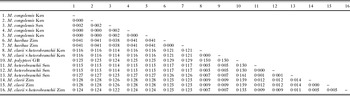
The pairwise genetic distances are summarized in Table 3. The distance between the 2 groups (M. clarii+M. heterobranchii versus M. karibae+M. congolensis) is about 12%, slightly less than the distance observed between the outgroup M. polypteri and all other species. M. polypteri is more closely related to the clade M. karibae+M. congolensis (distance of about 12·5%) than to the M. clarii+M. heterobranchii clade (distance of about 15·5%). The pairwise difference between M. clarii and M. heterobranchii is only 1·4%, while that between M. karibae and M. congolensis is 4%. The intraspecific/geographical differences range from 0·2% to 0·5%.
Genetic distances (Tamura Nei – gamma model) between the Macrogyrodactylus specimens sequenced in this study
(GB denotes specimen whose sequence was obtained from GenBank.)

The first 260 bp of the ITS1 sequence denoted in the tree as M. clarii×heterobranchii Zim is identical to M. clarii while the remainder of ITS1 and ITS2 is identical to M. heterobranchii with the exception of a single T/A substitution. This strongly suggests a hybrid origin of this sequence. The mutations in the sequence denoted as M. clarii×heterobranchii Ken are either shared with M. clarii (4 mutations) or with M. heterobranchii (6 mutations), with only a single unique T/A substitution, and are therefore also suggestive of an (albeit older) hybrid origin. These hypotheses are supported by the partial cox1 sequence. The cox1 sequence of M. clarii and M. clarii×heterobranchii Zim is identical with the exception of 2 transitions in a 705bp fragment (0·3% Kimura two parameter (K2P) distances, Kimura, Reference Kimura1980), while the distance between these 2 species and M. heterobranchii is 5·4% (K2P distances). The sequence of M. clarii×heterobranchii Ken is more closely related to M. heterobranchii (2·1% K2P distance), while the distance with M. clarii is about 5·5%.
DISCUSSION
Prior to this study, M. polypteri was the only species of Macrogyrodactylus that had been identified using molecular tools (Matějusová et al. Reference Matějusová, Gelnar, Verneau, Cunningham and Littlewood2003), while the other 8 species described on the African continent had been identified by morphology alone (Paperna, Reference Paperna1979; Douëllou and Chishawa, Reference Douëllou and Chishawa1995; Khalil and Mashego, Reference Khalil and Mashego1998; Přikrylová and Gelnar, Reference Přikrylová and Gelnar2008). This study therefore provides the first ITS1 and ITS2 rDNA and partial cox1 sequences of the species infesting the gills of clariid catfishes. In total, 15 specimens belonging to 4 Macrogyrodactylus species and 2 hybrids collected from 3 different African countries have been sequenced.
Hybridization
Parasites can provide much information about the hosts in which they live, such as the type of food they eat (Bush et al. Reference Bush, Fernández, Esch and Seed2001), their migration (Combes, Reference Combes2001), and also about the ecosystem in which they live. In this study, Macrogyrodactylus parasites provided evidence for historical distribution ranges of the catfish hosts. We showed that M. heterobranchii and M. clarii are very closely related sister taxa, differing only by 1·2–1·4% in the ITS rDNA region and 5·4% in the cox1 mtDNA fragment. Morphologically, they can be clearly differentiated from each other by the shape and size of the ventral bar and the shape of the marginal hook sickles. However, our sequence data suggest that they are able to interbreed, with specimens M. clarii×heterobranchii from Zimbabwe and Kenya being possible outcomes of such hybridization events.
The nuclear ribosomal RNA genes and the ITS region are very suitable markers to detect hybridization, because they can retain both parental copies for several generations before they are homogenized by concerted evolution (Sang et al. Reference Sang, Crawford and Stuessy1995). The rate or extent of homogenization after hybridization can, however, differ among species (Waters and Schaal, Reference Waters and Schaal1996). Biased homogenization towards one of the parental sequences can already occur in F2 hybrids or backcross generations, such that the signal of hybridization is blurred (Fuertes Aguilar et al. Reference Fuertes Aguilar, Rosselló and Nieto Feliner1999). In such cases, mitochondrial DNA can help to identify hybridization events, as this is a purely maternally inherited marker.
The signature of hybridization is still very clear in the specimen from Zimbabwe, where partial 18S rRNA and the beginning of the ITS region were identical to one parental sequence and the remaining part identical to the other parental species (except for 1 bp) as a result of crossing over. This together with the cox1 fragment that is nearly identical to the parental M. clarii, suggest it to be a ‘younger’ hybrid compared to the specimens from Kenya. However, the ITS and cox1 fragments already accumulated respectively 1 and 2 unique mutations in comparison to the parental species, suggesting it to be a stable hybrid. Parental backcrossing will complicate time estimations, as it will erase new mutations. Applying a standard molecular clock of 2% per million years (Avise, Reference Avise1994) for the cox1 fragment will therefore result in a timing of hybridization of at least 75 000 years ago, but it is probably much older. The specimens from Kenya appear to be the result of the reverse cross, with M. heterobranchii as the original female parent because these specimens had a cox1 sequence most similar to M. heterobranchii. The ITS profile was intermediate between both parental species, with alternating fixation of parental mutations. This pattern and the unique mutations in the cox1 fragment, suggest that these 2 specimens are the result of an older hybridization event, of at least 500 000 years according to the same calibration as above.
A prerequisite for hybridization to occur is sympatry. Normally M. heterobranchii is supposed to be specific to the vundu Heterobranchus longifilis Valenciennes, 1840 (N'douba and Lambert, Reference N'Douba and Lambert1999), while M. clarii is described on Clarias gariepinus (Khalil and Polling, Reference Khalil and Polling1997). Since Heterobranchus and Clarias are very closely related catfish genera with a very similar biology (Agnèse et al. Reference Agnèse, Teugels, Galbusera, Guyomard and Volckaert1997; Rognon et al. Reference Rognon, Teugels, Guyomard, Galbusera, Andriamanga, Volckaert and Agnèse1998; Agnèse and Teugels, Reference Agnèse and Teugels2005), host switching between both fish hosts might be facilitated. However, the current southern-most range of Heterobranchus longifilis is the Zambezi River system (Skelton, Reference Skelton1994), whose tributaries drain the northern parts of Zimbabwe (including Lake Chivero) (Marshall, Reference Marshall1999). One of the hybrids was found in the Save River System, a geographically distinct basin in southern Zimbabwe. One explanation for the presence of the parasite in this system could be that the Zambezi and Save systems used to be connected, but when they separated, Heterobranchus was restricted to the Zambezi system, while C. gariepinus remained in both systems, together with monogenean parasites contracted from the Zambezi. This is supported by the fact that the current geomorphological erosion surfaces of the Save-Runde system in Zimbabwe are connected to the Limpopo River basin in the south (Moore et al. Reference Moore, Cotteril, Broderick and Plowes2009), which in turn has been historically linked to the upper Zambezi system until the beginning of the Paleocene (Stankiewicz and de Wit, Reference Stankiewicz and de Wit2006). Our results might suggest a more recent exchange, which can possibly be linked with the Pleistocene cycling (Trauth et al. Reference Trauth, Larrasoan and Mudelsee2009); see below. The occurrence of the hybrids found in Lake Turkana (Kenya) could be explained by the fact that although only C. gariepinus is predominant in the lake, fossils of Heterobranchus sp. have been found (Trapani, Reference Trapani2008), which proves a historical connection with the Nile system (Goudie, Reference Goudie2005) where the latter fish species still occurs today (see also below).
Species complex
The observed hybridization events might lead to a species complex of closely related taxa that are difficult to identify based on morphology alone. Similar to this, the anisakid nematode Contracaecum rudolphii exists as a species complex comprising sibling species in different geographical regions (Shamsi et al. Reference Shamsi, Norman, Gasser and Beveridge2009). Future studies should focus on obtaining more molecular data on Macrogyrodactylus species and propose a sibling classification system of the M. clarii-heterobranchii complex, and our results pave the way for this possibility. It might well be that, with the current advances in molecular genotyping, previously described species-complexes turn out to be complexes of hybrid species. In that case, the biological species concept (Mayr, Reference Mayr1963) cannot be applied (no reproductive isolation) and the difficulties in species identification are not the result of incomplete lineage sorting but a result of the mixed, hybrid origin.
Host and parasite phylogeography
As suggested by Pariselle (personal communication; and also by Agnèse and Teugels, Reference Agnèse and Teugels2005), monogeneans can provide information on the evolution and zoogeography of fishes. This is because they have a close relationship with their host and they are mostly host specific (Kearn, Reference Kearn1998). Klassen and Beverly-Burton (Reference Klassen and Beverly-Burton1988) showed that the ecological association between ancyrocephalid monogeneans and their centrarchid fish host in North America could be attributed to the combined effects of host hybridization and the differential geographical distribution of hosts and parasites in response to Pleistocene glaciation events. Where hybridization between sympatric hosts takes place, changes in host ethology and ecology may be reflected by changes in their monogenean parasite specificity, prevalence and intensity (Le Brun et al. Reference Le Brun, Renaud, Berrebi and Lambert1992).
If we compare the 4 basins from where Macrogyrodactylus species were collected (Senegal River, Lake Turkana, Zambezi and Save Rivers), they seem distinctly separated spatially, but the parasite fauna suggests that they were once in sufficiently close contact to be able to exchange parasites. The above example of hybridization, together with the fact that M. karibae collected from C. gariepinus in the Save River basin in southern Zimbabwe (Barson et al. Reference Barson, Bray, Ollevier and Huyse2008) were morphologically identical to those collected from Lake Chivero (Zambezi system, this study), point to a historical connection between the two systems. Additionally, the close relationship between M. karibae from the Zambezi system and M. congolensis, which is absent from the Zambezi based on current data, was here confirmed phylogenetically, and might explain why Douëllou and Chishawa (Reference Douëllou and Chishawa1995) initially termed M. karibae as M. congolensis-karibae, although it has been found in South Africa (Khalil and Mashego, Reference Khalil and Mashego1998). This close relationship can be a result of the historical connection between the Congo and Zambezi River basins (ichthyofaunal regions 3 and 5), in which catfishes could have migrated across the wetland headwaters of these basins (Bell-Cross, Reference Bell-Cross and Snowball1965; Skelton, Reference Skelton1994). Geological evidence actually shows that a current tributary of the Zambezi River, the Luangwa River, used to be part of the Congo River before the east African rifting events in the Pliocene-Pleistocene periods (about 1–5 million years ago; see Goudie, Reference Goudie2005; Stankiewicz and de Wit, Reference Stankiewicz and de Wit2006).
There was a marked distinction (12·5% distance) between the M. congolensis+M. karibae clade and the M. heterobranchii+M. clarii clade although some catfish individuals were infected by representatives of the 2 clades, e.g. C. gariepinus in the Save River was infested by both M. clarii and M. karibae. This can probably be explained by the fact that this catfish has one of the widest geographical ranges (Rognon et al. Reference Rognon, Teugels, Guyomard, Galbusera, Andriamanga, Volckaert and Agnèse1998; Giddelo et al. Reference Giddelo, Arndt and Volckaert2002; Agnèse and Teugels, Reference Agnèse and Teugels2005; Jansen et al. Reference Jansen, Devaere, Weekers and Adriaens2006). The parasite M. congolensis shows a similarly wide distribution. Even though we currently have no sequence data of M. congolensis in southern African systemsFootnote *, its presence has been confirmed by morphology in the Limpopo system (Khalil and Mashego, Reference Khalil and Mashego1998; K. Christison, personal communication). Here we show that specimens from Senegal and Kenya (Lake Turkana) are very similar genetically (0·2% distance) in spite of the large geographical separation. Such a close relationship is also found for Clarias gariepinus populations from both regions and can be explained by the historical connection of the Nile system and West African basins which is currently lost because of climate changes (Trapani, Reference Trapani2008). This shows a common phylogeographical pattern for host and parasites, consistent with the savanna belt formation around the central African tropical forest shaped by Pleistocene glaciations (Hewitt, Reference Hewitt2004), which influenced many of Africa's fauna and flora (Trauth et al. Reference Trauth, Larrasoan and Mudelsee2009).
This study has shown that parasites can hold clues to the history of their hosts. Essential to this, is the simultaneous use of nuclear and mitochondrial markers, otherwise most hybridization events remain unnoticed (Huyse et al. Reference Huyse, Webster, Geldof, Stothard, Diaw, Polman and Rollinson2009). Parasite hybridization is the ultimate proof of (historical) close connection between host species, especially in case of host-specific parasites. Future ichthyological field studies of (African) fishes should therefore be matched by intensive parasitological studies.
ACKNOWLEDGMENTS
We are grateful to F. Volckaert and B. Hellemans for use of the DNA laboratory (K. U. Leuven) and Miloslav Jirků (Institute of Parasitology ASCR, České Budějovice, Czech Republic) who collected the material in Kenya. Zimbabwe fieldwork was logistically supported and assisted by Tamuka Nhiwatiwa, University of Zimbabwe technical staff and students, and the Save Valley Conservancy hosts (Chishakwe, Sango, Senuko and Humani ranches). Filip Volckaert, Luc Brendonck, Brian Marshall, Jos Odeurs and Frans Ollevier made valuable comments to an early draft of this manuscript. Comments by the two anonymous reviewers of this article have added value to its content.
FINANCIAL SUPPORT
The Flemish Interuniversity Council (VLIR-UOS) financed the Zimbabwe study through a scholarship grant to M.B. in the context of the UZ-VLIR IUC project. Collection of material in Kenya was supported by the grant KJB600960813 from the Grant Agency of the Academy of Sciences of the Czech Repulic; Ministry of Education, Youth and Sports of the Czech Republic (Ichthyoparasitology Research Centre – Project no. LC522). I.P. received financial support from the research project of Masaryk University (MSM 0021622416). Molecular analysis was funded through a ‘Krediet aan navorsers’ of the Research Foundation Flanders (FWO Vlaanderen) to T.H. M.P.M.V. and T.H. respectively are recipients of a Ph.D. and a post-doctoral fellowship of FWO Vlaanderen.