Advances in cardiac catheterisation techniques, miniaturisation and improvement in catheter design, equipment, and vascular access has significantly decreased the risk of complications related to the procedure, even in neonates and infants with critical congenital heart defect. Vascular complications have been reported between 3 and 6% of all cardiac catheterisation-related complications.Reference Vitiello, McCrindle, Nykanen, Freedom and Benson 1 , Reference Kobayashi, Sallaam and Aggarwal 2 Transient loss of pulse is reportedly more common than any major vascular injury.Reference Vitiello, McCrindle, Nykanen, Freedom and Benson 1 We report an infant with tricuspid atresia presenting with cardiogenic shock and coagulopathy related to progressive restriction of the ventricular septal defect and patent ductus arteriosus. She underwent an emergent transcatheter ductal stenting that successfully improved the pulmonary blood flow; however, the patient developed lower limb ischaemia following the procedure that necessitated below-knee limb amputation.
Case report
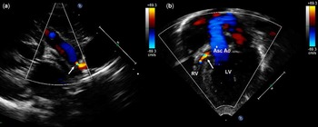
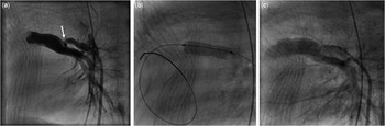
A 2-month-old girl born at 35 weeks gestation, weighing 3 kg with unrepaired tricuspid atresia (type 1b), small patent ductus arteriosus, and initially large ventricular septal defect providing adequate pulmonary blood flow, presented to the emergency room in cardiogenic shock and profound cyanosis from closing ventricular septal defect and tiny patent ductus arteriosus with severely decreased pulmonary blood flow. On arrival at the emergency room, she was seen to have severe metabolic acidosis with pH of 6.8, bicarbonate of 9, and O2 saturations were 40% on room air. She was immediately intubated and mechanically ventilated with 100% FIO2. Initial resuscitation included intubation with mechanical ventilation, intra-osseous venous access, fluid bolus, inotropic support, and prostaglandins was started at 0.2 μg/kg/minute to maintain and improve ductal patency. Bedside echocardiogram revealed moderately decreased left ventricular function, restrictive perimembranous ventricular septal defect, and tiny patent ductus arteriosus with small left-to-right shunt (Fig 1). Her haemoglobin was 16 gm%, decreased platelet count of 92,000 μl−1, coagulation profile revealed prolonged prothrombin time of 21 seconds (normal 9–12 seconds), partial thromboplastin time 51 seconds (normal 23–34 seconds), elevated D-dimer 18 mg/L (normal <0.6 mg/L), and decreased fibrinogen level of 125 mg/dl (normal 184–470 mg/dl), consistent with disseminated intravascular coagulation. Multiple blood products, including packed red blood cells, platelet infusion, fresh frozen plasma, and cryoprecipitate, were infused to correct the coagulopathy. Following transfer to the paediatric ICU, her oxygen saturation briefly improved to 50–60% on inspired oxygen of 100%; however, she had severe metabolic and lactic acidosis (pH 6.9, bicarbonate 12, lactate 10). After extensive discussions with the cardiovascular surgeon regarding emergency modified Blalock–Taussig shunt versus extracorporeal membrane oxygenation cannulation versus cardiac catheterisation, the decision was made to attempt to augment pulmonary blood flow by performing ductal stenting in the cardiac catheterisation lab. Left femoral arterial and venous access were obtained by percutaneous technique using a 4-Fr sheath in a single attempt. Intravenous heparin was not used because of the presence of coagulopathy. An angiogram in the distal arch of the aorta revealed tortuous patent ductus arteriosus with a severely restrictive pulmonary end, with minimal flow to the pulmonary arteries (Fig 2a). The extracorporeal membrane oxygenation team was activated as a precautionary measure in the event of failure to stent the patent ductus arteriosus in the catheterisation lab. Using a 4-Fr glide catheter, an 0.014 Hi-torque wire was advanced across the tortuous and stenotic patent ductus arteriosus into the distal right pulmonary artery. The left femoral artery short sheath was exchanged for a 4-Fr Mullins sheath to help control stent delivery and positioning. Further advancement of this sheath was met with resistance, and despite multiple attempts, we were unable to negotiate the passage beyond the left iliac artery. The sheath and dilator were withdrawn and exchanged for 4-Fr short Cordis sheath in the left femoral artery. There were two coronary stents, a 4×8 mm at the pulmonary end and a 4×8 mm at the aortic end, advanced and deployed across the patent ductus arteriosus with instantaneous improvement in O2 saturations (low 90s), metabolic acidosis (pH 7.2, bicarbonate 21), and increase in the blood pressure. Removal of the arterial sheath prompted manual compression at the left groin lasting for 45 minutes to stop the bleeding from severe coagulopathy. Progressive left lower limb discolouration with pulse loss at the popliteal and pedal level were detected 2 hours after catheterisation. The blood gases continued to show improvement in acid–base balance, and the diastolic blood pressures were maintained. With concern for ischaemia and venous occlusion, the central venous sheath was removed and intravenous heparin was started. Topical nitroglycerine and intravenous papaverine were administered for vasodilatory effect. At this point, skin discolouration with fixed mottling was noted in the area of the left thigh, calf, foot, and involved the toes. Emergency vascular exploration for critical left lower limb ischaemia was performed within 8 hours of the catheterisation procedure. Preoperative vascular ultrasound revealed no flow detected at the left common femoral artery, but there was a pulsatile flow in the proximal left external iliac artery. Significant findings on exploration included a thrombus within the common femoral artery extending proximally into the external iliac artery and dissection of the left external iliac artery, correlating the findings on the vascular ultrasound. A thrombectomy was performed, and the dissection of the left external iliac artery was repaired using a bovine pericardial patch. Intraoperative arteriogram showed widely patent left common, internal, and external iliac artery. There was no flow into the left superficial femoral artery or the profunda femoris artery, however. Over the course of the next few days, dry gangrene developed progressively with the demarcation level above the knee. The patient finally underwent above-knee amputation of her lower left extremity. Following prolonged hospital course and recovery she was discharged home. After 2 months, she underwent elective surgical modified Blalock–Tausig shunt as there was narrowing of the ductal stent related to intimal growth. She underwent palliative Hemi–Fontan surgery 1 year later.
Echocardiogram demonstrating restrictive VSD and PDA. ( a ) High parasternal short-axis view shows a tiny PDA (arrow) with small left-to-right shunt. ( b ) Apical four-chamber view shows a small restrictive VSD with left-to-right shunt (arrow) in systole. Asc Ao=ascending aorta; LV=left ventricle; PDA=patent ductus arteriosus; RV=right ventricle; VSD=ventricular septal defect.

Angiograms of a restrictive PDA with stent placement. ( a ) Angiogram of the DAO shows a small tortuous PDA with stenotic pulmonary end (arrow). There were two coronary stents positioned and deployed across the PDA ( b ) with significantly improved contrast flow ( c ). DAO=descending aorta; PDA=patent ductus arteriosus.

Discussion
Vascular injury is one of the most common complications associated with paediatric cardiac catheterisation.Reference Vitiello, McCrindle, Nykanen, Freedom and Benson 1 , Reference Kobayashi, Sallaam and Aggarwal 2 Vitiello et alReference Vitiello, McCrindle, Nykanen, Freedom and Benson 1 reported vascular complications in 189 procedures (3.8%), representing 34% of all catheterisation-related adverse events with major complications such as permanent arterial thrombosis in <0.1%. Data from our centre (n=527) showed vascular complications significantly higher in infants weighing <2.5 kg (6%) versus 2.5–3.5 kg (0.9%). Loss of arterial pulse was seen in 0.01% infants; however, no patient had limb ischaemia. Limb ischaemia is more commonly encountered as a complication in patients on extracorporeal membrane oxygenation following femoral artery cannulation approaching 50%.Reference Gander, Fisher and Reichstein 3 There is credible evidence suggesting that disseminated intravascular coagulation may play a role in half of the children who developed peripheral gangrene on extracorporeal membrane oxygenation support.Reference Alwi, Choo, Latiff, Kandavello, Samion and Mulyadi 4 Our patient presented with signs and symptoms of shock and disseminated intravascular coagulation related to progressive decrease in pulmonary blood flow. She was felt to be a high-risk surgical or extracorporeal membrane oxygenation cannulation candidate. As there was some flow noted through the tiny patent ductus arteriosus by echocardiogram (Fig 1), transcatheter ductal stenting was felt to be a prudent approach.
There could be several factors contributing to the vascular injury in our patient, despite our best efforts to minimise these risks. The smallest (4 Fr) available long delivery sheath to deploy ductal stents is the current standard of care in the neonatal period.Reference Alwi, Choo, Latiff, Kandavello, Samion and Mulyadi 4 Although our patient was 2 months old, she was relatively small for her age, weighing only 3 kg. There seemed to be a significant discrepancy in the size of a 4-Fr Mullins sheath and left iliac artery, possibly causing external iliac arterial dissection and injury, as noted during vascular surgery. In addition, the patient’s critical condition, shock, and prolonged post-catheterisation manual compression to stop bleeding because of the coagulopathy was speculated to have caused vascular thrombosis. Heparin was initiated after detection of pulse loss, despite the presence of coagulopathy with simultaneous use of blood products. Concerns of arterial run-off related to ductal stenting, as a cause of lower limb ischaemia, were negated with maintained and improving acid–base balance as well as diastolic blood pressures. As there was no improvement in perfusion, she was taken for an exploration surgery within 8 hours of the procedure.
A standard approach to treat decreased or absent pulse would include medical management within 6–8 hours of pulse loss, such as using leg warming, topical nitroglycerine, and intravenous heparin. If complete absence of pulse persists with no evidence of Doppler flow, further management involves using intravenous local papaverine, lidocaine, nitroglycerine, thrombolytics, and/or microvascular surgery.Reference Reed 5 – Reference Friedman, Fabre, Netscher and Jaksic 7 Thrombectomy, microvascular repair using venous patches, bypass grafts, and fasciotomies for the treatment of vascular injuries have been reported with complete limb recovery.Reference Filis, Arhontovasilis and Theodorou 6 – Reference Lin, Dodson and Bush 8 In our patient, the use of heparin and thrombolytic therapy was not an option in the presence of coagulopathy. Thrombectomy and patch repair of the external iliac artery improved flow to the femoral arteries, but were unable to restore perfusion distal to the femoral artery, requiring debridements and above-knee amputation. This case illustrates the complexity in the treatment of absent lower limb perfusion following catheterisation in the presence of coagulopathy.
Approaches to prevent severe leg ischaemia include early recognition of impending vascular damage, use of smallest arterial sheath, abort advancement of delivery sheath in the presence of any resistance across the femoral or iliac artery, and if necessary delivering a pre-mounted stent via the short sheath (Fig 2). There is a need for further miniaturisation of arterial delivery sheaths to deploy devices in smaller infants and neonates. Further research and treatment strategies for the use of anticoagulation in the setting of coagulopathy are warranted.
Our report also highlights the importance of early recognition of restrictive ventricular septal defect as well as unreliability of patent ductus arteriosus to maintain adequate pulmonary blood flow in single ventricle patients. Timely elective placement of a surgical systemic to pulmonary shunt would prevent catastrophic clinical presentation of shock with decreased perfusion. This may avoid the need for an emergent life-saving intervention and its associated risk for increased mortality and morbidity.
Acknowledgement
None.
Financial Support
This research received no specific grant from any funding agency, commercial, or not-for-profit sectors.
Conflicts of Interest
None.