Introduction
Relapsing polychondritis is a multi-system autoimmune disease characterised by inflammation and destruction of cartilaginous structures. The most common sites are the pinna, nose, laryngotracheobronchial tree and peripheral joints. In this article, we present our experience of performing laryngotracheal reconstruction in a patient with relapsing polychondritis and review the relevant literature.
Case report
Our patient was a 36-year-old gentleman who had been diagnosed with relapsing polychondritis 11 years previously. He had presented with auricular chondritis, subglottic stenosis and distal airway collapse. He had tested negative for an extensive panel of autoantibodies. He had initially been treated with prednisolone and methotrexate by his rheumatologist. In 2002, a year after he had been diagnosed with relapsing polychondritis, he unfortunately developed (human immunodeficiency virus negative) Kaposi's sarcoma in his limbs secondary to immunosuppression. As the laryngeal and tracheal architecture was destroyed, the laryngeal airway became more compromised and a decision was made by his otolaryngologist to fashion a surgical tracheostomy. He was referred to the oncology department, where he was started on various chemotherapy agents for Kaposi's sarcoma, including bleomycin, vincristine and liposomal doxorubicin (Caelyx®).
He subsequently developed bronchiectasis due to a recurrent cycle of respiratory infections caused by the combination of systemic immunosuppression and secretion retention. This was followed by the development of segmental atelectasis, mucus stasis and chronic Pseudomonas aeruginosa infection. At this stage, he required frequent hospital admissions, despite chemotherapy having been postponed for months.
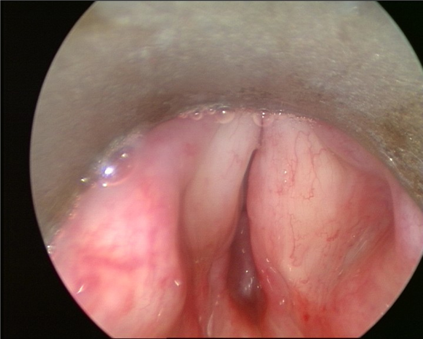
Computed tomography of his airway and thorax at this time confirmed severe collapse of the larynx with malacia, and a narrowing of his trachea and bronchi with bronchiectasis. The patient's oncologist decided that systemic therapy was no longer an option and the Kaposi's sarcoma was managed with palliative irradiation to the patient's limbs. He was referred to the Airway Reconstruction Unit at Imperial College in 2007 with a tracheostomy and no voice. He was managed by a multidisciplinary team (MDT), which included individuals from the rheumatology, interventional pulmonology and ENT departments. Endoscopic examination showed that the patient had reasonable movement of his vocal folds, but there was collapse of the subglottis, which did not respond to repeated laser treatment and dilatations (Figure 1). Attempts were also made to use stents in the tracheobronchial tree, but in the absence of cartilage to hold these stents in place, they quickly migrated and had to be removed.
Pre-operative view of patient's larynx.

A laryngotracheal reconstruction was proposed to restore the patient's voice as his difficulty in communicating was having a significant impact on his quality of life. This was performed in 2008.
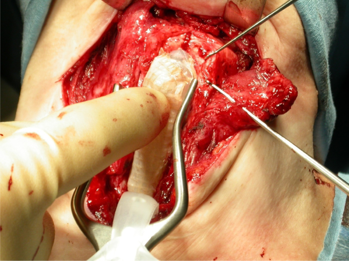
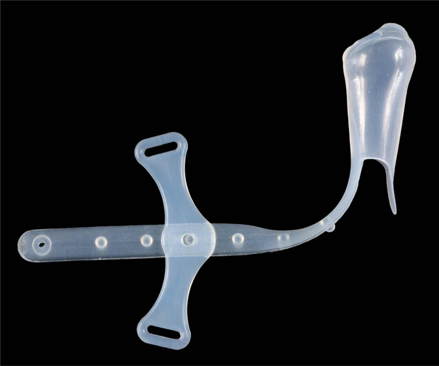
Computed tomography and suspension laryngoscopy indicated that damage and collapse of the cricoid were the cause of the complete subglottic stenosis. Cartilage as augmentation material could not be considered for obvious reasons. The patient therefore underwent an anterior laryngeal augmentation using hyoid bone pedicled on sternohyoid muscle (Figure 2). A standard laryngotracheal fissure procedure was performed, and the hyoid attached to the left sternohyoid was harvested in continuity and stitched to the edges of the cricoid using a non-absorbable monofilament. The reconstruction was supported endoluminally by an Eliachar laryngeal stent (Figure 3), which was covered with a superficial skin graft as a biological dressing. The patient retained the tracheostomy tube, and the stent was removed endoscopically two weeks later. Once the stent was removed, the patient had a reasonable quality of voice, which was maintained for the four years following this procedure. Unfortunately, he could not be decannulated because of the difficulty in clearing secretions, and because of the collapse of the trachea and bronchi. He has therefore been managed using a Silastic™ T-tube with a long lower limb, so that the majority of the trachea could be supported and the tube could be uncapped for suctioning.
Intra-operative view of anterior laryngeal augmentation using hyoid bone pedicled on sternohyoid muscle.

Eliachar laryngeal stent.

The histology from the excised fragments of the subglottis showed trabecular bone with foci of hyaline cartilage. There was no evidence of remodelling, infection, inflammation or granuloma.
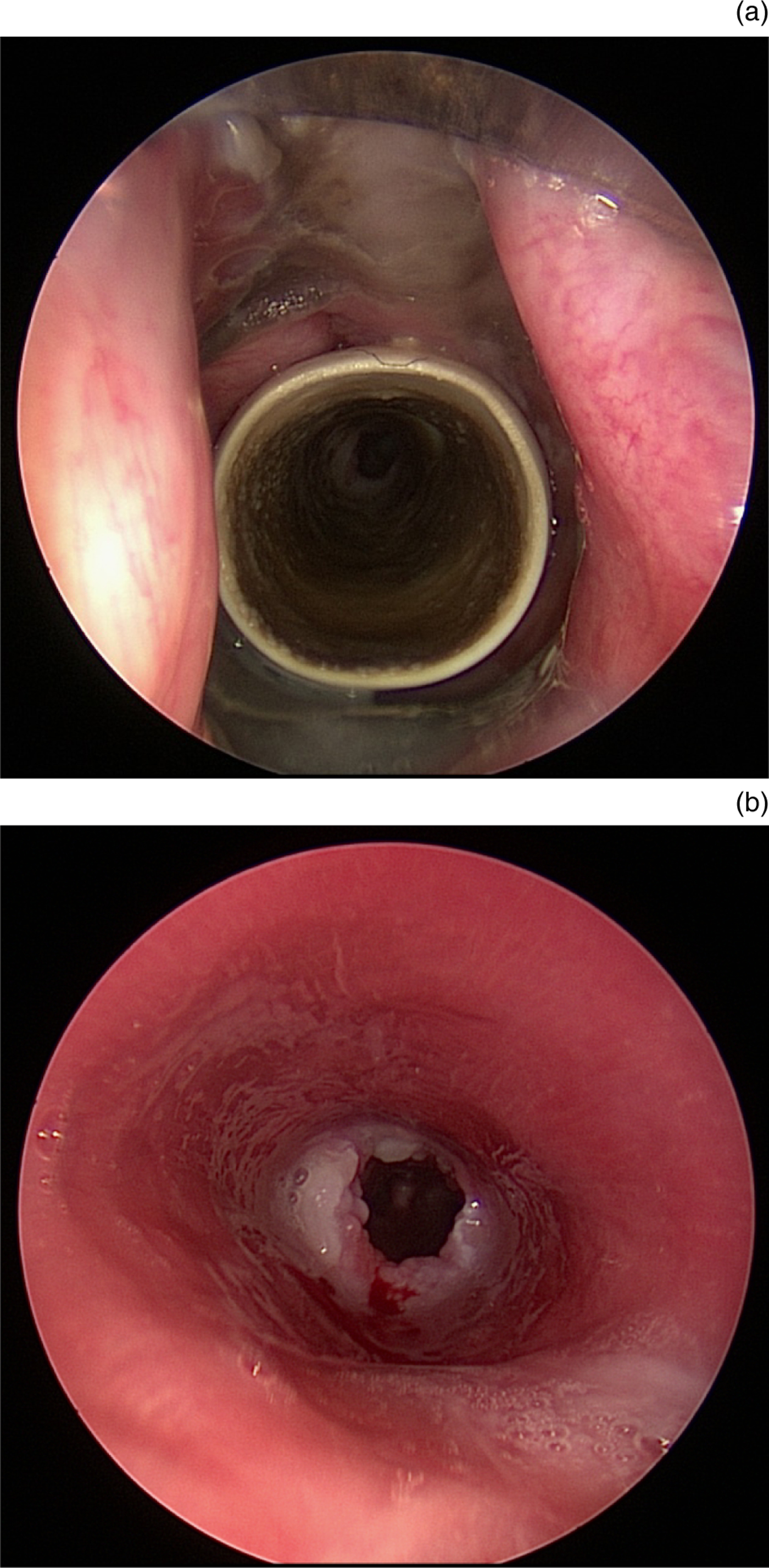
The patient was referred for consideration of a bone marrow transplant to halt the autoimmune process, which if successful was to be followed by a tissue engineered tracheobronchial replacement procedure. Unfortunately, this was not feasible; the MDT felt that his respiratory function was too compromised as a result of repeated infections. His last T-tube change was carried out in 2012, four years post-laryngotracheal reconstruction (Figure 4).
Laryngoscopic views from last T-tube change, showing (a) reconstructed larynx with T-tube in situ and (b) trachea with absence of normal tracheal rings.

Discussion
Relapsing polychondritis is a relapsing–remitting, multi-system autoimmune disease. It is characterised by the inflammation and destruction of cartilaginous structures. It can also involve non-cartilaginous structures such as the eyes, skin and cardiovascular system. This article focuses on respiratory tract involvement, which occurs in 20–50 per cent of patients.Reference Ernst, Rafeq, Boiselle, Sung, Reddy and Michaud1–Reference McAdam, O'Hanlan, Bluestone and Pearson3 Clinical features include stridor, wheeze, dyspnoea, cough, hoarseness and anterior laryngotracheal tenderness. Several mechanisms contribute to the pathophysiology of airway obstruction, including inflammatory oedema of the airway, and chondritic weakening of tracheal cartilage rings and replacement with fibrotic tissue. This causes tracheomalacia and collapse in the early stages, followed by cicatricial contraction and stenosis in the later stages.Reference Mohsenifar, Tashkin, Carson and Bellamy4 Histological findings show: loss of the basophilic staining property of the cartilage matrix, perichondrial round cell formation, polymorphonuclear leucocytes, lymphocytes, monocytes and plasma cell infiltration. Tracheal mucosa appears oedematous, and cartilage rings show changes ranging from mild inflammation to total resorption by granulation tissue.Reference Herman, Kelley, Harris, Ruddy and Sledge5 The result is airway collapse, obstruction and recurrent pneumonia.
Diagnostic criteria for relapsing polychondritis have been described (Table I),Reference McAdam, O'Hanlan, Bluestone and Pearson3, Reference Damiani and Levine6 but there are no specific investigations for the condition. Screening for anti-nuclear antibodies, anti-neutrophil cytoplasmic antibodies, rheumatoid factors, cryoglobulins, tuberculosis, viral hepatitis and syphilis is performed to exclude diseases that can follow a similar course. Furthermore, relapsing polychondritis can occur in conjunction with autoimmune disease.Reference Michet, McKenna, Luthra and O'Fallon2, Reference McAdam, O'Hanlan, Bluestone and Pearson3 Plain X-ray findings include tracheal stenosis and the calcification of cartilage. Computed tomography scans reveal tracheal and bronchial thickening, stenosis, and calcification. Smooth anterior and lateral wall thickening, with sparing of the posterior wall and main bronchi is virtually pathognomonic of this disease. Histological diagnosis is used when the clinical diagnosis is equivocal.Reference Damiani and Levine6
Relapsing polychondritis diagnostic criteria

*Three of six clinical features necessary for diagnosis. †One of three conditions necessary for diagnosis.
Surgical treatments for airway involvement in relapsing polychondritis include tracheostomy, dilatations, intraluminal stents, laser extirpation, local steroid injections and, rarely, laryngotracheal reconstruction. We found three papers that describe laryngotracheal reconstruction for relapsing polychondritis, two of which were written by the same authors. In their first paper, Cansiz and colleagues described a 25-year-old female with subglottic stenosis and no other clinical features of relapsing polychondritis.Reference Cansiz, Yilmaz and Duman7 They based the diagnosis of relapsing polychondritis on the cartilage histology, which alone does not fulfil the diagnostic criteria. In their second paper, these authors described the same patient, as well as two other female patients (who again did not fit the diagnostic criteria) with subglottic stenosis as their only manifestation.Reference Karaman, Duman, Cansiz, Yilmaz and Ibrahimov8 Furthermore, they described using nasal and auricular cartilage as grafts after tracheal resection, which could be affected by the natural disease process. The accuracy of their diagnoses is questionable as idiopathic subglottic stenosis in females is a well-recognised entity.Reference Blumin and Johnston9–Reference Dedo and Catten11
In the oldest paper, written by Spraggs et al., the authors describe two patients who received either laryngotracheoplasty or laryngotracheal reconstruction.Reference Spraggs, Tostevin and Howard12 The first patient received multiple previous surgical interventions before finally undergoing laryngotracheoplasty. They used a medial clavicular graft mobilised on the sternomastoid, which was inserted into a laryngofissure and midline tracheal incision above the tracheostomy. The patient was decannulated a year later. Unfortunately, she died three years later of pneumonia. Their second patient had previously had an emergency tracheostomy tube inserted. Laryngotracheal reconstruction was performed with a sternohyoid myocutaneous rotatory door flap. Although the procedure was complicated by flap prolapse into the airway, she was decannulated six months later.
In the case reported here, we performed an anterior laryngeal augmentation using hyoid bone pedicled on sternohyoid muscle. The use of the hyoid as an autologous graft for laryngotracheal reconstruction was first described by Looper in 1938.Reference Looper13 The use of muscle pedicled hyoid flaps has also been reported previously.Reference Ward, Canalis, Fee and Smith14, Reference Cansiz, Yener, Sekercioglu and Gunes15
A temporary tracheal stent was inserted during the operation and removed weeks later. The tracheostomy tube was then replaced with a Montgomery T-tube to allow a longer length of airway to be stented. The laryngotracheal reconstruction allowed our patient to vocalise again after a period of aphonia. However, distal airway disease secondary to disease progression prevented his quality of life from being significantly improved.
There is great discrepancy in the survival rates that have been reported over the years for patients with relapsing polychondritis, which probably reflects the diversity in the natural history of the disease. In 1986, the 10-year survival rate in 112 patients was 55 per cent.Reference Michet, McKenna, Luthra and O'Fallon2 In 1998, a study by Trentham et al. comprising 66 patients reported an 8-year survival rate of 94 per cent.Reference Trentham and Le16 In one of the largest recent case series comprising a total of 145 patients, in which 31 patients had airway involvement, the authors demonstrated that airway involvement could occur at any stage of the disease; they reported a 4-year survival rate of 97 per cent for relapsing polychondritis patients with airway involvement.Reference Damiani and Levine6 Although airway involvement has previously been regarded as a poor prognostic sign,Reference Michet, McKenna, Luthra and O'Fallon2 our report corroborates the more recent studies which show that airway measures are effective at slowing disease progression and can effectively palliate patients for substantial periods.Reference Ernst, Rafeq, Boiselle, Sung, Reddy and Michaud1, Reference Spraggs, Tostevin and Howard12
Conclusion
Laryngotracheal reconstruction for relapsing polychondritis can be feasible and beneficial to patients. However, despite aggressive surgical and medical therapy, many patients may experience disease progression. Subglottic or upper tracheal procedures, including tracheostomies, intraluminal stents and laryngotracheal reconstruction, may be complicated by frequent blockage, tube displacement and obstruction from more distal airways (as seen in our case).Reference Spraggs, Tostevin and Howard12, Reference Trentham and Le16–Reference Eng and Sabanathan18
Optimal medical management of relapsing polychondritis is yet to be established,Reference Rafeq, Trentham and Ernst19, Reference Lahmer, Treiber, Werder, Foerger, Knopf and Heemann20 but the availability of newer immunomodulating agents such as etanercept hold promise. As it is difficult to predict survival, we would recommend careful selection of patients for laryngotracheal reconstruction, taking into account disease severity, the extent of distal airways disease, medical fitness for surgery and the available facilities.