Introduction
Trichinella spiralis, the aetiologic agent of trichinosis, infects a wide range of mammalian hosts and is one of the most widespread parasitic diseases worldwide. The parasite can persist in muscle tissues for many years (Froscher et al., Reference Froscher, Gullotta, Saathoff and Tackmann1988) through its ability to produce a variety of biologically active proteins that suppress the host immune response, notably macrophage migration inhibitory factor (MIF) and cysteine protease inhibitors.
Mammalian MIF, capable of promoting and modulating the magnitude of the response, is a key regulatory cytokine in innate and adaptive immune responses (Bacher et al., Reference Bacher, Metz, Calandra, Mayer, Chesney, Lohoff, Gemsa, Donnelly and Bucala1996; Vermeire et al., Reference Vermeire, Cho, Lolis, Bucala and Cappello2008). The orthologues of host MIF have been elaborated in various parasitic nematodes (Pastrana et al., Reference Pastrana, Raghavan, FitzGerald, Eisinger, Metz, Bucala, Schleimer, Bickel and Scott1998; Tan et al., Reference Tan, Edgerton, Kumari, McAlister, Roe, Nagl, Pearl, Selkirk, Bianco, Totty, Engwerda, Gray and Meyer2001; Zang et al., Reference Zang, Taylor, Wang, Meyer, Scott, Walkinshaw and Maizels2002; Augustijn et al., Reference Augustijn, Kleemann, Thompson, Kooistra, Crawford, Reece, Pain, Siebum, Janse and Waters2007; Cho et al., Reference Cho, Jones, Vermeire, Leng, DiFedele, Harrison, Xiong, Kwong, Chen, Bucala, Lolis and Cappello2007; Cordery et al., Reference Cordery, Kishore, Kyes, Shafi, Watkins, Williams, Marsh and Urban2007; Kamir et al., Reference Kamir, Zierow, Leng, Cho, Diaz, Griffith, McDonald, Merk, Mitchell, Trent, Chen, Kwong, Xiong, Vermeire, Cappello, McMahon-Pratt, Walker, Bernhagen, Lolis and Bucala2008; Shao et al., Reference Shao, Han, Lin, Zhang, Zhong, Feng, Guo and Wang2008, Reference Shao, Zhong, Zhou, Han, Lin, Wang, Bu, Zhang, Su and Wang2010). Trichinella spiralis MIF (TsMIF) (Pennock et al., Reference Pennock, Behnke, Bickle, Devaney, Grencis, Isaac, Joshua, Selkirk, Zhang and Meyer1998), an orthologue of mammalian MIF, is expressed by all stages of the parasite, including adult worms, newborn larvae, pre-cyst muscle larvae and post-cyst muscle larvae, although not all stages express it at the same level (Wu et al., Reference Wu, Boonmars, Nagano, Nakada and Takahashi2003). The translated sequence shows 42% and 44% identity to human and mouse MIF, and TsMIF can modulate the host immune defence in infected tissue (Tan et al., Reference Tan, Edgerton, Kumari, McAlister, Roe, Nagl, Pearl, Selkirk, Bianco, Totty, Engwerda, Gray and Meyer2001; Wu et al., Reference Wu, Boonmars, Nagano, Nakada and Takahashi2003). Recombinant parasite MIFs of T. spiralis (TsMIF) (Tan et al., Reference Tan, Edgerton, Kumari, McAlister, Roe, Nagl, Pearl, Selkirk, Bianco, Totty, Engwerda, Gray and Meyer2001), Brugia malayi (BmMIF-1 and -2) (Zang et al., Reference Zang, Taylor, Wang, Meyer, Scott, Walkinshaw and Maizels2002) and Ancylostoma ceylanicum (AceMIF) (Cho et al., Reference Cho, Jones, Vermeire, Leng, DiFedele, Harrison, Xiong, Kwong, Chen, Bucala, Lolis and Cappello2007) can induce chemotaxis and, as with recombinant human MIF, prevent the random migration of human monocytes. It was also reported that Brugia MIF synergizes with interleukin (IL)-4 to induce the development of fully suppressive macrophages in vitro (Prieto-Lafuente et al., Reference Prieto-Lafuente, Gregory, Allen and Maizels2009). High levels of MIF might actually block activator protein-1 (AP-1)-dependent proinflammatory gene expression by binding to the transcription factor Jun activation domain-binding protein 1 (Jab1) (Kleemann et al., Reference Kleemann, Hausser, Geiger, Mischke, Burger-Kentischer, Flieger, Johannes, Roger, Calandra, Kapurniotu, Grell, Finkelmeier, Brunner and Bernhagen2000). By secreting MIF at the site of infection, helminths might induce further production of endogenous host MIF, creating a local or possibly systemic anti-inflammatory host environment (Vermeire et al., Reference Vermeire, Cho, Lolis, Bucala and Cappello2008; Shao et al., Reference Shao, Zhong, Zhou, Han, Lin, Wang, Bu, Zhang, Su and Wang2010) and thus beneficial to the persistence of helminths in the host.
Parasitic nematodes secrete a diverse group of cysteine protease inhibitors and important immunomodulatory factors (Hartmann & Lucius, Reference Hartmann and Lucius2003; Gregory & Maizels, Reference Gregory and Maizels2008) that interact significantly with the host immune system. Recently, a novel multi-cystatin-like protein (MCD-1) secreted by T. spiralis (TsMCD-1) was identified as a new member of the non-inhibitory cystatin-related proteins (Robinson et al., Reference Robinson, Massie and Connolly2007). Unlike other parasitic cystatins (Lustigman et al., Reference Lustigman, Brotman, Huima, Prince and McKerrow1992; Dainichi et al., Reference Dainichi, Maekawa, Ishii, Zhang, Nashed, Sakai, Takashima and Himeno2001; Newlands et al., Reference Newlands, Skuce, Knox and Smith2001; Murray et al., Reference Murray, Manoury, Balic, Watts and Maizels2005), recombinant TsMCD-1 is incapable of inhibiting the cysteine protease papain in vitro (Robinson et al., Reference Robinson, Massie and Connolly2007). Expression of TsMCD-1 is largely restricted to intracellular stages, predominantly in the adult worms, and acts as a proinflammatory factor (Nagano et al., Reference Nagano, Wu and Takahashi2009).
The importance of immunomodulatory molecules in immune responses and in parasitism has been demonstrated in cases where they have been used as antigens, or co-expressed as fusion antigens, to construct DNA vaccines against parasite helminths (LoVerde et al., Reference LoVerde, Carvalho-Queiroz and Cook2004; Beghetto et al., Reference Beghetto, Nielsen, Del Porto, Buffolano, Guglietta, Felici, Petersen and Gargano2005; Solis et al., Reference Solis, Ostoa-Saloma, Lugo-Martinez, Johnston and Laclette2005; Fallon & Alcami, Reference Fallon and Alcami2006; Kutzler & Weiner, Reference Kutzler and Weiner2008). Trichinella spiralis-derived MIF (TsMIF) and multi-cystatin-like protein (TsMCD-1) are both important modulators to the host immune response, yet their possible antigenicity and protection against T. spiralis infection are unclear. In the present study, we assessed the immune response and protective immunity induced by vaccination with recombinant plasmids expressing/co-expressing TsMIF and/or TsMCD-1 in BALB/c mice.
Materials and methods
Proteins, antiserum, animals and parasites
Recombinant TsMIF-TsMCD-1 protein expressed in Escherichia coli BL21 (DE3) and mouse antiserum against TsMIF-TsMCD-1 were provided by the Laboratory of Veterinary Molecular and Immunological Parasitology, Nanjing Agricultural University, China. Female 6-week-old BALB/c mice were purchased from the Experimental Animal Centre of Nanjing Medical University and were maintained in plastic microisolator cages before and after immunization. Trichinella spiralis strain ISS 534 was maintained in BALB/c mice. Adult (AD) parasites were recovered from the small intestines of Sprague–Dawley rats on day 5 post-infection (Zhu et al., Reference Zhu, Long, Xu and Gui1993). All parasites were washed extensively several times in phosphate-buffered saline (PBS) and stored at − 80°C for use.
Construction of expression plasmids
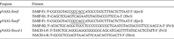
To amplify the open reading frames (ORFs) of Tsmif and Tsmcd-1, total RNA was isolated from adult parasites and amplified by reverse transcription-polymerase chain reaction (RT-PCR) using primers designed from the published sequences (GenBank AY050661 and DQ777102, respectively). As shown in table 1, two different DNA fragments of Tsmif and Tsmif″ were amplified to include different restriction sites with or without translation stop codons. An artificial Kozak sequence (GCCACC) was included in sense primers to permit efficient initiation of translation (Kozak, Reference Kozak1987). Amplicons were then ligated into the expression vector pVAX1 (Invitrogen, Carlsbad, California, USA) and transformed into E. coli DH5α (Invitrogen). A fusion expression plasmid, pVAX1-Tsmif-Tsmcd-1 was constructed from recombinant plasmids pVAX1-Tsmif″ and pVAX1-Tsmcd-1. A flexible peptide linker (SerGly4Ser) was introduced into the fusion protein between the two coding sequences to facilitate protein folding in their native conformation (Frigerio et al., Reference Frigerio, Jolliffe, Di Cola, Felipe, Paris, Neuhaus, Lord, Ceriotti and Roberts2001; Hu et al., Reference Hu, Li, Yang, Li, Xia, Li, Wang and Zhang2004). Clones containing inserts of the expected size were selected by restriction digestion and subjected to DNA sequencing using T7 and BGH primers by Invitrogen (Invitrogen Co. Ltd, Shanghai, China). DNA and predicted amino acid sequences were analysed using Lasergene 7.1 software (DNASTAR Inc., Madison, Wisconsin, USA).
Primers for PCR amplification.
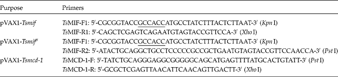
Shaded, restriction site; underlined, Kozak sequence; bracketed, name of restriction site; framed, flexible peptide linker gene.
Immunization and challenge of mice
Six-week-old female BALB/c mice were divided into groups of 20 animals. Three groups were immunized with the recombinant plasmids pVAX1-Tsmif, pVAX1-Tsmcd-1 and pVAX1-Tsmif-Tsmcd-1, respectively, and two control groups were immunized with pVAX1 plasmid or PBS (pH 7.4). Plasmid DNA for immunization was prepared using Classic Endo-free Plasmid DNAout (Tiandz, Beijing, China) and quantified using a Hitachi U-2800 spectrophotometer. Plasmid DNA was diluted to 1 μg/μl in PBS and 100 μg aliquots were injected into the quadriceps femoris muscle of mice twice at an interval of 2 weeks. Two weeks following the second immunization, challenge was carried out by oral infection with 200 L1 infectious T. spiralis larvae per animal.
Detection of in vivo target gene expression by Western blotting
The detection protocol was as described previously (Wallach et al., Reference Wallach, Mencher, Yarus, Pillemer, Halabi and Pugatsch1989; Xu et al., Reference Xu, Song, Xu, Yan, Shah and Li2008). Ten days post-vaccination, five mice in each group were sacrificed and injected muscles were homogenized in RIPA solution (50 mmol/l Tris–HCl, 150 mmol/l NaCl, 1% Nonidet P-40, 0.1% sodium dodecyl sulphate (SDS)) for 3 h, and supernatants were collected after centrifugation at 12,000 rpm. Contralateral uninjected muscles from the same animals were used as controls. Western blotting used a 1:40 dilution of mouse antiserum directed against recombinant TsMIF-TsMCD-1 fusion protein as first antibody and horseradish peroxidase (HRP)-conjugated goat anti-mouse IgG as second antibody (Sigma-Aldrich, St. Louis, Missouri, USA). Blots were developed using diaminobenzidine HRP substrate and a chromogenic reagent kit (Boster, WuHan, China).
Analysis of antibody responses
Blood was drawn from the ophthalmic venous plexus of ten mice in each group at 0, 10, 20 and 28 days following the first vaccination. Serum was isolated by centrifugation and stored at − 20°C until use. Serum antibody (IgG, IgG1, IgG2a, IgG2b, IgA, IgM and IgE isotypes) levels were measured in triplicate by indirect enzyme-linked immunosorbent assay (ELISA). Briefly, 96-well plates were coated with 5 μg/ml recombinant TsMIF-TsMCD-1 proteins (100 μl/well) and incubated for 18–24 h at 4°C. Serum sample were serially diluted (twofold dilutions) in buffer (PBS with 0.05% Tween20), added at 100 μl/well and incubated for 1.5 h at 37°C. Sera from non-immunized mice were used as a negative control. After washing three times, plates were incubated with a 5000-fold dilution of HRP-conjugated goat anti-mouse IgG, IgG1, IgG2a, IgG2b, IgA, IgM or IgE (Santa Cruz Biotechnology, Santa Cruz, California, USA) for 1 h at 37°C, respectively. After a final washing, 100 μl of substrate solution (3,3′,5,5′-tetramethylbenzidine, TMB; Amresco, Solon, Ohio, USA) was added and incubated for 10 min at 37°C. Reactions were halted by the addition of 50 μl 2 m sulphuric acid, and optical densities at 450 nm (OD450) and 570 nm (OD570) were determined using an automated microplate reader (Rayto, Shenzhen, China). Data were analysed after subtraction of the background OD570 reading from the OD450 value (OD450 − 570). The cutoff titre was defined as the lowest antibody concentration at which the OD450 − 570 of sample (S) was equal to that of the negative control (N) (P < 0.05).
Analysis of serum cytokines
Sandwich ELISA (Boster, WuHan, China) performed according to the manufacturer's instructions was used to determine the concentrations of five cytokines (interferon (IFN)-γ, IL-4, IL-5, transforming growth factor (TGF)-β1 and IL-17). Serum samples were isolated from ten mice of each group at 0, 10, 20 and 28 days following the first vaccination. Briefly, biotin-conjugated anti-cytokine, avidin-HRP conjugate and TMB substrate solution were successively employed; plates were read at 450 nm.
Flow cytometric analysis of CD4+/CD8+ T cells
Two weeks following the last immunization, whole blood was drawn from five mice of each group for analysis by flow cytometry. Peripheral blood lymphocytes were analysed using fluorescein isothiocyanate (FITC)-labelled mouse monoclonal anti-CD3 antibody or phycoerythrin (PE)-labelled mouse monoclonal anti-CD4 or -CD8 antibodies (MultiSciences, HangZhou, China). Labelled cells were analysed using a FACScan flow cytometer with CellQuest software (BD Biosciences, Franklin Lakes, New Jersey, USA). Results were analysed using FCS Express V3 software (version 3.00.0316; De Novo Software, Los Angeles, California, USA). The percentages of CD4+ T cells and CD8+ T cells in peripheral blood lymphocytes were determined and the CD4+/CD8+ ratio was calculated.
Detection of larvae in muscle
Five weeks after challenge infection, ten mice in each group were sacrificed. Trichinella spiralis muscle larval burden was evaluated as larvae per gram (LPG) in muscles of the diaphragm, anterior limb and neck following treatment with acid-pepsin digestion solution (Zhu et al., Reference Zhu, Long, Xu and Gui1993) containing 0.6% w/v pepsin (800–2500 units/mg; Sigma-Aldrich); 37% w/v HCl was added to give a pH of between 1.5 and 2. Muscles were mixed with the digestion solution (25 ml/g tissue) and incubated at 38°C for 6 h. Larvae in each digested sample were counted using light microscopy at 40 × magnification. The reduction of muscle larval burden was calculated using the following formula: reduction rate of muscle larvae = [(mean of LPG in PBS control − mean of LPG in DNA vaccination)/mean of LPG in PBS control] × 100%.
Statistical analysis
Statistical significances of inter-group differences were determined by analysis of variance (ANOVA) using SPSS Statistical Software (version 16.0; SPSS Inc., Chicago, Illinois, USA); P < 0.05 was taken to indicate statistical significance.
Results
Sequence analysis of recombinant expression plasmids
Sequence analysis indicated that the amplified fragment of Tsmif consisted of 345 bp, and the predicted ORF encoded a protein of 115 amino acid residues with a molecular mass of 12.5 kDa and 99.7% identity to the published sequence (GenBank AY050661). The amplified fragment of Tsmcd-1 consisted of 1221 bp and the predicted ORF encoded a protein of 407 amino acid residues with a molecular mass of 45.9 kDa and 99% identify to the published sequence (GenBank DQ777102).
Detection of in vivo target gene expression
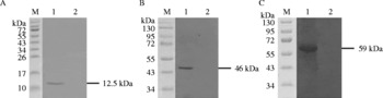
Western blot analysis revealed specific immunoreactive bands at ~12.5 kDa in pVAX1-Tsmif-injected muscle (fig. 1A, lane 1), ~46 kDa in pVAX1-Tsmcd-1-injected muscle (fig. 1B, lane 1), and ~59 kDa in pVAX1-Tsmif-Tsmcd-1-injected muscle (fig. 1C, lane 1). No immunoreactive bands were detected in contralateral non-injected muscle (fig. 1A–C, lane 2).
Detection of in vivo target gene expression by Western blotting was carried out with mouse antiserum against recombinant TsMIF-TsMCD-1 protein. Ten days following the first vaccination, five mice in each group were sacrificed and injected muscles of mice vaccinated with pVAX1-Tsmif (A), pVAX1-Tsmcd-1 (B) and pVAX1-Tsmif-Tsmcd-1 (C), respectively, were homogenized and supernatants were collected for Western blotting; contralateral uninjected muscles from the same animals were used as negative controls. Lanes: M, PageRuler™ prestained protein ladder (SM0671, Fermentas, Burlington, Canada); 1, the protein extract of injected muscle; 2, the contralateral uninjected muscle (control).

Antibody response induced by recombinant plasmids
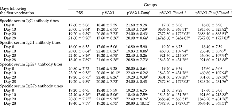
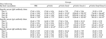
Sera collected 0, 10, 20 and 28 days following the first vaccination were assayed for the presence of specific antibodies against recombinant TsMIF-TsMCD-1 protein. As shown in tables 2 and 3, compared to PBS control, no antibody (IgM, IgG, IgA or IgE) response was detected in the pVAX1 control group (P>0.05) or in mice vaccinated with pVAX1-Tsmif (P>0.05). A clear IgM, IgG and IgA antibody response was observed in both pVAX1-Tsmcd-1 and pVAX1-Tsmif-Tsmcd-1 immunized animals (P < 0.05 compared to PBS or pVAX1 control groups in both cases), whereas IgE antibody was not significantly induced in any group.
ELISA titres of specific serum IgG, IgG1, IgG2a and IgG2b antibodies against the recombinant TsMIF-TsMCD-1 protein in mice.
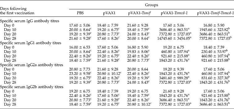
Blood was drawn from ten mice in each group at 0, 10, 20 and 28 days following the first vaccination. Serum was isolated and the production of specific antibodies (IgG, IgG1, IgG2a and IgG2b isotypes) was measured in triplicate by indirect enzyme-linked immunosorbent assay (ELISA). Values are shown as mean ± standard deviation (n = 10 mice/group). Data bearing a different superscript letter (a–c) within a line differ significantly from one another (P < 0.05).
ELISA titres of specific serum IgM, IgA and IgE antibodies against the recombinant TsMIF-TsMCD-1 protein in mice.

Blood was drawn from ten mice in each group at 0, 10, 20 and 28 days following the first vaccination. Serum was isolated and the production of specific antibodies (IgM, IgA and IgE isotypes) was measured in triplicate by indirect enzyme-linked immunosorbent assay (ELISA). Values are shown as mean ± standard deviation (n = 10 mice/group). Data bearing a different superscript letter (a–c) within a line differ significantly from one another (P < 0.05).
Compared to the pVAX1-Tsmcd-1 vaccine, levels of IgG antibody induced in mice vaccinated with pVAX1-Tsmif-Tsmcd-1 were significantly lower (P < 0.05). IgG antibodies were detected at 10 days following the first immunization, and increased progressively to titres of 14 745.60 ± 3454.05 (pVAX1-Tsmcd-1) and 7372.80 ± 1727.03 (pVAX1-Tsmif-Tsmcd-1) before challenge (28 days following the first immunization). Further analysis of IgG isotypes indicated that both IgG2a and IgG2b had higher titres than IgG1 in these two groups (P < 0.05 in all cases). We also observed that IgM antibody levels in mice vaccinated with pVAX1-Tsmif-Tsmcd-1 were lower than that in animals receiving pVAX1-Tsmcd-1 (P < 0.05). IgM antibodies were detected 10 days following the first immunization and reached peak titres of 2252.80 ± 647.63 (pVAX1-Tsmcd-1) and 1126.40 ± 323.82 (pVAX1-Tsmif-Tsmcd-1) 20 days following the first immunization. IgA antibodies were detected with lower titres of 230.40 ± 53.97 (pVAX1-Tsmcd-1) and 121.60 ± 20.24 (pVAX1-Tsmif-Tsmcd-1) 20 days following the first immunization, and decreased thereafter.
Cytokine production
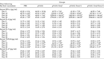
Concentrations of five cytokines (IFN-γ, IL-4, IL-5, TGF-β1 and IL-17) were measured in sera collected 0, 10, 20 and 28 days following the first vaccination. As shown in table 4, serum IL-4, IL-5 and IL-17 levels in vaccine immunized groups were not significantly different from those in control groups (P>0.05 versus controls in all cases).
Concentrations of serum cytokines in mice.

Blood was drawn from ten mice in each group at 0, 10, 20 and 28 days following the first vaccination. Serum was isolated and cytokine (IFN-γ, TGF-β1, IL-4, IL-5 and IL-17) concentrations were measured in triplicate using ELISA kits. Values represent the mean ± standard deviation (n = 10 mice/group). Data bearing a different superscript letter (a–c) within a line differ significantly from one another (P < 0.05).
Compared to the PBS control, the same amount of serum IFN-γ was observed in the pVAX1 control group (P>0.05). More serum IFN-γ was detected in animals vaccinated with recombinant plasmids 10 days following the first immunization (P < 0.05 versus controls in all cases) and reached levels of 1790.00 ± 137.74 pg/ml (pVAX1-Tsmif), 2035.00 ± 140.53 pg/ml (pVAX1-Tsmcd-1) and 3540.00 ± 162.53 pg/ml (pVAX1-Tsmif-Tsmcd-1) before challenge (28 days following the first immunization). The elevation of serum IFN-γ following vaccination with pVAX1-Tsmif-Tsmcd-1 was significantly greater than the level obtained by vaccination with pVAX1-Tsmif (P < 0.05) or pVAX1-Tsmcd-1 (P < 0.05), and there was no notable difference between the pVAX1-Tsmif and pVAX1-Tsmcd-1 immunized groups (P>0.05).
Similarly, the same amount of serum TGF-β1 was observed in the pVAX1 group versus PBS control (P>0.05). More serum TGF-β1 was detected in the three groups immunized with recombinant plasmids (P < 0.05 versus PBS control or pVAX1 control) 10 days following the first immunization, and reached 151.30 ± 10.13 pg/ml (pVAX1-Tsmif), 152.60 ± 7.57 pg/ml (pVAX1-Tsmcd-1) and 158.40 ± 5.19 pg/ml (pVAX1-Tsmif-Tsmcd-1) before challenge, and there was no notable difference among these three groups (P>0.05 in all cases).
Flow cytometric analysis of CD4+/CD8+ T cells
Two weeks after the last immunization, the percentages of CD4+ and CD8+ T cells in peripheral blood lymphocytes were determined and the CD4+/CD8+ ratio was calculated. As shown in table 5, there was no significant difference between PBS control and pVAX1 control in CD4+%, CD8+% or CD4+/CD8+ ratio (P>0.05 in all cases). Compared to PBS control or pVAX1 control, a higher percentage of CD4+ cells was observed in animals receiving pVAX1-Tsmif (P < 0.05), pVAX1-Tsmcd-1 (P < 0.05) or pVAX1-Tsmif-Tsmcd-1 (P < 0.01). The level of CD4+ cells in animals treated with pVAX1-Tsmif-Tsmcd-1 was significantly higher than in animals receiving pVAX1-Tsmif (P < 0.05) or pVAX1-Tsmcd-1 (P < 0.01), and there was a significant difference between the latter two groups (P < 0.05).
Flow cytometric analysis of CD4+ and CD8+ T cells in peripheral blood lymphocytes of vaccinated mice 14 days following the second immunization.

Values are shown as mean ± standard deviation. Data bearing a different superscript letter (a–d) within a line differ significantly from one another (P < 0.05).
Moreover, vaccination with either pVAX1-Tsmif (P < 0.05) or pVAX1-Tsmif-Tsmcd-1 (P < 0.05) gave rise to increased levels of CD8+ T cells compared to controls, and levels of CD8+ T cells were significantly higher in animals vaccinated with pVAX1-Tsmif-Tsmcd-1 than in animals receiving pVAX1-Tsmif (P < 0.05). In contrast, the levels of CD8+ T cells detected in mice vaccinated with pVAX1-Tsmcd-1 were same as controls (P>0.05), but less than that in animals receiving pVAX1-Tsmif (P < 0.05) or pVAX1-Tsmif-Tsmcd-1 (P < 0.01).
CD4+/CD8+ ratios were then calculated. The CD4+/CD8+ ratios in animals immunized with recombinant plasmids were significantly elevated versus controls (P < 0.05 in all cases).
Worm burden after challenge infection
Five weeks after challenge infection, T. spiralis muscle larval burden was evaluated as larvae per gram (LPG) in each group. There was no significant LPG difference among the following groups: PBS (2398.09 ± 187.57), pVAX1 (2375.98 ± 131.91), pVAX1-Tsmif (2328.08 ± 186.22) and pVAX1-Tsmcd-1 (2272.87 ± 215.09) (P>0.05 in all cases). In contrast, LPG was significantly reduced in mice vaccinated with pVAX1-Tsmif-Tsmcd-1 (1842.47 ± 234.93) compared to controls (P < 0.05), representing a reduced muscle larval burden of 23.17%.
Discussion
Trichinella spiralis produces two important modulators of the host immune response: TsMIF, a cytokine homologue, and TsMCD-1, a protease inhibitor. To evaluate their potential antigenicities, in this research three recombinant plasmids expressing/co-expressing TsMIF and TsMCD-1 were constructed and injected intramuscularly in BALB/c mice. DNA vaccinations showed preferentially activated Th1 immune responses and, importantly, a partial protection against T. spiralis infection was induced by vaccination with the fusion plasmid co-expressing TsMIF-TsMCD-1. Trichinella spiralis excretory–secretory products possessing immunomodulatory functions have been investigated as candidate vaccines with some success (J. Yang et al., Reference Yang, Yang, Gu, Li, Wei, Wang, Boireau and Zhu2008, Reference Yang, Gu, Yang, Wei, Wang, Cui, Pan, Li and Zhu2010; Wang et al., Reference Wang, Zhu, Yang, Yang, Gu, Wei, Hao, Boireau and Cui2009; Wei et al., Reference Wei, Gu, Yang, Yang, Wang, Cui and Zhu2011). This report may provide additional evidence for the new immune therapeutic strategy against trichinellosis.
Immunization with pVAX1-Tsmif induced scarcely specific ELISA antibodies without a significant difference from controls, although the expression of target gene product was confirmed by Western blotting using anti-TsMIF-TsMCD-1 antibodies. This indicates that the Tsmif did not produce a strong antibody response. However, it has been reported that a significant amount of MIF complexes existing in human serum do not bind to the MIF capture antibody in ELISA experiments (Meyer-Siegler et al., Reference Meyer-Siegler, Leifheit and Vera2004), although the molecule of parasite-derived MIF and its antibody existed in infected humans (Zang et al., Reference Zang, Taylor, Wang, Meyer, Scott, Walkinshaw and Maizels2002; Cordery et al., Reference Cordery, Kishore, Kyes, Shafi, Watkins, Williams, Marsh and Urban2007; Shao et al., Reference Shao, Han, Lin, Zhang, Zhong, Feng, Guo and Wang2008). In the present study, the absence of specific antibodies probably also indicates that the TsMIF antibodies are inaccessible to the coated protein in the indirect ELISA.
Vaccination with pVAX1-Tsmcd-1 induced specific antibody (IgG, IgA, IgM) responses, suggesting the good immunogenicity of TsMCD-1. It has been shown that different delivery systems of vaccines influence the profile of immune responses. For example, oral immunization with Ts87 DNA vaccine (Y. Yang et al., Reference Yang, Zhang, Yang, Chen, Cui and Zhu2010) elicited a balanced Th1/Th2 antibody response, while subcutaneous immunization with recombinant proteins of T. spiralis induced mainly Th2 antibody responses (J. Yang et al., Reference Yang, Yang, Gu, Li, Wei, Wang, Boireau and Zhu2008, Reference Yang, Gu, Yang, Wei, Wang, Cui, Pan, Li and Zhu2010; Wang et al., Reference Wang, Zhu, Yang, Yang, Gu, Wei, Hao, Boireau and Cui2009; Wei et al., Reference Wei, Gu, Yang, Yang, Wang, Cui and Zhu2011). DNA delivery by intramuscular injection induced a predominant Th1-like response (mainly of IgG2a and IgG2b) in addition to an inferior Th2-like response (IgG1) (Sykes, Reference Sykes2008). It was also reported that the intramuscular route was the best one among five delivery routes, including oral administration (Song et al., Reference Song, Xu, Yan, Huang, Shah and Li2009). In this research, we only used the intramuscular route to deliver the vaccine. However, further investigation is needed to determine which delivery method is best for the TsMCD-1 DNA vaccine.
IgA provides intestinal protection in a non-inflammatory manner (Macpherson et al., Reference Macpherson, McCoy, Johansen and Brandtzaeg2008). It has been demonstrated that non-specific and specific mucosal IgA responses, as a part of the overall immune response against the parasite, act against the infective larval and adult worms of T. spiralis in the mouse intestine (Robinson et al., Reference Robinson, Bellaby and Wakelin1995; Inaba et al., Reference Inaba, Sato and Kamiya2003a, Reference Inaba, Sato and Kamiyab; Martinez-Gomez et al., Reference Martinez-Gomez, Santiago-Rosales and Ramon Bautista-Garfias2009; Y. Yang et al., Reference Yang, Zhang, Yang, Chen, Cui and Zhu2010). In the present study, the level of specific serum IgA antibodies also increased with lower levels and slower dynamics than IgM or IgG antibodies, suggesting the existence of specific mucosal immune responses.
IgM antibody is the first specific antibody detected in T. spiralis infected mice (Reiterova et al., Reference Reiterova, Antolova and Hurnikova2009). Its role in host defence against T. spiralis is under intense investigation (Kolodziej-Sobocinska et al., Reference Kolodziej-Sobocinska, Dvoroznakova and Dziemian2006). Recently, increasing evidence has suggested that IgM could also provide a component of protective immunity against some intracellular bacteria and other intracellular pathogens (Fulop et al., Reference Fulop, Mastroeni, Green and Titball2001; Salinas-Carmona & Perez-Rivera, Reference Salinas-Carmona and Perez-Rivera2004; Racine & Winslow, Reference Racine and Winslow2009; Savitt et al., Reference Savitt, Mena-Taboada, Monsalve and Benach2009). In this research, increased specific IgM responses may be effective in the immune response against T. spiralis infection.
Vaccination with pVAX1-Tsmif-Tsmcd-1 also elicited specific antibody (serum IgG, IgA and IgM) responses versus controls, but with lower levels compared to pVAX1-Tsmcd-1 vaccine. This decreased specific antibody response might suggest that TsMIF down-regulated or interfered with the antibody responses to TsMCD-1.
In the present study, specific IgE antibody was absent in all groups. This result was consistent with the previous investigation of vaccination with total soluble antigen of T. spiralis that anti-T. spiralis specific IgE did not increase until challenge compared to controls (Deville et al., Reference Deville, Pooter, Aucouturier, Laine-Prade, Cote, Boireau and Vallee2005).
Vaccination with pVAX1-Tsmif elicited obvious cellular immune responses versus controls, including moderate Th1 cytokines (serum IFN-γ) but no Th2 cytokines (IL-4 or IL-5), as well as increases of both CD4+ and CD8+ T cells. MIF secreted by host T-lymphocytes has been shown to be essential for T-cell-receptor-stimulated proliferation (Bacher et al., Reference Bacher, Metz, Calandra, Mayer, Chesney, Lohoff, Gemsa, Donnelly and Bucala1996). However, it has also been shown that the recombinant TsMIF protein had no significant effect on murine T-cell proliferation in vitro, nor were the levels of IL-2, IL- 4 or IFN-γ in the cell supernatants significantly altered (Tan et al., Reference Tan, Edgerton, Kumari, McAlister, Roe, Nagl, Pearl, Selkirk, Bianco, Totty, Engwerda, Gray and Meyer2001). According to an idealized model (Kutzler & Weiner, Reference Kutzler and Weiner2008), it can be speculated that the TsMIF expressed by DNA vaccine is mainly presented on major histocompatibility complex (MHC) class I molecules to produce peptide–MHC complexes, and thus to activate T cells and to enhance cytokine synthesis. The expressed TsMIF in the present study may also act as a circulating factor, like that secreted by the host, while the recombinant TsMIF protein acting on the murine T cells in vitro in Tan's research (Tan et al., Reference Tan, Edgerton, Kumari, McAlister, Roe, Nagl, Pearl, Selkirk, Bianco, Totty, Engwerda, Gray and Meyer2001) appears to be just an exogenous factor. This may explain the inconsistency between our results and those of previous research (Tan et al., Reference Tan, Edgerton, Kumari, McAlister, Roe, Nagl, Pearl, Selkirk, Bianco, Totty, Engwerda, Gray and Meyer2001), but further research is still required.
Vaccination with pVAX1-Tsmcd-1 also elicited Th1 cytokines (serum IFN-γ) and an increase of CD4+ T cells versus controls. These results reveal that TsMCD-1 exerts an influence on cellular immune responses in vivo. Since IFN-γ is widely regarded as the major macrophage-activating cytokine (Nathan et al., Reference Nathan, Murray, Wiebe and Rubin1983; Suzuki et al., Reference Suzuki, Orellana, Schreiber and Remington1988), our results also support the speculation that TsMCD-1 acts as proinflammatory factor during T. spiralis infection (Nagano et al., Reference Nagano, Wu and Takahashi2009).
Moreover, vaccination with pVAX1-Tsmif-Tsmcd-1 elicited stronger Th1 cellular immune responses than a pVAX1-Tsmif or pVAX1-Tsmcd-1 single vaccine, suggesting a magnified proinflammatory effect on murine immune responses.
TGF-β1 is produced by regulatory T (Treg) cells with the negative regulation of differentiation of both Th1 and Th2 cells (Gorelik et al., Reference Gorelik, Fields and Flavell2000, Reference Gorelik, Constant and Flavell2002). In the present study, the same levels of TGF-β1 were detected in the three groups of recombinant plasmid vaccination, but no TGF-β1 was observed in controls. These results suggest that this cytokine acts as an immunosuppressive cytokine to attenuate the predominant cytokine (IFN-γ) response. Moreover, since TGF-β is implicated in the IgG2b-switching process (McIntyre et al., Reference McIntyre, Klinman, Rothman, Lugo, Dasch, Mond and Snapper1993; Lettesjo et al., Reference Lettesjo, Ridderstad and Moller1997), it might play an additional role in the production of IgG2b in mice vaccinated with pVAX1-Tsmcd-1 and pVAX1-Tsmif-Tsmcd-1.
IL-17 is a proinflammatory cytokine produced distinctively by Th17 inflammatory cells. It has been demonstrated that Th17 cells influenced intestinal smooth muscle contractility during intestinal infection with T. spiralis (Fu et al., Reference Fu, Wang, Tong, Pan, Long, Qian and Hou2009). The production of IL-17 is tightly controlled. Both IFN-γ and IL-4 can inhibit Th17 cell differentiation (Harrington et al., Reference Harrington, Hatton, Mangan, Turner, Murphy, Murphy and Weaver2005; Park et al., Reference Park, Li, Yang, Chang, Nurieva, Wang, Wang, Hood, Zhu, Tian and Dong2005; Chen & O'Shea, Reference Chen and O'Shea2008). In the present study, serum IL-17 was detected neither in mice vaccinated with recombinant DNA plasmids (in which various levels of IFN-γ were induced), nor in controls (in which Th1 and Th2 cytokines were both absent), suggesting the lack of this inflammatory response after DNA vaccination.
With regard to the efficacy of these DNA vaccines, T. spiralis muscle larval burden was evaluated as larvae per gram (LPG) in each group 5 weeks after challenge infection. No significant reduction of LPG was produced by pVAX1-Tsmif or pVAX1-Tsmcd-1 single vaccine versus controls, but, importantly, vaccination with the construct expressing both polypeptides, pVAX1-Tsmif-Tsmcd-1, elicited partial protection against T. spiralis infection, with the muscle larval burden reduced by 23.17%. This indicates that co-expressing Tsmif and Tsmcd-1 can improve immune protection, coincident with the immune responses.
MCD-1 is expressed predominantly in the adult worm, which is an intestinal parasite (Robinson et al., Reference Robinson, Massie and Connolly2007; Nagano et al., Reference Nagano, Wu and Takahashi2009). In the present study, muscle worm burden, but not the adult burden, was measured following the challenge infection. Vaccination with pVAX1-Tsmcd-1 induced no reduction in muscle worm burden. Further research is needed to determine whether the current immune response could reduce the adult burden and the functional mechanism of the response. Also, more studies are still needed to confirm whether or not the MCD-1 vaccine delivered orally or by other routes elicits a stronger response directed against the adult.
In conclusion, immunogenicities of two modulators of T. spiralis were investigated by DNA vaccination. TsMCD-1 showed better immunogenicity than did TsMIF. Co-expression of both molecules improved the immunogenicity and resulted in partial protection against the challenge of T. spiralis in BALB/c mice. Evaluations of antibody, T cells and cytokines suggested that the decreased muscle larval burden in immune mice post-challenge was related to Th1 responses. However, the exact mechanism of immune protection against T. spiralis needs to be investigated further.