Introduction
Colca Canyon, the deepest canyon in the world, located in the Arequipa Department in southern Peru, is an important geological, natural and cultural site. It is subject to intensive research by a group of Polish scientists, to facilitate the creation of the Colca Canyon and the Valley of the Volcanoes National Park (Paulo & Gałaś Reference Paulo and Gałaś2008; Paulo et al. Reference Paulo, Galaś and Galaś2014). Research at the Rio Colca Valley has focused mainly on its geology but preliminary lichenological, bryological and ornithological investigations have also been carried out in this region dominated by arid and semi-arid habitats (Flakus et al. Reference Flakus, Kukwa and Czarnota2006; Flakus & Kukwa Reference Flakus and Kukwa2007; Cykowska & Flakus Reference Cykowska and Flakus2008; Sobiech-Matura & Węgrzyn Reference Sobiech-Matura and Węgrzyn2008; Wilk Reference Wilk, Mirek, Flakus, Krzanowski, Paulo and Wojtusiak2010; Wilk & Flakus Reference Wilk and Flakus2017). Nevertheless, the lichens of the Colca Canyon region remain poorly known, particularly members of the genus Caloplaca s. lat., which are frequently found here due to the dry climate and richness of the various rocky substrata. Currently, there are c. 1000 species of lichenized fungi known from Peru, of which only 12 species belong to Caloplaca s. lat. (Soukup Reference Soukup1965; Ramos Reference Ramos2014; Nuñez-Zapata et al. Reference Nuñez-Zapata, Divakar, Huallparimachi, Holgado, Vela Clavo, Pavlich and Crespo2015; Rodríguez et al. Reference Rodríguez, Ramírez, Alvítez, Pollack, Leiva and Aguirre2017; Wilk & Flakus Reference Wilk and Flakus2017). This genus is similarly very poorly known in many other South American countries, the exceptions being Argentina, Brazil, Chile and Uruguay, from where between 30 and 60 Caloplaca species have been reported (Feuerer Reference Feuerer2015).
The family Teloschistaceae has been the subject of intensive phylogenetic studies in recent years (e.g. Søchting & Lutzoni Reference Søchting and Lutzoni2003; Gaya et al. Reference Gaya, Navarro-Rosinés, Llimona, Hladun and Lutzoni2008, Reference Gaya, Redelings, Navarro-Rosinés, Llimona, De Cáceres and Lutzoni2011, Reference Gaya, Högnabba, Holguin, Molnar, Fernández-Brime, Stenroos, Arup, Søchting, van den Boom and Lücking2012; Arup Reference Arup2009; Federenko et al. Reference Fedorenko, Stenroos, Thell, Kärnefelt and Kondratyuk2009; Vondrák et al. Reference Vondrák, Říha, Arup and Søchting2009; Kondratyuk et al. Reference Kondratyuk, Jeong, Yu, Kärnefelt, Thell, Elix, Kim, Kondratiuk and Hur2013). In 2013, a new classification of Teloschistaceae based on phylogenetic data was published (Arup et al. Reference Arup, Søchting and Frödén2013); as a result, the former Caloplaca Th. Fr., the most species-rich genus within the family, was divided into many genera. The number of genera in Teloschistaceae increased from 11 to 39 (Kärnefelt Reference Kärnefelt1989; Arup et al. Reference Arup, Søchting and Frödén2013) and more have been added subsequently (e.g. Kondratyuk et al. Reference Kondratyuk, Jeong, Yu, Kärnefelt, Thell, Elix, Kim, Kondratiuk and Hur2014, Reference Kondratyuk, Lokös, Upreti, Nayaka, Mishra, Ravera, Jeong, Jang, Park and Hur2017; Søchting et al. Reference Søchting, Søgaard, Elix, Arup, Elvebakk and Sancho2014a, Reference Søchting, Garrido-Benavent, Seppelt, Castello, Pérez-Ortega, de los Ríos Murillo, Frödén and Arupb). Currently, four subfamilies are proposed: Caloplacoideae, Teloschistoideae, Xanthorioideae (Gaya et al. Reference Gaya, Högnabba, Holguin, Molnar, Fernández-Brime, Stenroos, Arup, Søchting, van den Boom and Lücking2012; Arup et al. Reference Arup, Søchting and Frödén2013), and most recently Brownlielloideae (Kondratyuk et al. Reference Kondratyuk, Kärnefelt, Thell, Elix, Kim, Kondratiuk and Hur2015a).
The recently described genus Huriella S. Y. Kondr. & Upreti, belonging to Xanthorioideae (Kondratyuk et al. Reference Kondratyuk, Lokös, Upreti, Nayaka, Mishra, Ravera, Jeong, Jang, Park and Hur2017), is represented by three species known from South Korea, H. loekoesiana S. Y. Kondr. & Upreti, H. pohangensis S. Y. Kondr. et al. and H. salyangiana S. Y. Kondr. & J.-S. Hur (Kondratyuk et al. Reference Kondratyuk, Lokös, Upreti, Nayaka, Mishra, Ravera, Jeong, Jang, Park and Hur2017, Reference Kondratyuk, Lokös, Halda, Farkas, Upreti, Thell, Woo, Oh and Hur2018). For the last two species, however, descriptions were based only on morphological features and their systematic position needs to be confirmed by molecular data, as indicated by the authors (Kondratyuk et al. Reference Kondratyuk, Lokös, Halda, Farkas, Upreti, Thell, Woo, Oh and Hur2018). Huriella is closely related to Amundsenia Søchting et al. and Squamulea Arup et al., which form a strongly supported, monophyletic clade in the subfamily phylogeny. The members of this clade, including the species described here as new, are superficially very similar to each other. They are characterized by their small, squamulose or areolate thalli containing anthraquinones and their saxicolous habit. Anatomically, Huriella is most similar to Squamulea since both taxa produce apothecia with a paraplectenchymatous proper margin and hypothecium. In addition, the distribution patterns are quite different: Amundsenia occurs in polar regions, Huriella in eastern Asia and South America, and Squamulea occurs worldwide with a clear centre of distribution in south-western North America (Wetmore Reference Wetmore2003; Arup et al. Reference Arup, Søchting and Frödén2013; Søchting et al. Reference Søchting, Garrido-Benavent, Seppelt, Castello, Pérez-Ortega, de los Ríos Murillo, Frödén and Arup2014b; Kondratyuk et al. Reference Kondratyuk, Lokös, Upreti, Nayaka, Mishra, Ravera, Jeong, Jang, Park and Hur2017, Reference Kondratyuk, Lokös, Halda, Farkas, Upreti, Thell, Woo, Oh and Hur2018).
Huriella flakusii was discovered during taxonomic and phylogenetic surveys focused on South American members of Teloschistaceae, mainly from Bolivia and Peru. Morphological and molecular studies presented herein have independently confirmed the status of this taxon as a new species within Xanthorioideae. The results of the studies on subfamilies Caloplacoideae and Teloschistoideae will be published separately.
Materials and Methods
Measurements were made of morphological characters from dry material using a dissecting microscope, and anatomical characters were determined from hand-cut sections and squash preparations mounted in water. For ascospores measurements are provided as follows: (minimum value) x̄ ±SD (maximum value) (n). The granulation/crystals of anatomical structures were observed in polarized light; only a granulate anthraquinone pigment was observed, and non-anthraquinone crystals were absent in the thallus and apothecia cross-sections. Spot test reactions of thalli, apothecial margins and discs were made with 25% KOH (K). Hydrochloric acid (HCL) was used to check the presence of calcium carbonate (CaCO3) in the substratum. Additional freezing microtome sections of apothecia of the holotype stained in lactophenol cotton blue were examined using transmitted differential interference contrast (DIC) microscopy. Photographic documentation was made with a Nikon DS-Fi2 digital camera combined with the imaging software NIS-Elements D v.4.30 (Nikon Corporation, Japan). Additional photographic documentation of the examined material is available at: https://botany.pl/albumy/Teloschistaceae_en.html (W. Szafer Institute of Botany, Polish Academy of Sciences). Terminology of thallus and apothecium structures follows Bungartz (Reference Bungartz, Nash, Ryan, Gries and Bungartz2002) and Ryan et al. (Reference Ryan, Bungartz, Nash, Nash, Ryan, Gries and Bungartz2002, Reference Ryan, Bungartz, Hagedorn and Rambold2012). The term hypothecium is used here as the lower part of the apothecial proper margin following Ryan et al. (Reference Ryan, Bungartz, Hagedorn and Rambold2012). Voucher specimens are available from KRAM.
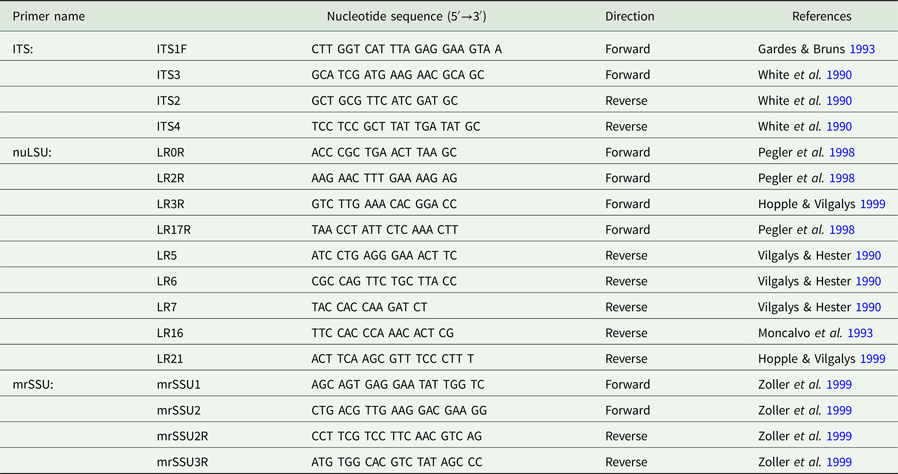
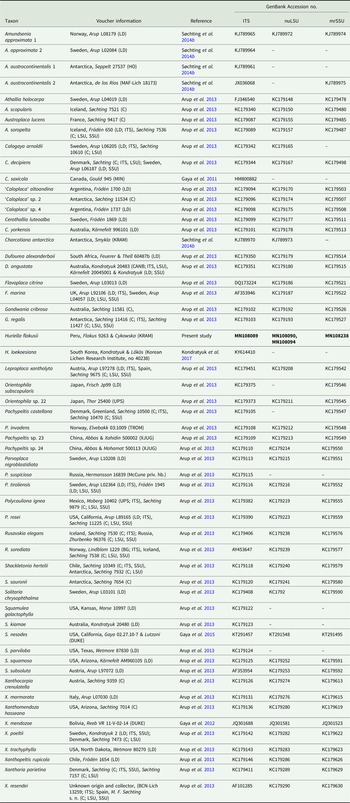
DNA was extracted using modified CTAB procedures of Särkinen et al. (Reference Särkinen, Staats, Richardson, Cowan and Bakker2012) and Healey et al. (Reference Healey, Furtado, Cooper and Henry2014). Primer sequences used in this study for amplifying and sequencing genomic regions are listed in Table 1; they are also available at http://www.lutzonilab.org/nuclear-ribosomal-dna/ and http://www.lutzonilab.org/mitochondrial-ribosomal-dna/. All PCR reactions were carried out in an Eppendorf Mastercycler Nexus GSX1 or Nexus SX1e and each 50 µl reaction mix contained 0.05 U μl−1 RedTaq DNA polymerase (Sigma-Aldrich), 1× RedTaq PCR reaction buffer (Sigma-Aldrich), 0.2 mM dNTPs in equimolar ratio (Sigma-Aldrich), 0.04 mg ml−1 bovine serum albumin (New England BioLabs), 0.2 µM of each of the primers and 1.5 µl template DNA. For the ITS rDNA, thermal cycling parameters for the PCR reaction were: 3 min at 94 °C, 35 cycles of 30 s at 94 °C, 30 s at 60 °C (with a decrease of 1 °C per cycle and a constant temperature of 50 °C starting from cycle 11) and 1 min at 72 °C, and a final extension step of 7 min at 72 °C. For the nuLSU rDNA and mrSSU rDNA, the parameters were: 5 min at 80 °C, 30 cycles of 1 min at 95 °C, 1 min at 50 °C, ramp of 0·3 °C/s to 65 °C and 4 min at 65 °C, and a final extension step of 5 min at 65 °C. PCR products were visualised on 1% agarose gels and successful amplifications were purified using ExoSAP (ThermoFisher Scientific) following the manufacturer's instructions. Purified PCR products were sequenced using the amplification primers and BigDye Terminator v.3.1 (ThermoFisher Scientific) chemistry together with BDX64 Sequencing Enhancement Buffer (Nimagen, at 32× dilution), and run on an ABI 3130 automated sequencer (Applied Biosystems).
Primer names and sequences, together with associated references, used in this study for amplifying and sequencing genomic regions.
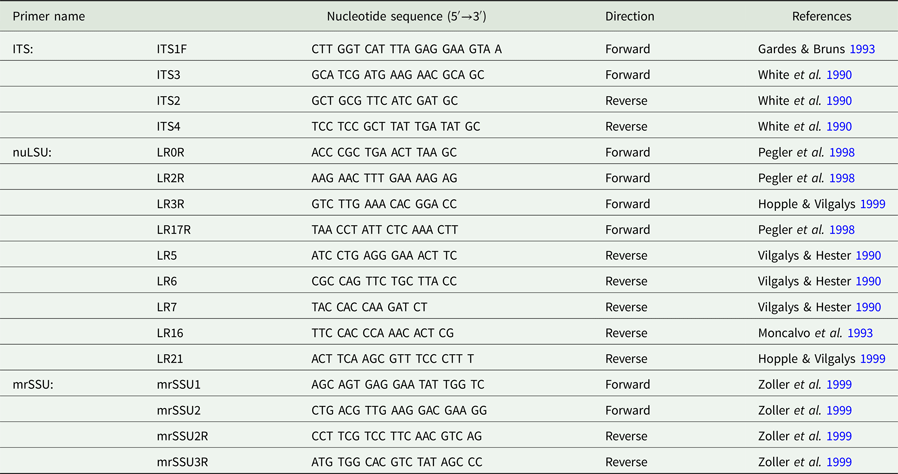
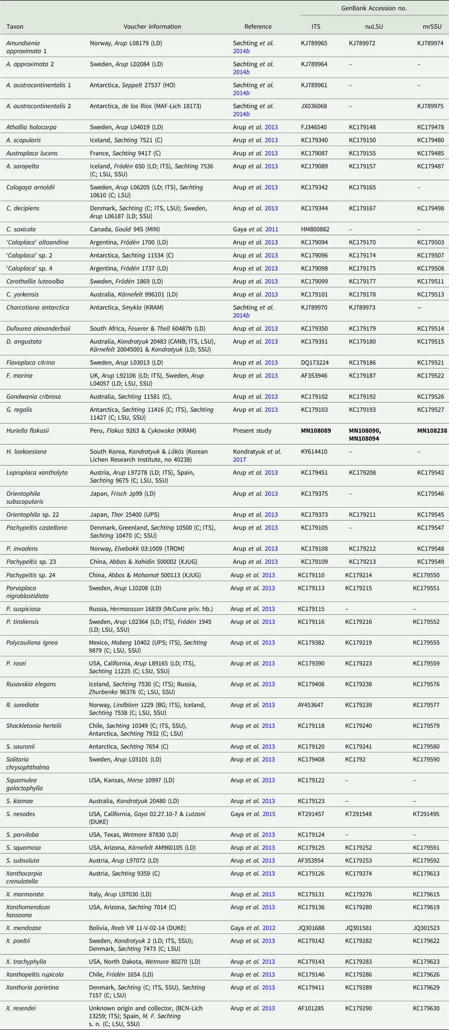
Sequences were assembled and edited using CodonCode Aligner v.6.0.2. Additional sequences (from Gaya et al. Reference Gaya, Redelings, Navarro-Rosinés, Llimona, De Cáceres and Lutzoni2011, Reference Gaya, Högnabba, Holguin, Molnar, Fernández-Brime, Stenroos, Arup, Søchting, van den Boom and Lücking2012, Reference Gaya, Fernández-Brime, Vargas, Lachlan, Gueidan, Ramírez-Mejía and Lutzoni2015; Arup et al. Reference Arup, Søchting and Frödén2013; Søchting et al. Reference Søchting, Garrido-Benavent, Seppelt, Castello, Pérez-Ortega, de los Ríos Murillo, Frödén and Arup2014b; Kondratyuk et al. Reference Kondratyuk, Lokös, Upreti, Nayaka, Mishra, Ravera, Jeong, Jang, Park and Hur2017) were downloaded from GenBank. Voucher information and GenBank Accession numbers are provided in Table 2. Sequences generated for this study were deposited in GenBank.
Voucher information and GenBank Accession numbers for specimens used in the phylogenetic analyses. New sequences are indicated in bold. Numbers after names help to distinguish specimens in the phylogenetic tree (Fig. 1).
A dataset was constructed, encompassing DNA sequences from representatives of all major lineages within the family Teloschistaceae, to identify the clades to which the newly collected specimen is most closely related. New alignments were then prepared for the subfamily Xanthorioideae only, plus a closely related outgroup (Leproplaca xantholyta (Nyl.) Hue). Separate alignments were prepared for each marker using the MUSCLE algorithm in MEGA 6.0 (Tamura et al. Reference Tamura, Stecher, Peterson, Filipski and Kumar2013) and then ambiguously aligned regions were eliminated manually in AliView 1.17.1 (Larsson Reference Larsson2014), resulting in alignments of 601 bp for ITS (58 taxa), 772 bp for nuLSU (46 taxa) and 830 bp for mrSSU (47 taxa). The final concatenated alignment contained 2203 bp, for 58 taxa. This dataset was subjected to maximum likelihood (ML) analysis in RAxML v. 8.2.10 (Stamatakis Reference Stamatakis2014) under the GTR-Gamma model using raxmlGUI 1.5b1 (Silvestro & Michalak Reference Silvestro and Michalak2012). Branch support was assessed by bootstrapping (1000 replicates). PartitionFinder v.2.1.1 (Lanfear et al. Reference Lanfear, Calcott, Ho and Guindon2012) was applied with the linked branch lengths option and the corrected Akaike Information Criterion (AICc) to select the optimum partitioning scheme consisting of gene-specific partitions evolving under the separate GTR + I+G (ITS, nuLSU, mrSSU) nucleotide substitution models. These parameters were specified in a Bayesian phylogenetic analysis (BI) in MrBayes v.3.2 (Ronquist et al. Reference Ronquist, Teslenko, van der Mark, Ayres, Darling, Höhna, Larget, Liu, Suchard and Huelsenbeck2012). The analysis was run for 107 generations, with 4 chains sampled every 103 generation; 25% of the generations was discarded as burn-in.
Results
Phylogenetic analyses
The combined analysis of the ITS, nuLSU and mrSSU rDNA datasets included 58 terminal species and a total of 2203 positions (601 positions of ITS, 772 of nuLSU and 830 of mrSSU). The obtained phylogenetic tree (Fig. 1) represents the subfamily Xanthorioideae, with Leproplaca xantholyta as the outgroup following Arup et al. (Reference Arup, Søchting and Frödén2013). The ingroup was split into three moderately (in the ML analysis) to strongly (in the BI analysis) supported major clades, but the relationship between them is unclear. The tree topology is generally congruent with previously published results (Arup et al. Reference Arup, Søchting and Frödén2013; Søchting et al. Reference Søchting, Garrido-Benavent, Seppelt, Castello, Pérez-Ortega, de los Ríos Murillo, Frödén and Arup2014b), with the exception of the relationship between subclades Amundsenia-Squamulea and Pachypeltis-Parvoplaca-Xanthomendoza which, unlike the aforementioned studies, formed a monophyletic group, strongly supported by the BI analysis (PP = 0.99). In the tree, Huriella flakusii is nested within a strongly supported (BS = 97/ PP = 1) Amundsenia-Squamulea subclade, and has a fully supported sister position to the Asian taxon, Huriella loekoesiana. The two Huriella species form a clade that is sister to Squamulea with moderate to strong support (BS = 77/ PP = 0.99).
Phylogenetic position of Huriella flakusii in the subfamily Xanthorioideae, based on maximum likelihood (RAxML) and Bayesian (MrBayes) analyses of the combined ITS, nuLSU and mrSSU dataset (Table 2). Topology is derived from RAxML analysis. Leproplaca xantholyta was used as an outgroup. Only bootstrap values ≥70% or posterior probabilities ≥0.70 are indicated. Thickened branches denote both posterior probabilities ≥0.70 and bootstrap support values ≥70%. The subclade Amundsenia-Squamulea is indicated with a black rectangle. The samples of this subclade form two groups characterized by the structure of their apothecial proper margins.
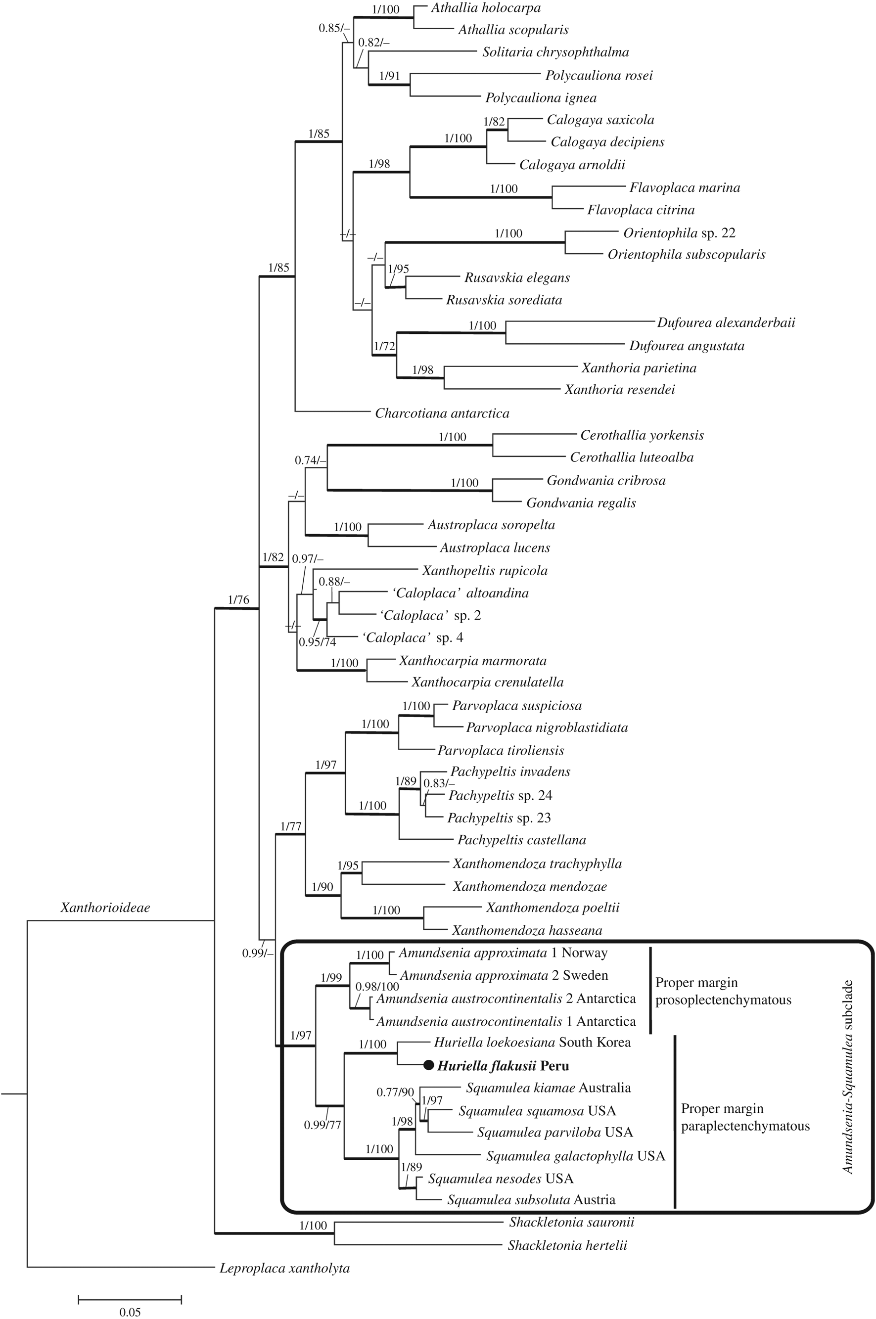
In the reconstructed Teloschistaceae phylogeny (ML analysis; unpublished data), the Amundsenia-Huriella-Squamulea group always forms a monophyletic subclade with moderate to strong support. However, Huriella changes position from a sister lineage to either Amundsenia or Squamulea, whereas in the Xanthorioideae phylogeny (Fig. 1) it has a constant sister relationship to Squamulea with moderate to strong support (BS = 77/ PP = 0.99).
It should be noted that sequences of Caloplaca inconnexa ssp. nesodes (Poelt & Nimis) Cl. Roux (=Squamulea nesodes (Poelt & Nimis) S.Y. Kondr.) used in the ML and BI analyses clearly nested within the Squamulea clade. However, the phylogenetic position of this species requires clarification since it was recently included in two genera, Squamulea and Athallia Arup et al., based on two independent studies by Kondratyuk et al. (Reference Kondratyuk, Kim, Yu, Jeong, Jang, Kondratiuk, Zarei-Darki and Hur2015b) and Vondrák et al. (Reference Vondrák, Halici, Güllü and Demirel2016), respectively. According to Nimis (Reference Nimis2016), the name Athallia nesodes (Poelt & Nimis) Halıcı & Vondrák is invalid and it is clear that the taxonomy and nomenclature of the Caloplaca inconnexa complex requires further study.
Taxonomy
Huriella flakusii Wilk sp. nov.
MycoBank No.: MB 831528
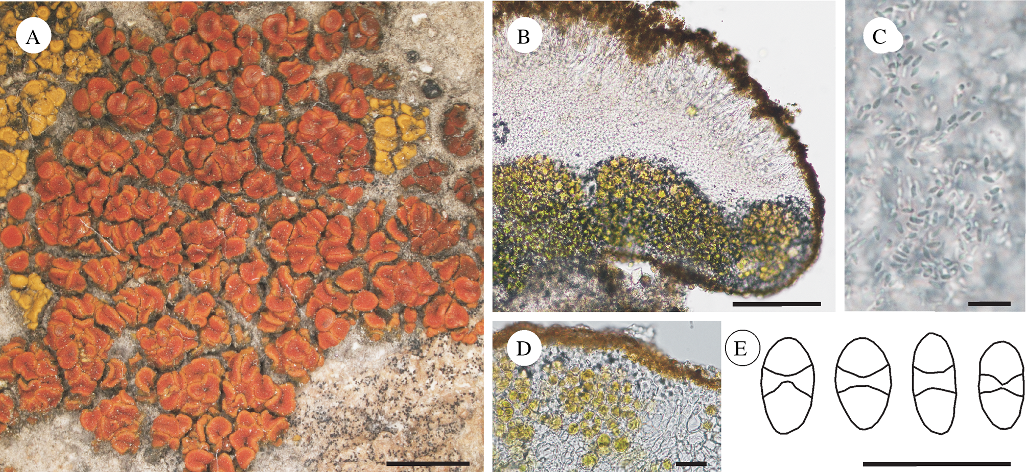
Thallus squamulose, orange. Apothecia abundant, crowded and aggregated, zeorine, with a paraplectenchymatous proper margin and hypothecium. Ascospores polarilocular, broadly ellipsoid with rather thin septa, 10‒15 × 5.0‒9.5 µm, septum 2‒4 µm.
Type: Peru, Dept. Arequipa, Prov. Caylloma, Valle del Colca, near Achoma Village, open semi-desert montane area, 15°39′26″S, 71°43′38″W, elev. 3500 m, 2 July 2006, Flakus 9263 & Cykowska (KRAM-L-70242—holotype).
Huriella flakusii (holotype, A. Flakus 9263, KRAM). A, thallus habit; B, transverse section of apothecium showing paraplectenchymatous proper margin and hypothecium; C, pycnoconidia; D, thallus section showing paraplectenchymatous upper cortex; E, ascospores. Scales: A = 2 mm; B = 100 µm; C = 10 µm; D & E = 20 µm. In colour online.
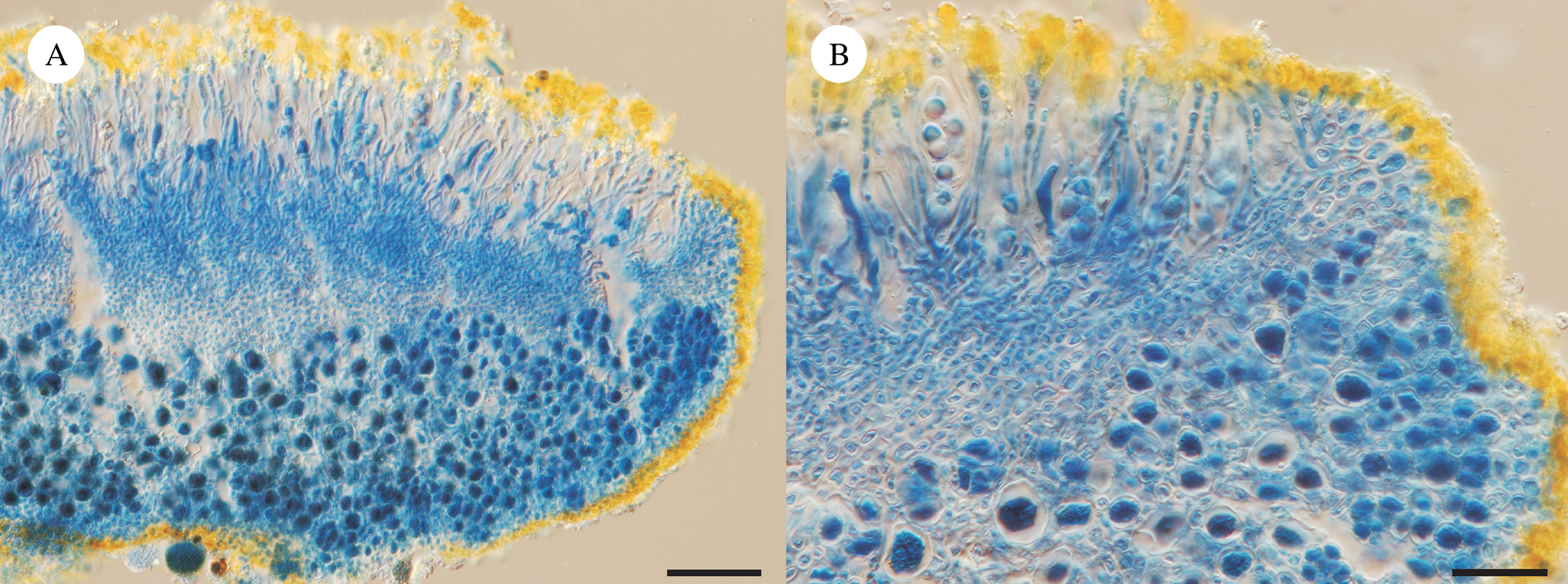
Huriella flakusii (holotype, A. Flakus 9263, KRAM). Freezing microtome sections of apothecia in lactophenol cotton blue showing paraplectenchymatous proper margin and hypothecium. Scales: A = 50 μm; B = 25 μm. In colour online.
Thallus squamulose or strongly reduced and almost invisible, 120‒300 µm thick, orbicular to irregular in outline, orange, K+ purple, epruinose, smooth; vegetative propagules absent; prothallus absent; cortex thin to thick (in well-developed thallus), 15‒70 µm thick, paraplectenchymatous, even, anthraquinone pigments present; algal layer continuous; photobiont trebouxioid, cells more or less globose, 7‒15 µm diam.
Apothecia abundant, crowded and aggregated, rounded, angular or flexuous when compressed, old apothecia distinctly undulate, adnate, zeorine, 0.2‒0.8(‒1.0) mm wide; disc first concave then plane to slightly convex, reddish, K+ purple, epruinose, often cracked (especially in old apothecia); proper margin thin, 50‒75 µm thick, slightly prominent in young apothecia, then level with disc, slightly paler than disc, epruinose, paraplectenchymatous, anthraquinone pigments present; thalline margin 50‒110 µm thick, conspicuous, partly to much reduced, algal cells abundant, forming a continuous layer, cortex 15‒25 µm thick, paraplectenchymatous, anthraquinone pigments present; epihymenium brownish yellow, granular inspersed, K+ purple; hymenium 70‒85 µm thick; hypothecium 100‒170 µm thick, paraplectenchymatous, hyaline or in the upper parts yellowish, oil droplets abundant, stipe absent; paraphyses rather robust, simple to slightly branched, 1.5‒2.0 µm broad at base, with upper cells slightly thickened, up to 5 µm, oil droplets absent; asci clavate, 40–55 × 11–19 µm, 8-spored; ascospores polarilocular, usually broadly ellipsoid, (10.0‒)12.6 ± 1.6 (‒15.0) × (5.0‒)6.7 ± 0.8(–9.5) μm, ascospore septa (2.0‒)3.0 ± 0.7(‒4.0) μm (n = 39).
Pycnidia totally immersed in the thallus or red ostiole visible; conidia bacilliform, 3–4 × 1 µm.
Chemistry
The thallus cortex, epihymenium and outer portions of the proper margin K+ purple.
Etymology
Named after Adam Flakus, the Polish lichenologist, in appreciation of his major contribution to the knowledge of tropical lichens.
Distribution and ecology
On siliceous rock in arid montane habitats, at elevations of c. 3500 m in the Colca Canyon region in southern Peru.
Remarks
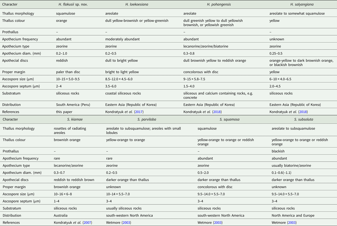
Huriella flakusii is characterized by squamulose, orange thalli and abundant, crowded and aggregated, usually flexuous when compressed, apothecia with double margins and red discs. The thalline cortex, proper margin and hypothecium are paraplectenchymatous, and the ascospores are broadly ellipsoid with rather thin septa. All species of the Huriella-Squamulea group, including the new taxon, are phenotypically very similar. They produce small, areolate or mostly squamulose thalli (except S. galactophylla (Tuck.) Arup et al., which has lobed thalli) containing anthraquinones, usually small apothecia, and a paraplectenchymatous thalline cortex and proper margin. All species grow on siliceous rock substrata. Morphological and anatomical differences between the species of the Huriella-Squamulea group are presented in Table 3. Huriella flakusii differs from the other Huriella species in certain features, such as the clearly squamulose type of thallus, the colour of the thallus and apothecia which are orange and red respectively, broadly ellipsoid spores, and habitat preferences associated with arid high mountainous regions. The new species appears to be more similar to species of Squamulea, in particular S. squamosa (B. de Lesd.) Arup et al. The latter, however, differs in having a better developed thallus and narrower spores, mostly with thicker spore septa. In the case of the closely related genus Amundsenia, H. flakusii resembles A. austrocontinentalis Garrido-Benavent et al., which differs from H. flakusii in its areolate type of thallus, usually dispersed apothecia, prosoplectenchymatous proper margin and smaller ascospores (Søchting et al. Reference Søchting, Garrido-Benavent, Seppelt, Castello, Pérez-Ortega, de los Ríos Murillo, Frödén and Arup2014b).
Comparison of Huriella flakusii with other species of the genera Huriella and Squamulea.
The morphology of apothecia in H. flakusii resembles that in Calogaya saxicola (Hoffm.) Vondrák, but the latter usually produces distinct marginal lobes, larger apothecia (up to 4.0 mm in diam.), narrowly ellipsoid spores, and a prosoplectenchymatous proper margin (Gaya Reference Gaya2009).
The saxicolous (sub-)squamulose species of Caloplaca s. lat. occurring in South America are not rare and include, for example, Caloplaca americana (Malme) Zahlbr., C. brachyloba (Mull. Arg.) Zahlbr., C. chapadensis (Malme) Zahlbr., C. malmeana Zahlbr. (=Callopisma brachysporum Malme), C. muelleri (Vain.) Zahlbr., C. rubina Zahlbr., C. tucumanensis H. Magn. and C. xanthobola (Kremp.) Zahlbr. (Malme Reference Malme1926; Wetmore Reference Wetmore2003, Reference Wetmore, Nash, Gries and Bungartz2007). Of these, only C. americana produces a paraplectenchymatous proper margin. This species was studied in detail by Wetmore (Reference Wetmore2003) and was synonymized with C. subsoluta (Nyl.) Zahlbr. (currently known as Squamulea subsoluta (Nyl.) Arup et al., see Table 3). Another species, which was also synonymized with C. subsoluta (Wetmore Reference Wetmore2003, Reference Wetmore, Nash, Gries and Bungartz2007), is C. sipeana Magn. described from North America (Magnusson Reference Magnusson1944). This taxon differs from H. falkusii in its scattered squamules, distinctly smaller apothecia (up to 5 mm) which are concolorous with the thallus, and thinner thalline cortex (up to 30 µm) (Magnusson Reference Magnusson1944).
Following the new classification of Teloschistaceae (based mainly on North Hemisphere representatives), species producing a squamulose thallus are generally known in Squamulea and Wetmoreana, and also in Flavoplaca Arup et al. and Gyalolechia A. Massal. Of these species only Squamulea, produces a paraplectenchymatous proper margin (Arup et al. Reference Arup, Søchting and Frödén2013). A preliminary molecular study (KW, unpublished) of South American squamulose Caloplaca s. lat. collections, including only species with a prosoplectenchymatous proper margin, showed closest affinity to Wetmoreana. These species are currently being assessed and the results will be published separately. The morphology of the squamulose species is highly variable which makes the taxonomy of this group very problematic.
Additional specimens examined
Peru:Dept. Arequipa: Prov. Caylloma: Valle del Colca, near Achoma Village, open semi-desert montane area, 15°39′26″S, 71°43′38″W, elev. 3500 m, 2006, Flakus 9262 & Cykowska (KRAM); by Colca Canyon, near Cabanaconde Village, open semi-desert montane area, 15°37′49″S, 71°59′39″W, elev. 3310 m, 2006, Flakus 9574 & Cykowska (KRAM).
Selected material investigated for comparison
Caloplaca brachyloba. Paraguay:Distrito Capital: Asunción, in pascuo aperto, 1893, Malme 1423 (S).
Caloplaca chapadensis. Brazil:Mato Grosso: Serra da Chapada, 1894, Malme 2318 (S, holotype).
Caloplaca malmeana. Brazil:Minas Gerais: São João d'el Rey, 1892, Malme 267 (S, sub Callopisma brachysporum, holotype).
Caloplaca rubina. Chile:Coquimbo: Alto Talinay, 1960, Follmann 12765 (B).
Caloplaca tucumanensis. Uruguay:Dept. Montevideo: Punta Shannon, 1984, Ranta (MIN 876802).
Caloplaca xanthobola. Uruguay:Dept. Durazno: Rio Negro, 1974, Osorio 7015 (MIN).—Brazil:Rio Grande do Sul: inter São Martinho et Santa Maria, 1893, Malme 1304 (S).
Squamulea subsoluta. Peru:Prov. Abancay: Casinchihua, SW of Abancay, 1962, Iltis et al. 2351 (MIN).—Argentina:Buenos Aires: Sierra de la Ventana, Cerro Ceferino, 1975, Osorio 7071 (MIN).
Discussion
Molecular phylogenetic analyses inferred that Huriella flakusii is nested in the Amundsenia-Squamulea subclade and is closely related to H. loekoesiana. All species clustered in this subclade are morphologically similar but there is one important feature regarding the structure of the proper margin that divides the species into two separate groups (see also Fig. 1). The first group is distinguished by a paraplectenchymatous type of proper margin and includes Huriella and Squamulea species; the second includes Amundsenia species, which form a prosoplectenchymatous type of proper margin.
The structure of the fungal plectenchyma is an important diagnostic character in lichens (e.g. Hale Reference Hale1983; Gaya Reference Gaya2009). In general, two basic types of plectenchyma are distinguished, namely paraplectenchyma and prosoplectenchyma, but gradual transitions between these two types are common and have been studied and classified (Yoshimura & Shimada Reference Yoshimura and Shimada1980; Ryan et al. Reference Ryan, Bungartz, Nash, Nash, Ryan, Gries and Bungartz2002; Gaya Reference Gaya2009; Vondrák et al. Reference Vondrák, Frolov, Arup and Khodosovtsev2013). The structure of the thalline cortex varies from prosoplectenchymatous to paraplectenchymatous, with paraplectenchymatous-like forms occurring more frequently in members of the family Teloschistaceae (Kärnefelt Reference Kärnefelt1989). The true paraplectenchymatous cortex, however, is probably rather rare in these lichens, as was shown for example in the Caloplaca saxicola group (Gaya Reference Gaya2009). The structure of the thalline cortex is a useful character in species delimitation but is not important at the generic level (for example, both basic types of thallus cortex occur in the genus Flavoplaca; Arup et al. Reference Arup, Søchting and Frödén2013). The structure of the proper margin of zeorine apothecia in Teloschistaceae is commonly prosoplectenchymatous, usually with the uppermost part almost paraplectenchymatous and the lower part prosoplectenchymatous (see Fig. 2 in Vondrák et al. (Reference Vondrák, Frolov, Arup and Khodosovtsev2013)). A true paraplectenchymatous proper margin is also rare, but in Squamulea it is a very good diagnostic character for this genus (Arup Reference Arup1992; Wetmore Reference Wetmore2003; Arup et al. Reference Arup, Søchting and Frödén2013). A detailed description of this margin for Squamulea is presented by Wetmore (Reference Wetmore2003), who characterized it as a cellular area occurring like a cone or cup under the hypothecium.
The current study demonstrates that the true paraplectenchymatous proper margin may also be characteristic for genera other than Squamulea, and the proper margin similarity in both Huriella and Squamulea is probably the result of convergent morphological evolution. The phylogenetic analyses of the subfamily Xanthorioideae (ML and BI) show that Huriella and Squamulea are well separated from each other and form distinct sister taxa (Fig. 1). In the reconstruction of the Teloschistaceae phylogeny (based on ML; unpublished data), the phylogenetic position of Huriella tends to change and the genus was sister to Amundsenia instead of Squamulea in some cases. The observed genetic divergence between the taxa, combined with the results of Xanthorioideae phylogeny, suggests that Huriella and Squamulea should be kept as separate taxa. According to Kondratyuk et al. (Reference Kondratyuk, Lokös, Upreti, Nayaka, Mishra, Ravera, Jeong, Jang, Park and Hur2017), Huriella was separated from Squamulea on the basis of a different thallus type (areolate instead of squamulose), smaller apothecia, narrower ascospores and its position on the phylogenetic tree. However, the type of thallus is a subordinate feature in these genera, since both thallus types may change gradually from one type to the other; furthermore, the size of the apothecia and ascospores may also be similar (see Table 3). To summarize, anatomical and morphological features are not very useful for the differentiation of these genera. However, in the current Teloschistaceae systematics, based on molecular data (Arup et al. Reference Arup, Søchting and Frödén2013), such situations are common as phenotypic features often fail in taxa delimitation. According to Kondratyuk et al. (Reference Kondratyuk, Lokös, Upreti, Nayaka, Mishra, Ravera, Jeong, Jang, Park and Hur2017), the phylogenetic position of Huriella was clearly nested within the Amundsenia-Squamulea subclade but the relationship between Amundsenia, Squamulea and Huriella, has not been resolved.
Acknowledgements
The curators of herbaria B, MIN and S, are gratefully thanked for the loan of material. Adam Flakus (IB PAS, Kraków) is acknowledged for providing herbarium specimens for investigation and sequencing as well as preparing extra microtome sections of the holotype, Marta Saługa (IB PAS, Kraków) for providing sequences of studied species, Daniel Ramos (HSP herbarium) for literature support, Maciej Pabijan (IZ UJ, Kraków) and Marcin Piątek (IB PAS, Kraków) for comments on the manuscript, and Mark R. D. Seaward (University of Bradford) for linguistic revision of the text. I am very grateful to the anonymous reviewers and Mats Wedin for valuable comments. This research was supported by the National Science Centre (grant no. N N303 821740) and statutory funds of the W. Szafer Institute of Botany, Polish Academy of Sciences, Kraków.