Implications
Reproductive technologies with cooled and frozen semen have unquestionable advantages in animal breeding; however, semen preservation results in important injuries in the sperm cells, compromising their viability and hence the pregnancy rates. In the last decade, the scientific community has increased its efforts to improve current cryopreservation protocols. The results of the current study demonstrate that LC3, a protein involved in autophagy, is activated under stressful conditions during cryopreservation, and plays a role in regulating viability. Therefore, this study is of interest not only from a scientific point of view, but also for the improvement of semen quality, which could result in high fertility and finally increased economic gain.
Introduction
Reproductive technologies such as artificial insemination and in vitro production of embryos with cooled and frozen semen have been used extensively and improved in the last decade. The use of cooled and frozen semen has unquestionable advantages in animal breeding, as it facilitates the international trade in equine semen, increases the ability to select a stallion with the desired pedigree and/or performance traits and avoids the risk of exposure to contagious diseases during breeding (Loomis and Graham, 2008). However, semen preservation results in important injuries in sperm cells that compromises their viability and hence the quality and the resulting pregnancy rates.
Storing semen at 5°C to 8°C does not have significant effects on motility or viability in the first 2 days of storage; however, from 48 h on, chilling of semen has effects on the spermatozoa. These effects result in a decrease in the viability rate, plasma membrane integrity, decreased motility and conception rates (Watson, 2000; Medeiros et al., 2002; Aurich, 2008). The main advantage of freezing semen is the possibility of storing it indefinitely at −196°C in liquid nitrogen, but during cryopreservation spermatozoa are exposed to a variety of physical and chemical stresses that cause irreversible damage to the sperm cell. The drastic change in temperature during freezing results in membrane bound phospholipid reorientation, which affects membrane functionality and permeability, and causes serious damage to the cell. Other damage occurring during this process includes an increase in reactive oxygen species (ROS), lipid peroxidation, DNA fragmentation, pH changes and ATP depletion (Watson, 2000; Neild et al., 2005; Ortega Ferrusola et al., 2009a and 2009b).
Because of the important loss of cell viability during these processes, several approaches have been tried for increasing the duration of storage without compromising fertility. Efforts have been made to improve the composition of extenders used for both refrigeration and cryopreservation, by adding antioxidants, cryoprotectants, and other similar reagents (Ball and Vo, 2001; Ball et al., 2001; Guthrie et al., 2002; Aboagla and Terada, 2003; Pena et al., Reference Pena, Garcia, Samper, Aparicio, Tapia and Ferrusola2011; Lagares et al., Reference Lagares, Martins, Carvalho, Oliveira, Souza, Penna, Cruz, Stahlberg and Henry2012; Morillo-Rodriguez et al., Reference Morillo-Rodriguez, Macias-Garcia, Tapia, Ortega-Ferrusola and Pena2012; Karimfar et al., Reference Karimfar, Niazvand, Haghani, Ghafourian, Shirazi and Bakhtiyari2015). More recently, the study of molecular mechanisms controlling cell survival or cell death has emerged as a successful approach to improve semen conservation and to extend the lifespan of the sperm in vitro (Watson, 2000; Ortega Ferrusola et al., Reference Ortega Ferrusola, Gonzalez Fernandez, Salazar Sandoval, Macias Garcia, Rodriguez Martinez, Tapia and Pena2010; Cordova et al., Reference Cordova, Strobel, Vallejo, Valenzuela, Ulloa, Burgos, Menarim, Rodriguez-Gil, Ratto and Ramirez-Reveco2014). One of the intracellular pathways most studied during refrigeration and cryopreservation is apoptosis (programmed cell death type I). Apoptosis is activated by stressful conditions or extrinsic stimuli inducing cell death. Typical markers of apoptosis (caspase activation, externalization of phosphatidylserine, alteration of the mitochondrial membrane and DNA fragmentation) have been found to be increased in frozen/thawed semen from different mammalian species (Martin et al., 2004; Ortega-Ferrusola et al., 2008; Sokolowska et al., 2009; Said et al., Reference Said, Gaglani and Agarwal2010; Zeng et al., Reference Zeng, Tang, He, Peng, Ding, Fang and Zhang2014). Moreover, it has been demonstrated that spermatozoa subjected to preservation protocols also die through necrosis (programmed cell death type III) (Januskauskas et al., 2003; Chaveiro et al., 2007; Rovegno et al., Reference Rovegno, Feitosa, Rocha, Mendes, Visintin and D’Avila Assumpcao2013).
Apart from apoptosis and necrosis, cells also die through a third mechanism named programmed cell death type II or autophagy. Autophagy is physiologically activated under starvation and stressful conditions, and its activation contributes to maintaining cytoplasmic homoeostasis and provides an energy source when it is demanded by the cell. Although the primordial role of autophagy is to confer cell protection, it also has been shown to facilitate, antagonize or cooperate with apoptosis, serving either a pro-survival or pro-death function (Nikoletopoulou et al., Reference Nikoletopoulou, Markaki, Palikaras and Tavernarakis2013).
When autophagy is activated, a membrane cisterna (called a phagophore) encloses a portion of cytoplasm, resulting in the formation of the autophagosome. Next, the outer membrane of the autophagosome fuses with the membrane of a lysosome resulting in a degradative structure called the autolysosome or autophagolysosome, where hydrolytic enzymes supplied from the lysosome degrade the cytoplasm-derived materials together with the inner membrane of the autophagosome (Kraft and Martens, Reference Kraft and Martens2012). Products resulting from the degradation are released back into the cytosol in order to recycle the macromolecular constituents and to generate energy to maintain cell viability. Phagophore, autophagosome and autophagolysosome formation is finely regulated by at least 30 autophagy-related proteins (Atg). One of the mammalian homologues of yeast Atg8 is the microtubule-associated protein light chain 3 (LC3) and exists in two forms, LC3-I and LC3-II. LC3-I is an 18-kDa polypeptide normally found in the cytosol, whereas the product of its proteolytic maturation (LC3-II, 16 kDa) resides in the autophagosomal membranes (Li et al., Reference Li, Hou, Wang, Chen, Shao and Yin2011). LC3-II has been used widely to study autophagy and it has been considered an autophagosomal marker in mammals (Tanida et al., 2004).
Among the numerous proteins involved in the regulation of autophagy, mammalian target of rapamycin (mTOR) is a key component (Jung et al., Reference Jung, Ro, Cao, Otto and Kim2010). Under steady-state conditions, mTOR inhibits autophagy. Starvation conditions and environmental stress inactivate mTOR, which results in the activation of autophagy. Other important regulators of autophagy include class I and class III phosphoinositide 3 kinases (PI3Ks) and AMP-activated protein kinase (AMPK) (Shang and Wang, Reference Shang and Wang2011; O’Farrell et al., Reference O’Farrell, Rusten and Stenmark2013).
Previous results from our group have shown that both LC3-I and LC3-II are present in stallion spermatozoa (Gallardo Bolanos et al., Reference Gallardo Bolanos, Miro Moran, Balao da Silva, Morillo Rodriguez, Plaza Davila, Aparicio, Tapia, Ortega Ferrusola and Pena2012; Bolanos et al., Reference Bolanos, Moran, da Silva, Davila, Munoz, Aparicio, Tapia, Ferrusola and Pena2014). Equine semen filtration through single-layer centrifugation significantly increased the conversion of LC3-I to LC3-II. Moreover, we observed an increase in LC3-II after 5 days of cooled storage at 5°C, independent of the type of extender used (Gallardo Bolanos et al., Reference Gallardo Bolanos, Miro Moran, Balao da Silva, Morillo Rodriguez, Plaza Davila, Aparicio, Tapia, Ortega Ferrusola and Pena2012; Bolanos et al., Reference Bolanos, Moran, da Silva, Davila, Munoz, Aparicio, Tapia, Ferrusola and Pena2014). Together, these results indicate that LC3-I lipidation is an active process in stallion spermatozoa. It is not known, however, whether this activation is a pro-death or pro-survival mechanism. Therefore, the aim of this study was to determine the effect of several conditions that are known to cause stress in spermatozoa during preservation protocols. These conditions include cooling, freezing/thawing, osmotic and oxidative stress, UV radiation and temperature change. Further, it was determined if this LC3-I to LC3-II conversion can be modified and if, through the modulation of LC3, the quality of stored semen may be improved.
Material and methods
The following chemicals were purchased from Sigma Aldrich (St. Louis, MO, USA): NaCl, KCl, MgSO4, KPO4, 2-[4-(2-hydroxyethyl)piperazin-1-yl]ethanesulfonic acid (HEPES), polivinilpirrolidone (PVP), poly(vinyl alcohol) (PVA), sodium pyruvate, sodium lactate, CaCl2, NaHCO3, formaldehyde, Triton X-100, deoxycholate, ethyleneglycol-bis(β-aminoethyl)-N,N,Nʹ,Nʹ-tetraacetic acid (EGTA), ethylenediaminetetraacetic acid (EDTA), Na3VO4, glucose, bovine serum albumin (BSA), phosphate-buffered saline, chloroquine diphosphate salt (cat no. C6628), and anti-LC3B (cat no. L7543). YO-PRO-1, Hoechst 33342 and ethidium homodimer-1 (EthD-1) were obtained from Molecular Probes (Eugene, OR, USA). Complete, EDTA-free, protease inhibitor cocktail was purchased from Roche Diagnostics (Penzberg, Germany). Bradford reagent, Tris/glycine/SDS buffer (10X) and Tris/glycine buffer (10X) were purchased from Bio-Rad (Richmond, CA, USA). 3-MA (cat no. 189490), STF-62247 (cat no. 189497) and rapamycin (cat no. 553211) were purchased from MERCK (Billerica, MA, USA). Anti-rabbit IgG horseradish peroxidase-conjugated secondary antibody and enhanced chemiluminescence detection reagents were obtained from Pierce (Rockford, IL, USA). Hyperfilm ECL was from Amersham (Arlington Heights, IL, USA), and nitrocellulose membrane was obtained from Schleicher & Schuell (Keene, NH, USA).
Ethics
Stallions were maintained according to institutional and European regulations, and were collected on a regular basis (two collections per week) during the 2014-15 breeding season. Animal semen collection protocols were reviewed and approved by the Animal Ethics Committee of the University of Extremadura and were performed in accordance with Spanish and European guidelines for research on animals.
Semen collection
Semen was obtained from a total of 25 Andalusian horses individually housed at the Veterinary Teaching Hospital of the University of Extremadura, Caceres, Spain. All animals were healthy donors and no selection of the ejaculates was performed for incubation. Ejaculates from stallions (in the age range of 4 to 20 years) were collected using a Missouri model artificial vagina with an inline filter to eliminate the gel fraction, and were lubricated and warmed from 45°C to 50°C. Semen was immediately evaluated and processed.
Freezing and thawing
The ejaculate was diluted 1 : 2 in INRA96® (IMV, L’Aigle, France) and centrifuged at 600×g for 10 min. Spermatozoa were resuspended in Cáceres® freezing medium (Rodriguez et al., Reference Rodriguez, Ferrusola, Garcia, Morrell, Martinez, Tapia and Pena2011). Samples at a density of 100×106 spermatozoa/ml (spz/ml) were packed in 0.5 ml straws (IMV), ultrasonically sealed with UltraSeal 21® (Minitube of America MOFA®, Verona, WI, USA) and immediately placed in an IceCube® 14S (SY-LAB, Neupurkersdorf, Austria) for freezing according to the following freezing curve: equilibration for 20 min at 22°C, temperature ramped down from 22°C to 5°C at a rate of 0.5°C/min, equilibrated to 5°C for 10 min, temperature ramped down from 5°C to −140°C at a rate of 30°C/min. Then, the straws were introduced in liquid nitrogen and analysed in the next 2 weeks. Thawing was performed by placing the straws in a 37°C water bath for 30 s. When it was necessary, chloroquine (50 µM) and 3-MA (5 mM) were added to samples before packaging in straws.
Incubation media
To perform the incubation of semen samples, aliquots of each stallion’s ejaculate were centrifuged at 600×g for 10 min at room temperature (RT). Seminal plasma was removed and the sperm pellet resuspended at 100×106 spz/ml in modified Biggers, Whitten and Whittingham (BWW) media (91.6 mM NaCl, 4.6 mM KCl, 2.44 mM MgSO4, 1.2 mM KPO4, 20 mM HEPES, 5.6 mM glucose (anhydrous), 0.27 mM sodium pyruvate, 44 mM sodium lactate, 1.7 mM CaCl2, 25 mM NaHCO3 and 7 mg/ml BSA, pH 7.4) or INRA96®.
Treatments
Incubation
Samples resuspended in BWW were incubated for 3 h at 37°C in the presence of inducers or inhibitors including STF-62247 (50 µM), rapamycin (250 nM), chloroquine (50 µM) and 3-MA (5 mM). STF-62247 is a selective autophagy activator (Anbalagan et al., Reference Anbalagan, Pires, Blick, Hill, Ferguson, Chan and Hammond2012). Rapamycin was used as an mTOR inhibitor (Zhou et al., Reference Zhou, Luo and Huang2010). 3-MA is an inhibitor of PI3K class III (O’Farrell et al., Reference O’Farrell, Rusten and Stenmark2013). Chloroquine inhibits autophagy because it raises the lysosomal pH, which leads to inhibition of both fusion of the autophagosome with the lysosome and lysosomal protein degradation (Long et al., Reference Long, Yang, Southwood, Lu, Marciniak, Dunmore and Morrell2013). Samples resuspended in INRA96® were stored at 4°C for 5 days (0, 24, 48, 72, 96 h). When necessary, chloroquine (50 µM) and 3-MA (5 mM) were added to fresh samples, before refrigeration.
Stressful conditions
A quantity of 1 ml of equine semen at 100×106 spz/ml was exposed to UV radiation (100 J/m2) for 1 h at RT. Control samples were kept in the dark for 1 h. To induce oxidative stress, 1 ml of equine semen at 100×106 spz/ml was incubated in the presence of H2O2 (100 µM) for 1 h at 37°C. Osmotic stress was induced by incubating spermatozoa (100×106 spz/ml) for 15 min at 37°C in isotonic (300 mOsmol/kg), hypotonic (75 mOsmol/kg) or hypertonic (1500 mOsmol/kg) solutions. Hypotonic solution (75 mOsmol/kg, pH 7.4) was prepared by adding distilled water to the BWW medium (described above) and the hypertonic solution (1500 mOsmol/kg) was made with BWW five times concentrated. The osmolalities of the solutions were measured using freezing point depression (digital micro-osmometer Type 5 R, Hermann Roebling Messtechnik, Berlin, Germany). The pH of the solutions was adjusted to pH 7.4. Finally, spermatozoa were subjected to drastic changes in the temperature of incubation. For this purpose, semen was centrifuged at 800×g for 10 min and the precipitate was resuspended in INRA96® and refrigerated at 4°C (100×106 spz/ml). After 3 h of refrigeration, spermatozoa were transferred directly to a 37°C water bath and incubated for 30 min. In all treatments, proteins were extracted and resolved by SDS-PAGE (see below).
Assessment of subtle membrane changes and viability by flow cytometry
A quantity of 1 ml of sperm suspension (5×10⁶ spz/ml) was stained with 3 µl of Hoechst 33342 (1.62 mM) and 3 µl of Yo-Pro-1 (25 µM) for 25 min at 37°C. Then, 1 µl of ethidium homodimer-1 (1.167 mM) was added and incubated for 5 min at 37°C. This staining distinguishes four sperm subpopulations related to sperm viability, as described by Ortega-Ferrusola (2008). Flow cytometric analyses were conducted using a MACSquant Analyzer 10 (Miltenyi Biotech, Bergisch Gladbach, Germany) flow cytometer equipped with three lasers emitting at 405, 488 and 635 nm and 10 photomultiplier tubes. Hoechst 33342, Yo-Pro-1 and EthD-1 were detected in V1 (excitation (Ex) 405, emission (Em) 450/50), B1 (Ex 488 filter 525/50) and B3 (Ex 488 filter 655 to 730 (655LP+split 730)), respectively. The system was controlled with the MACSquantify software. Forward and sideways light scatter were recorded for a total of 10 000 events/sample. Non-sperm events were discarded by gating the sperm population after Hoechst 33342 (0.5 µM) staining for 30 min at 37°C.
Western blotting
Proteins were separated according to their apparent molecular masses as previously described (Caselles et al., Reference Caselles, Miro-Moran, Morillo Rodriguez, Gallardo Bolanos, Ortega-Ferrusola, Salido, Pena, Tapia and Aparicio2014). A quantity of 15 µg of protein extract of spermatozoa was loaded and resolved by SDS-PAGE on a 12% polyacrylamide gel. Immunoblotting was performed by incubating the membranes in blocking buffer overnight at 4°C with anti-LC3 antibody (1/2000). Membranes were then washed in tris-buffered saline-tween 20 (TBS-T) and incubated with goat anti-rabbit horseradish peroxidase-conjugated secondary antibody for 45 min at RT.
Statistical analysis
Data were first examined using the Kolmogorov–Smirnov test to determine their distribution. Multivariate ANOVA was performed followed by a post hoc Tukey’s test. If the data were not normally distributed, the non-parametric Mann–Whitney U-test was used to compare pairs of values directly. All analyses were performed using SPSS version 17.0 for Windows (SPSS Inc., Chicago, IL, USA). Statistical significance was set at P<0.05.
The statistical model used was as follows:
 $$Yijk{\rm \,{\equals}}\,\mu {\rm {\plus}}Si{\rm {\plus}}Mj{\rm {\plus}}SMij{\rm {\plus}}eijk$$
where Yijk is the observation; µ the overall mean; Si the semen storage and its related stresses; Mj the LC3 modulation; SMij the LC3 modulation during semen storage; eijk the experimental error associated with the experimental determinations (random error).
Results
Effect of cooling and freezing/thawing on the conversion from LC3-I to LC3-II
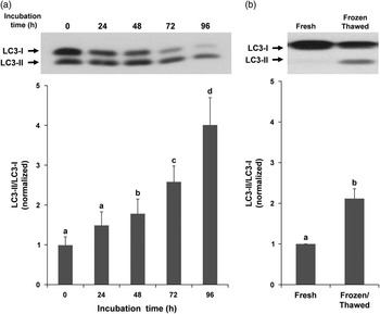
Semen refrigeration for 5 days at 4°C induced an increase in the LC3-II/LC3-I ratio in a time-dependent manner. This increase was statistically significant after 48 h of refrigeration, compared with fresh samples (time=0) (Figure 1a). After 96 h (5 days) this increase was four times greater than in fresh samples. The LC3-II/LC3-I ratio was also studied in fresh and frozen/thawed semen. As shown in Figure 1b, cryopreservation induced a significant increase in the LC3-II/LC3-I ratio as compared with fresh samples.
Light chain (LC)3 processing during cooling and freezing/thawing. Proteins were extracted every day during cooling (a) and before freezing and after thawing (b). A quantity of 15 µg of proteins were loaded and separated by SDS-PAGE. Immunoblotting was performed with an antibody that recognized both LC3-I and LC3-II proteins. LC3 processing was expressed as ratio of LC3-II/LC3-I and data were normalized with respect to time 0 (a) and fresh samples (b). Columns with different letters indicate significant differences (P<0.05, n=4).

Autophagy activation under stressful conditions
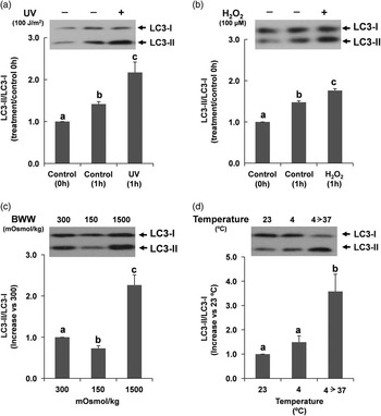
Cell death during preservation protocols is primarily caused by changes in temperature, osmotic variation, increased ROS production and DNA fragmentation. In order to investigate which of these changes is responsible for the increase in the LC3-II/LC3-I ratio during cooling and freezing, stallion spermatozoa were subjected to several stressful conditions. Exposure of semen samples to UV radiation (100 J/m2) or the presence of H2O2 (100 µM) significantly increased the LC3-II/LC3-I ratio, compared with its control (1 h) (Figure 2a and b, respectively). Osmotic stress was induced by incubation of spermatozoa in isotonic (320 mOsmol/kg), hypotonic (75 mOsmol/kg) or hypertonic (1500 mOsmol/kg) solutions. The hypotonic condition induced a significant decrease in the LC3-II/LC3-I ratio whereas this ratio was significantly increased in hypertonic media, compared with spermatozoa incubated in an isotonic solution (Figure 2c). Finally, to assess whether changes in incubation temperature had an effect on LC3-I conversion, spermatozoa were refrigerated for 3 h at 4°C and immediately transferred to a bath at 37°C for 30 min. After refrigeration there was an increase in the LC3-II/LC3-I ratio, although it was not statistically significant. However, the change in temperature from 4°C to 37°C induced a significant increase in the LC3-II/LC3-I ratio (Figure 2d).
Light chain (LC)3 processing under stressful conditions. Spermatozoa were exposed to UV radiation (a); incubated in the presence of H2O2 (100 µM) (b); incubated in isotonic (320 mOsmol/kg), hypotonic (75 mOsmol/kg) or hypertonic (1500 mOsmol/kg) solutions (c); or refrigerated for 3 h at 4°C and immediately transferred to 37°C bath for 30 min (d). A quantity of 15 µg of protein was loaded and separated by SDS-PAGE. Immunoblotting was performed with an antibody that recognized both LC3-I and LC3-II proteins. Data were represented as a ratio of LC3-II/LC3-I and normalized with respect to control (a, b), isotonic solution (c) or 23°C (d). Columns with different letters indicate significant differences (P<0.05, n=4). BWW=Biggers, Whitten and Whittingham.

Modulation of LC3-I conversion
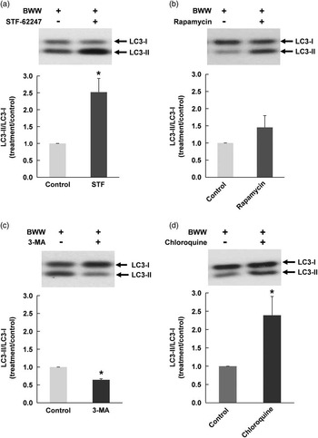
Once we had determined that LC3-I processing took place in stallion spermatozoa, our next objective was to assess whether the drugs used as autophagy activators (STF-62247 and rapamycin) and inhibitors (3-MA and chloroquine) were able to modify the LC3-II/LC3-I ratio. As shown in Figure 3a and b, both activators induced an increase in the LC3-II/LC3-I ratio, however, the increase observed after treatment with STF-62247 was significantly higher compared with untreated cells (containing dimethyl sulfoxide (DMSO) as vehicle). Incubation with 3-MA induced a significant decrease in the LC3-II/LC3-I ratio (Figure 3c), whereas chloroquine incubation for 3 h at 37°C induced a clear increase in the LC3-II/LC3-I ratio as compared with control samples (Figure 3d).
Activation and inhibition of light chain (LC)3 processing in equine sperm cells. Spermatozoa were incubated in the presence of STF-62247 (50 µM) (a), rapamycin (250 nM) (b), 3-MA (5 mM) (c) or chloroquine (50 µM) (d). After incubation, proteins were extracted and further resolved in SDS-PAGE. Immunoblotting was performed with an antibody that recognized both LC3-I and LC3-II proteins. LC3 processing was calculated as the ratio of LC3-II/LC3-I and data were normalized with respect to control (containing vehicle) in all treatments. Columns with asterisk indicate significant differences (P<0.05, n=5). BWW=Biggers, Whitten and Whittingham.

Effect of autophagy modulators on equine sperm viability
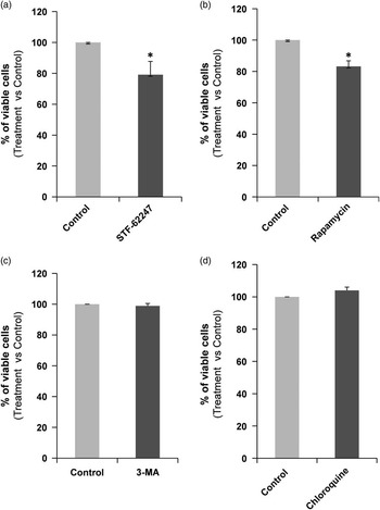
Once we observed that the LC3 ratio can be modulated in sperm cells, our next objective was to investigate whether this modulation may have a role in viability. Viability was assessed by flow cytometry after 3 h of incubation at 37°C in BWW in the presence of STF-62247, rapamycin, 3-MA or chloroquine (Figure 4a to d). The results revealed that incubation with STF-62247 and rapamycin reduced the percentage of viable cells (Yo-Pro-1 and ethidium negative) by 21±8.6% and 17±3.52%, respectively (Figure 4a and b). On the other hand, treatment with 3-MA and chloroquine did not modify the percentage of viable sperm cells (Yo-Pro-1 and ethidium negative) (Figure 4c and d).
Effect of modulation of light chain 3 conversion on equine sperm viability. Spermatozoa were incubated in presence of STF-62247 (50 µM) (a), rapamycin (250 nM) (b), 3-MA (5 mM) (c) or chloroquine (50 µM) (d). Viability was represented as a percentage of EthD-1 and YO-PRO-1 negative cells normalized with respect to control (containing vehicle). Columns with asterisks indicate significant differences (P<0.05, n=5).

Effect of 3-MA and chloroquine during refrigeration
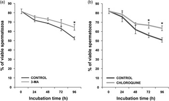
Percentage of viable cells during refrigeration in the presence of 3-MA was comparatively higher than control samples in all times studied, reaching statistical significance at 96 h (Figure 5a). On the other hand, refrigeration of sperm samples at 4°C in the presence of chloroquine induced an increase in sperm viability from 24 h on; being statistically significant at 72 and 96 h (Figure 5b), compared with control in all times tested.
Effect of inhibition of light chain 3 conversion on equine sperm viability during refrigeration. Stallion spermatozoa were cooled for 96 h in the presence of 3-MA (5 mM) (a) or chloroquine (50 µM) (b). Viability was represented as percentage of EthD-1 and YO-PRO-1 negative cells. Arrows with asterisks indicate significant differences with respect to control at the same time point (P<0.05, n=6).

Effect of 3-MA and chloroquine during freezing
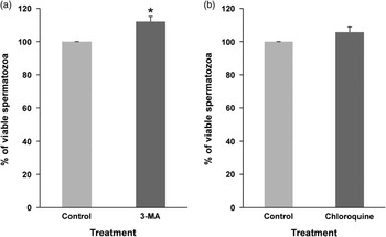
Stallion spermatozoa were frozen in the presence of 3-MA and chloroquine and stored at −196°C. After thawing, cell viability was measured by flow cytometry. The percentage of viable cells in the presence of both 3-MA and chloroquine was higher compared with controls samples, which contained only the vehicle (Figure 6a and b). However, the only statistically significant preservation of viability after thawing was observed when the spermatozoa were frozen in the presence of 3-MA (Figure 6a).
Effect of inhibition of light chain 3 conversion on equine sperm viability during freezing. Stallion spermatozoa were frozen at −196°C in the presence of 3-MA (5 mM) (a) or chloroquine (50 µM) (b). Viability was assessed after thawing and represented as a percentage of EthD-1 and YO-PRO-1 negative cells normalized with respect to control (containing vehicle). Columns with asterisks indicate significant differences (P<0.05, n=4).

Discussion
In somatic cells, the primary role of autophagy is to confer cell protection under stressful conditions. However, autophagy also has been linked to apoptosis, by facilitating, antagonizing or cooperating with it; serving either a pro-survival or pro-death function (Nikoletopoulou et al., Reference Nikoletopoulou, Markaki, Palikaras and Tavernarakis2013). Previous results from our group have demonstrated a relationship between LC3 processing and viability (Gallardo Bolanos et al., Reference Gallardo Bolanos, Miro Moran, Balao da Silva, Morillo Rodriguez, Plaza Davila, Aparicio, Tapia, Ortega Ferrusola and Pena2012; Bolanos et al., Reference Bolanos, Moran, da Silva, Davila, Munoz, Aparicio, Tapia, Ferrusola and Pena2014). Semen filtration and extenders used for cooling storage influenced the processing of LC3. Specifically, those samples with higher motility and viability had a higher LC3-II/LC3-I ratio. These findings may indicate that an ‘autophagy-like’ phenomena may exist in sperm and may be related to sperm survival. Having in mind these findings, in the present study, we investigated LC3 processing in depth during short- and long-term semen storage.
Fertility and quality of cooled semen is maintained for 24 to 48 h (Freitas-Dell’Aqua et al., Reference Freitas-Dell’Aqua, Monteiro, Júnior and Papa2012), whereas a longer storage duration causes damage to the cells affecting viability, motility, DNA repair, phosphatidylserine exposition and membrane integrity (Linfor and Meyers, 2002; Dondero et al., 2006; Freitas-Dell’Aqua et al., Reference Freitas-Dell’Aqua, Monteiro, Júnior and Papa2012; Bolanos et al., Reference Bolanos, Moran, da Silva, Davila, Munoz, Aparicio, Tapia, Ferrusola and Pena2014). Cooling semen at 4°C for 96 h induced an increase in the LC3-II/LC3-I ratio in a time-dependent manner; however, this increase began to be statistically significant from 48 h forward.
The reduced lifespan of spermatozoa during freezing and thawing procedures is due to increases in ROS, osmotic changes, DNA damage/fragmentation and high variations in the temperature (Watson, 2000); all of which are stressful conditions that have been described to stimulate autophagy in somatic cells (Kroemer et al., Reference Kroemer, Marino and Levine2010). In order to investigate which of these stresses are responsible for the observed increase in the LC3-II/LC3-I ratio during the freezing/thawing process, we subjected spermatozoa to different stressors. Oxidative stress was induced through incubation of spermatozoa with H2O2 (Ball et al., 2001), DNA damage/fragmentation was induced by exposure of spermatozoa to UV radiation (Dietrich et al., 2005; Pruski et al., 2009), osmotic stress was induced through incubation of spermatozoa in incubation media at different osmolalities and, finally, cold shock was mimicked through drastic changes in the incubation temperature. All of these conditions induced a significant increase in the LC3-II/LC3-I ratio, except for the decrease in temperature from 23°C to 4°C and the incubation in hypotonic solution, which caused a significant decrease in the ratio as compared with control. This difference in the LC3-II/LC3-I ratio after osmotic shock may be due to the fact that stallion spermatozoa have a higher tolerance to hypotonic v. hypertonic solutions in terms of viability or motility (Glazar et al., 2009; Burnaugh et al., Reference Burnaugh, Ball, Sabeur, Thomas and Meyers2010). As all of these situations are known to affect cell lifespan, these results indicate that an ‘autophagy-like’ phenomena may be activated in order to control sperm viability during semen storage.
Autophagy is thought to promote cell survival because the products resulting from degradation in the autophagolysosomes can be recycled and reused for macromolecular synthesis and ATP generation. However, autophagy has also been related to the death process and it is often referred to as type II programmed cell death (Bursch et al., 2000). Furthermore, crosstalk between autophagy and apoptosis has been shown to exist (Eisenberg-Lerner et al., 2009; Zhang et al., Reference Zhang, Jiang, Bi, Zhu, Zhou and Sha2012). Therefore, our next objective was to investigate whether autophagy was activated as a survival mechanism or, in opposition, was induced to produce cell death. To address this question, we used two modulators of autophagy known for their effects in the final steps in the process of autophagy: STF-62247 as an activator (Anbalagan et al., Reference Anbalagan, Pires, Blick, Hill, Ferguson, Chan and Hammond2012) and chloroquine as an inhibitor (Long et al., Reference Long, Yang, Southwood, Lu, Marciniak, Dunmore and Morrell2013) of autophagy. We used rapamycin as an mTOR inhibitor (Zhou et al., Reference Zhou, Luo and Huang2010), and 3-MA as a PI3K class III inhibitor (O’Farrell et al., Reference O’Farrell, Rusten and Stenmark2013), to modulate two proteins considered the major regulators of this process (Jung et al., Reference Jung, Ro, Cao, Otto and Kim2010; O’Farrell et al., Reference O’Farrell, Rusten and Stenmark2013). In stallion spermatozoa, STF-62247 and rapamycin induced a significant decrease in viability, whereas chloroquine and 3-MA had no effect on this parameter. Our results are in accordance with those published by Zhang et al. (Reference Zhang, Jiang, Bi, Zhu, Zhou and Sha2012), where autophagy-induced germ cell death after heat stress in mice.
Taking into account the results obtained, our final objective was to investigate whether the inhibition of this ‘autophagy-like’ process would improve the viability of spermatozoa in short- and long-term storage. Refrigeration of spermatozoa at 4°C in the presence of 3-MA and chloroquine had a positive effect on viability, resulting in 12% to 13% more viable cells after 96 h of refrigeration. Freezing and thawing in the presence of both inhibitors also increased the percentage of viable cells, whereas samples treated with 3-MA had 12% more viable cells than control (containing vehicle), chloroquine-treated samples had 5% more viable cells than control (not statistically significant).
Cell metabolism and biochemical reactions are more active at 37°C than at low temperatures; however, whereas chloroquine and 3-MA preserved cell viability at low temperatures, no effect on this parameter was observed at 37°C. This result may be explained by the stress that refrigeration and freezing cause on the sperm cell. As showed in the Figure 2, and same as occurring in somatic cells, stressful conditions cause an increase in the ratio of LC3-II/LC3-I. At 37°C, although we find an increase in the ratio between both proteins, the loss of viability and the stress suffered by the cells is minimal compared with low-temperature storage. Moreover, the increase in LC3-II/LC3-I at 37°C is lower than when stress is induced. Therefore, the significant effect that we observe on viability with the inhibitors at low temperatures might be due to a higher cell death in control samples. If the autophagy inhibitors are preserving or maintaining viability and the percentage of dead cells is higher in controls, the differences observed between the treated group and the control group would be higher.
Our results show that stressful conditions that occur during cryopreservation procedures, known to stimulate autophagy in somatic cells, induced an increase in the conversion of LC3-I to LC3-II. Modulators of autophagy, widely used in somatic cells, also modified the conversion of LC3 and cell viability. When autophagy inhibitors were added to the extenders in refrigeration and in freezing, the viability of the samples was improved. These results may indicate that an autophagy-related process could be regulating sperm viability during short- and long-term storage. However, a more detailed study of this pathway would be necessary to clarify whether autophagy, as described in somatic cells, exists in equine sperm.
In recent years, the scientific community has increased its efforts to understand the different stresses that stallion spermatozoa experience during freezing and thawing, in order to define potential methods that can be used to improve current cryopreservation protocols. The results of the current study could help to decipher the intracellular pathways involved in the regulation of equine sperm viability during cryopreservation.
Acknowledgements
This work was supported by Junta de Extremadura-FEDER (GR10010, PCE1002 and DE12006) and by Ministerio de Ciencia e Innovación-FEDER, grants BFU2011-30261 and AGL 2010-2075. The authors want to thank Ana M. Moreno Garcia and Mercedes Gomez Blazquez for their technical support.
References (Before 2010) given in Supplementary Material S1.
Supplementary Material
To view supplementary material for this article, please visit http://dx.doi.org/10.1017/S1751731116000240