INTRODUCTION
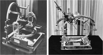
Radiosurgery (RS) developed by Lars LeksellReference Murphy, Chang and Gibbs 1 and later stretched to include fractionated stereotactic radiotherapy (SRT), are well-established modalities for the treatment of benign and malignant intracranial lesions including brain metastases, vestibular schwannomas (VS) and arteriovenous malformations.Reference Verbakel, Lagerwaard, Verduin, Heukelom, Slotman and Cuijpers 2 RS delivers an ablative dose and therefore requires a high accuracy in dose alignment, typically using 1 mm multileaf collimator margin to provide adequate dose coverage to tumour volume as well as avoiding injury to surrounding organs at risk.Reference Tanyi, Doss and Kato 3 Historically, the accuracy of stereotactic treatment has been highly dependent on patient positioning and immobilisation and achieved using invasive fixed frames (Figure 1). 4 These frames were, however, associated with an inaccuracy in the order of several millimetres, as well as patient discomfort, increased risk of infection and inhibition of fractionation.Reference Verbakel, Lagerwaard, Verduin, Heukelom, Slotman and Cuijpers 2 Over recent years there has been a growing interest in extracranial SRT which has boosted the development of high-precision image-guided radiotherapy (RT), and soon after several techniques for frameless RS/SRT emerged.
Example of invasive fixed frames: L-R Riechert Stereotactic Frame (1955) Todd-Wells Stereotactic Guide (1960). 4

One such system is the BrainLAB (Heimstetten, Germany) frameless mask combined with ExacTrac (ET) X-ray 6 degrees of freedom (6D) Novalis. The manufactures report a submillimetre precision, however, supporting evidence is limited.Reference Murphy, Chang and Gibbs 1 As the treatment site is not fully immobilised, intrafractional movement of the patient’s head inside the mask is a concern, and at this level of accuracy, even small movements can result in geographic miss of the target or overdosage of adjacent critical structures.Reference Guckenberger, Baier and Guenther 5 As such, frameless RS/SRT is not clinically feasible without appropriate target localisation and verification during treatment, therefore the aim of this review is to investigate localisation and verification accuracy performance of ET and Novalis Tx System.
THE EQUIPMENT
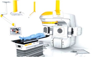
The Novalis Tx is a purpose-built linear accelerator used for the delivery of RS/SRT and is characterised by its flexibility in dose planning and delivery.Reference Lo, Yang, Huang, Chen, Liao and Huang 6 BrainLAB ET 6D system is comprised of ET and a 6D robotic couch controlled by a robotic tilt module mounted underneath the Varian couch top (Varian Medical Systems, Inc., Palo Alto, California, USA). This allows for 6D online setup corrections to be made before, and during treatment.Reference Badakhshi, Barelkowski, Wust, Budach, Boehmer and Graf 7 ET consists of two infrared (IR) cameras, one video detector and two dual diagnostic kV X-ray tubes mounted under the floor with two corresponding amorphous silicon digital detectors mounted on the ceiling (see Figure 2). 8 IR reflecting markers placed on the body (for extracranial sites) or, a cranial array attached to the treatment couch over the patients head, are located by the IR cameras and can be used to pre-position patients to the treatment isocentre and monitor the patient thereafter.Reference Chang, Wang, Ma, O’Daniel, Kirkpatirck and Yin 9 After positioning, a pair of oblique high–quality radiographic images are acquired and fused, using a 6D algorithm, with various sets digitally reconstructed radiographs obtained from simulation computed tomography (CT) dataset. This allows position variations and positioning shift parameters to be calculated in both three translational and three rotational directions.Reference Jin, Yin, Tenn, Medin and Solberg 10 Typical clinical settings for cranial cases to acquire X-ray images at Edinburgh Cancer Centre are 80 kV, 80 mA and 80 ms, however, this can vary with settings up to 100 Kv, 100 mA and 100 ms used elsewhere.Reference Gevaert, Verellen and Tournal 11 The couch shift and rotation parameters are transferred to the 6D robotic couch and once enabled, using the robotic enable bar on the control pendent, the couch moves to reposition the patient and thus corrects the setup error with the guidance of the IR system. 6D movements include standard left-right, craniocaudal and anteroposterior translations, and rotations around these axes, resulting in pitch, roll and yaw, respectively.Reference Gevaert, Verellen and Tournal 11 Repeat acquisition and fusion is required to verify patient position and correct for intrafraction motion before each treatment arc is delivered.
Diagram illustrating Novalis Tx with ExacTrac X-ray degrees of freedom (6D) System showing floor-mounted X-ray tubes, corresponding detectors and infrared (IR) camera. 8

OVERALL ACCURACY
As with many technological advancements in RT, increasing dependence is placed on the equipment and associated software. As a result radiographers rely heavily on the information output being accurate and fit for purpose. The manufactures of ET 6D report submillimetre accuracy, but with little clinical evidence it is important to review the current literature, therefore the current study seeks to investigate the overall accuracy and performance of ET and Novalis Tx System.
Calibration of the ET system
First, it is important to note that the accuracy of ET is highly dependent on regular calibration as the isocentre of the ET system is linked to the isocentre defined by the treatment room lasers. Laser alignment to the megavoltage isocentre accuracy is measured via Winston–Lutz tests and should record better than 0·5 mm with systematic and random deviations not 0·25 mm. A 10×10 cm2 flat BrainLAB calibration phantom with five IR markers is used to link the IR tracking system to the isocentre defined by the lasers. The stereoscopic X-ray system is calibrated using a second well-defined BrainLAB phantom which house internal structures and uses the IR tracking system to position the phantom relative to the isocentre.Reference Verbakel, Lagerwaard, Verduin, Heukelom, Slotman and Cuijpers 2 , Reference Ohtakara, Hayashi and Tanaka 12
Further to the deviation seen between external laser isocentre and the isocentre of the ET system is the deviation between the external laser isocentre (to which ET isocentre is linked to) and the Linac radiation isocentre. Tideman and CarlReference Tideman and Carl 13 evaluated the geometric accuracy of the isocentre of ET relative to the Linac radiation isocentre and found that the deviation between external laser isocentre and the isocentre of ET varied from 0·21 to 0·42 mm and the deviation between the external laser isocentre and the Linac radiation isocentre ranged from 0·37 to 0·83 mm, as is standard. A further three-dimensional (3D) deviation between the radiation isocentre and the ET isocentre ranged from 0·31 to 1·07 and although the authors state that results are complicated due to routine maintenance of the Linac, including laser calibration, the combined deviations are of an order that may have clinical relevance.Reference Tideman and Carl 13
Hidden Target Tests (HTT)
In order to determine the accuracy of ET for frameless intracranial RT, two main components need to be measured and evaluated. First, the accuracy of patient positioning and the linkage between this automated system and the Linac isocentre, and second; the accuracy of the imaging (verification) and couch corrections.
Evidence to support the manufactures claim is limited but a few studies have investigated the overall accuracy, involving both phantoms and patients. Although results from phantom studies give us a measurement of the random error of couch positioning, data from patients provide a more accurate estimation of the random imaging matching accuracy as each patient is unique and therefore provides different anatomical matches as well as measuring intrafractional patient motion.
From the literature, it is evident HTT are the gold standard when testing for localisation accuracy whereby hidden markers are randomly positioned within a phantom and the automated couch positioning preformed according to ET. The deviation of the IR markers with the reference to the Linac’s isocentre is then measured indicating the accuracy. The importance of this test was highlighted in a study dual phantom/patient study by Verbekel et al.Reference Verbakel, Lagerwaard, Verduin, Heukelom, Slotman and Cuijpers 2 who recorded an initial systematic deviation of 1·5±0·29 mm in the vertical direction based on 31 repeated measurements. This large deviation was due to the deterioration of the reflective surface of the IR spheres and only eliminated after recalibration with a new phantom and subsequently reduced to 0·3 mm based on five measurements. Although the phantom aspect of the study is based on low numbers, their patient study was based on 46 patients over 135 fractions and demonstrated an accuracy of 0·3 mm in each direction measured from setup and verification images which is well within a stereotactic tolerance.Reference Verbakel, Lagerwaard, Verduin, Heukelom, Slotman and Cuijpers 2 Similar results have been achieved by several studies. In a 5-year experience performance study of ET (March 2005 to March 2010) an excellent result of <1 mm was recorded for positional accuracy in a total of 94 patients. Deviations in head position within the mask required shifts of up to 3 mm in 26% which highlights the need for image verification before each treatment arc, something that is now common practice.Reference Jani, Weinsyein and Shah 14 In further support of this observation, a 2012 quantitative analysis of 849 images was carried out by Lesiuk et al.Reference Lesiuk, Spencer, Chan, Voroney and Lau 15 with the objective to measure setup and intrafraction variability using ET. The averaged results were a setup offset of 1·98 mm and post-treatment offset of 0·66 mm. Although no consideration was given to intrafraction motion during treatment, their results show patient position can be reproduced within 1·0 mm.
In a theoretical world a higher positioning accuracy is achieved. Verellen et al.Reference Verellen, Soete, Linthout, Van Acker, De Roover and Ving-Hung 16 assessed the uncertainty of the IR tracking device on 40 repeated phantom observations, using a comparison made with the gold standard HTT. The average deviation between the IR tracking measurements of the iso-position and the actual position of the HT with respect to the treatment isocentre was 0·24, 0·45 and 0·49 mm in the vertical, longitudinal and lateral directions, respectively, and a deviation vector of 0·7°. In a separate phantom-based study by Gevaert et al.,Reference Gevaert, Verellen and Tournal 11 the overall accuracy was tested again using HTTs repeated ten times using a phantom, and then furthered his investigation by repeating the HTT for seven different locations in the brain to test its accuracy in situations with varying amounts of bony anatomy. Their study showed a high detection accuracy for initial positioning as well as a subdegree accuracy for subsequent verification. This study is more relevant to a clinical situation as it takes into account varying anatomical tumour locations. Wurm et al.Reference Wurm, Erbel and Schwenkert 17 repeat the precise results achieved using ET, recording an average setup error of 0·31 mm for translations and 0·26° for rotations after verification and correction. These figures support the manufactures claims of the high accuracy of positioning achieved by ET.
Overall survival
Although the above figures are positive and demonstrate ET’s accuracy, it is important to note patient outcomes. In a 4-year retrospective study involving 23 patients, 26 VS (three patients suffered from neurofibromatosis Type II) were treated with Novalis TX ET X-ray 6D. After a follow-up over a range of 17–87 months ultimate radiological tumour control rate was 88·5% which is comparable with 87·3–95·98% growth control rate via Gamma Knife in previous reports. Encouragingly, 20 patients retained their pre-irradiation hearing functionReference Lo, Yang, Huang, Chen, Liao and Huang 6 and is comparable with results of study involving 29 patients who underwent partial surgical resection followed by Gamma Knife surgery demonstrating 22/29 patients retaining same levels of function hearing.Reference Iwai, Ishibashi, Wantanabe, Uemura and Yamanaka 18 Ultimately, a direct comparison study would give a more robust conclusion, however, the results demonstrate ET as a reliable tool. As it is currently unclear if SRT stabilises hearing or actually accelerates hearing loss, further comfort in using SRT as a treatment modality for VS is found in a retrospective study by Elliott et al.Reference Elliott, Hebb, Walling, Morris and Bance 19 who found no difference between surgery (n=98) and SRT (n=25), albeit patient numbers are low for the SRT arm of the study.
Overall, few clinical studies have made direct comparisons between Linac-based and Gamma Knife stereotactic radiosurgery (SRS) due to the complex nature of the difference in dose distribution characteristics and cost considerations which influence the use of each modality.Reference Mathis 20 However, encouragingly, Pinkham et al.Reference Pinkham, Whitfield and Brada 21 found that overall survival in patients with brain metastases were equivalent when comparing Gamma Knife against Linac-based treatment.
INFLUENCING FACTORS
Value of 6D and secondary counteractive movement
The value of 6D can be questioned as rotational movement of the couch has the potential of introducing secondary counteractive patient motion. Even with tilt and roll rotations commonly limited to 3·0° for safety reasons, large couch movements are still required because the centre of rotation is located at the foot end of the treatment couch. For example, a tilt rotation of 2·5° results in a vertical movement of the treatment couch of about 4·5 cm at isocentre level. This being said, results of a large retrospective study (n=104), 6D setup corrections did not induce significant secondary motion in the majority of patients.Reference Linthout, Verellen, Tournel, Reynders, Duchateau and Storme 22
In addition, several studies conclude that better translational accuracy is achieved with a 6D algorithm as well optimal positioning when rotations are corrected for.Reference Jin, Ryu and Faber 23 , Reference Verellen, Soete and Erbel 24 Yin et al.Reference Jin, Ryu and Faber 23 studied the localisation accuracy of 6D and 3D fusion methods for cranial and spinal lesions. Results showed up to a 4 mm difference between each method. When target coverage and the volume of healthy tissues irradiated was analysed by Gevaert et al.Reference Gevaert, Verellen and Engeld 25 the mean conformity index was increased for the 6D plans in comparison with 4D plans, and that the entire target volume was covered by the prescribed dose. Furthermore, it appears that it is a fairly simple and effective method, fitting well within a clinical setting with no additional workload.
Reliance on bony anatomy
Concerns have been raised over the appropriateness of using bony anatomy for image guidance because of the time period between planning and treatment.Reference Guckenberger, Baier and Guenther 5 Changes in tumour shape or size may not be accounted for resulting in a possible geographical miss. Guckenberger et al.Reference Guckenberger, Baier and Guenther 5 carried out a small study with 18 patients with 20 brain metastases (ten patients with 17 metastases treated in a single fraction and two patients with three lesions with fractionated SRT), investigating the accuracy of cranial SRT with image guidance based on bony anatomy and evaluated whether the position of brain metastases remains stable relative to the bony anatomy. Their results showed excellent correlation (p<0·01) between bone and soft tissue registration in all three directions. For left/right, superior/inferior and anterior/posterior directions the correlation coefficient (r) was 0·94, 0·93 and 0·88, respectively. In addition, the mean difference between bone and soft tissue match was <1 mm in all three directions and the 3D difference between isocentre based on bone and based on soft tissue registration was 1·7±0·7 mm. All of which indicate the safe reliance of bony anatomy for image guidance. A limitation of this study for the purpose of this review is that it did not involve ET. This is because a soft tissue match is required for study evaluation which can only be achieved through use of cone beam computed tomography (CBCT). Although further investigation is required, results can be applied to all image guidance, providing the system itself is accurate.
COMPARISON WITH CBCT
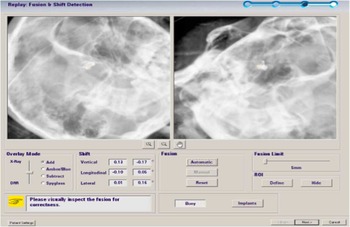
CBCT is often favoured in image-guided RT due to its ability to create a 3D view with better visualisation of anatomical structures and soft tissues over 2D imaging. However, in a RS/SRT setting its practise is limited by several practical concerns including a relatively high concomitant patient dose and long acquisition time.Reference Chang, Wang, Ma, O’Daniel, Kirkpatirck and Yin 9 In a comparative study by Ma et al.Reference Ma, Chang, Wang, Wu, Kirkpatrick and Yin 26 the setup accuracy between ET and CBCT was compared by determining the residual setup errors after initial corrections were determined and corrected for via ET. Their measurements were essentially consistent, leading the authors to conclude that ET provides accurate positioning and represents a potential alternative to CBCT. Potential sources for the negligible discrepancies include patient motion between image acquisition, differences in image fusion algorithms, visibility of anatomical structures and a discrepancy of geometric accuracy. Although the authors have provided an in-depth analysis of their results, the study is limited by their modest number of patients used (18). In contrast, a residual translational and rotational discrepancy of 1·3 mm and 0·8° was recorded in a separate comparison study comparing ET with CBCT involving larger patient numbers (100). Due to the precise nature of RS/SRT this is a considerable discrepancy.Reference Chang, Wang and Ma 27 A potential reason for the discrepancy could be the nature of the radiographs taken at oblique angles rather than the standard orthogonal parameters which can be challenging for users to interpret (Figure 3)Reference Tanyi, Casey, Kato, Marquez, Laub and Fuss 28 . Further conclusions can be drawn from a large study involving 245 paired clinical ET/CBCT acquisitions post pre-positioning alignment via automatic detection of IR marker configuration on each patient’s thermoplastic mask using ET system. Image guidance and repositioning was based on online 6D stereoscopic X-ray imaging (ET) and online 4D CBCT, and subsequently offline with 6D CBCT. For the purpose of their study, CBCT was considered the gold standard for data comparison. A slight difference in residual errors was found between ET and 4D CBCT in vertical, longitudinal and lateral translations; 0·40±0·72 mm, 0·24±0·56 mm and −0·06±0·67 mm, respectively. In addition, rotational setup differences between ET and CBCT were minor, however, due to the precision required, not negligible with offline rotational assessments in lateral, longitudinal and vertical axis being 0·08±0·70°, −0·06±0·46° and 0·05±0·38°. The results of this large study indicate comparable shifts between the two modalities giving confidence in ET for setup and image guidance.Reference Tanyi, Casey, Kato, Marquez, Laub and Fuss 28
Example of cranial oblique images using ExacTrac taken at Edinburgh Cancer Centre.

ADDITIONAL BENEFITS
In addition to ET’s ability for automated positioning and quantitative rotational errors, it offers the important benefit of reduced concomitant dose to the patient. When comparing concomitant dose given to a patient over a fractionated course by ET with that of CBCT, a total dose of 2·0 and 14·0 mSv (1 mGy=1 mSv), respectively, is absorbed by the patient.Reference Jin, Ryu and Faber 23 Further to the reduction in concomitant dose is faster set and image acquisition time of ET. The time taken to acquire a CBCT is in the region of a few minutes in comparison to just a few seconds for ET, making ET the more superior and efficient image guidance modality for SRS/SRT.
Despite the obvious benefits from frameless SRS/SRT, thermoplastic masks cannot fully eliminate intrafraction motion thereby introducing a varying degree in magnitude and probability of patient movement between each arc dose delivery, as such intrafraction motion may significantly impact target coverage.Reference Hoogeman, Nuyttens, Levendag and Heumen 29 ET 6D and Novalis Tx System also offers motion tracking, real-time tracking and online adjustment. Tanyi et al.Reference Tanyi, Casey, Kato, Marquez, Laub and Fuss 28 recorded ET monitored intratreatment patient motion-based errors of 0·04±0·58 mm (lateral), 0·00±0·73 mm (longitudinal) and −0·19±0·53 mm (vertical), demonstrating the importance of ETs ability in intrafraction monitoring and online adjustment, thereby significantly contributing to the precision of frameless intracranial SRS/SRT.
CONCLUSION
Stereotactic treatment for patients with intracranial lesions have undergone major technological advancements since its conception. The need for a more patient friendly frameless technique coupled with growing interest in image-guided RT has seen the development of BrainLABs frameless mask and ET X-ray 6D Novalis Tx System. The manufactures report submillimetre accuracy, and although evidence within the literature is limited it supports these claims. However, limitations do exist and are not negligible. Most pertinent is the need for regular calibration to prevent systematic errors and potential geographic miss. Overall, due to ET’s additional benefits, including reduced concomitant dose and faster imaging time, ET is the superior image guidance modality for RS/SRT in the treatment of intracranial lesions.
Acknowledgements
The authors thank to Edinburgh Cancer Centre and Joanna Henderson, Advanced Practitioner in SRT, for giving the authors support and time for writing this review for publication.